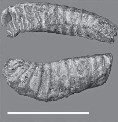
The Woolly Mammoth, Mammuthus
primigenius, is thought to have diverged from the earlier Steppe Mammoth Mammuthus trogontherii in northeast Asia
around 700 000 years ago and by 200 000 years ago spread across Asia and into
Europe and across the Bering Strait into North America, where they hybridized
with the Columbian Mammoth Mammuthus columbi.
They are believed to have been the last surviving species of Mammoth,
persisting to as late as 3700 years ago on Wrangel Island, off the northeast
coast of Siberia (with some claims of even more recent specimens). As a
widespread and apparently numerous species living in the recent past they have
left an extensive fossil record, primarily of isolated teeth and bones, but
also including a number of mummified and frozen specimens, trapped within the
Arctic permafrost. These have allowed a number of detailed anatomical and
molecular studies of the Woolly Mammoth, although many were excavated in the
nineteenth and early twentieth centuries, and have subsequently degraded due to
poor storage facilities, preventing the application of the most modern methods
to these specimens.
In a paper published in the Journal of Paleontology in July 2014, a
team of scientists led by Daniel Fisher of the Museum of Paleontology at the
University of Michigan describe the results of a series of X-ray Computed
Tomography studies of two recently discovered Woolly Mammoth calves from permafrost
in the Siberian Arctic.
The first specimen, named Lyuba, was found in May 2007 by on a bank
of the Yuribei River on the Yamal Peninsula, where it is believed to have been
deposited by an ice-melt flood the previous spring. When found it was almost
intact, having lost only its hair and nails, however it was transported to a
nearby village where it was partially scavenged by domestic Dogs, losing part
of the tail and right ear, before being acquired by the Shemanovskiy Museum and Exhibition Center in Salekhard in the Yamalo-Nenets Autonomous Okrug of the
Russian Federation. Here the specimen was found to be partially dehydrated,
having lost approximately half of its expected water content, and found to be
female by examination of the urogenital tract, and subsequent DNA analysis.
Lyuba’s body was found to have been acidified, probably by
colonization of the corpse by lactic acid-producing Bacteria, leading to the
degradation of much of the connective tissue (collagen). The facial region was
found to contain numerous masses of vivianite (hydrated iron phosphate).
Examination of Lyuba’s teeth was able to find a neonatal line (produced at the
time of birth) followed by 30-35 daily growth increment lines, setting her age
at the time of death at 30-35 days. She appears to have been healthy and well
fed at the time of her death. An isotopic analysis of the age of the body
suggested an age of about 41 800 years.
Lyuba was subjected to a full CT scan, then examined endoscopically
through two holes drilled in her left side. She then underwent two necropsy
sessions, in which her body was thawed, partially dissected then refrozen. In
the first the teeth were removed from the left side of the face, and portions
of the large and small intestine were also extracted. In the second the pleural
and abdominal cavities were examined. At this point it was determined by the Shemanovskiy
Museum that the body would need to be treated chemically to prevent further
decomposition, and allowed to dehydrate fully.
Fisher et al. were unable
to access the original CT scans of Lyuba made at the Shemanovskiy Museum before
chemical treatment. However they were able to take the body to the GE Healthcare Institute in Waukesha, Wisconsin, where it was possible to scan the
Mammoth’s head and neck and a portion of the right forelimb and (separately)
her pelvic region and left hind limb (the entire Mammoth was too large to fit
into the scanning equipment at Waukesha). A complete scan of the Mammoth’s body
was later made at the Nondestructive Evaluation Laboratory of the Ford Motor Company in Livonia, Michigan (as far as Fisher et al. are aware this is the first time a Mammoth has undergone a
complete full-body scan in this way). Unfortunately industrial scanners like
the one at Livonia are slower than medical models, and only seventeen hours
were available for the scans on Lyuba, so the resolution achieved was not as
high as hoped. Micro CT scans of the extracted teeth were carried out at the
University of Michigan Dental School in Ann Arbor, Michigan.
Lyuba was already known to have significant sediment lodged within
her trunk. Her oral cavity (mouth) was also filled with sediment, although this
matched the bank sediment at the site by the Yuribei River where she was found,
so it is assumed that this was emplaced during the recent transportation of the
body. The second necropsy carried out on Lyuba was able to determine that her
lungs had collapsed, and that larger bronchial cavities were filled with a
bright blue powder identified as vivianite with some clay minerals. The CT
scans revealed that Lyuba’s trachea was also filled with material which had the
same density as the material in both the trunk and lungs, suggesting that all
three are the same.
Fisher et al. suggest that
Lyuba died after inhaling mud which blocked her trachea and the front part of
the bronchial system in her lungs, preventing her from breathing and leading to
suffocation; this matches the distribution of sediment seen in the trachea and
lungs and the collapse of the other lung tissue (in the alternative scenario,
drowning, sediment would have spread throughout the lungs, which would not have
collapsed. It is impossible to assess where this happened, as the body had been
transported prior to its discovery, but fine-grained vivianite is typical of
lake-bottom sediments.
Aspirated sediment in the Mammoth calf Lyuba, sediment
with a radiodensity in the range of bone can be traced from the pharyngeal
region,through the trachea, and into the lung bronchi (from the Ford scan).
Fisher et al. (2014).
The first vivianite detected on Lyuba was found on her left side,
which was the side she was laying on when discovered, wher a number of circular
pit where filled with bright blue material. This is interpreted to have been
caused by fungal growth, which has previously been documented on a number of
other specimens of similar age, including the ‘Blue Babe’ Steppe Bison mummy
and the Tyrolian iceman ‘Otzi’. However vivianite was also found in nodules
throughout the facial region of Lyuba, and within the diaphyses of her long
bones (the growing ends of the long bones in a young mammal), where a better
explanation was needed.
Fisher et al. suggest that
this is connected closely to the manner of Lyuba’s death. If she did asphyxiate
on lake bottom sediments, then it is likely that she was in a cold, wet
environment suffering from oxygen deprivation immediately prior to death.
Mammals in such circumstances have a ‘diving reflex’, whereby blood is
withdrawn from the skin and extremities, but the supply increased to the face
and brain, thereby keeping the animal alive as it tries to escape its
predicament. Vivianite is an iron-phosphorous mineral, and needs a supply of
both elements to form. In the case of the facial nodules the iron comes from
blood pumped to the head as Lyuba struggled for her life, while the phosphorus
comes from the dissolution of bone tissue by the action of lactic-acid forming
Bacteria after her death. In the case of the long bone diaphyses the iron would
have come from bone marrow, which is particularly rich in iron in these areas
of bone growth.
Larger elements of Lyuba’s appendicular skeleton
(without maniand pedes) extracted from the Ford scan: (1) radiodense nodules
withindeveloping trabecular spaces in long bones are probably vivianite
crystalsformed from bone-derived phosphate and blood- and marrow-derived
iron;bones show radiodensity disparity between diaphyses and epiphyses;
rightlateral aspect, hind limbs on left (right ahead of left) and forelimbs on
right(right ahead of left); (2) left humerus in anterior aspect,diaphysis
(green) segmented separately from epiphyseal ossifications(labeled); (3)
segmented radiodense nodules show through cortical bone of diaphysis (humerus unsegmented
in this image so that nodules show through);common scale for 2 and 3. Fisher et al. (2013).
Lyuba’s ribcage had been laterally compressed (squashed from the
sides) after death, with the greatest amount of deformation occurring on the
left side. Her backbone is intact, and appears to be in life position. Her
skull is also slightly deformed and compressed, which may have been aided by
partial dissolution of the bone by lactic acid.
The second Mammoth calf, Khroma, was found preserved in situ,
upright in permafrost near the Khroma River in northern Yakutia in October
2008, and subsequently excavated and shipped to the Mammoth Museum at the
Institute of Applied Ecology of the North at North-East Federal University in Yakutsk
in the Sakha Republic. At the time of discovery it had been partially eroded
from the sediment, and parts of the head trunk and shoulders scavenged by
Ravens and (possibly) Arctic Foxes. Thus, while in generally good condition,
the body had lost much of the trunk, the flesh from the head, the fatty tissue
from the back of the neck (where a fatty hump would be expected) and the heart
and lungs. DNA analysis showed Khroma to be female, which was subsequently
confirmed by CT scanning of the urogenital tract. A necropsy revealed she had
abundant subcutaneous fat, and undigested milk in her stomach. It was not
possible to determine the age of Khroma isotopically, suggesting she died more
than 45 000 years ago.
Khroma was also subjected to two rounds of CT scanning, first at the
Centre Hospitalier Universitaire de Clermont-Ferrand, then at the CentreHospitalier Emile Roux in Le Puy-en-Velay, both in France. Her teeth were also
extracted and micro CT scanned at the at the University of Michigan Dental
School in Ann Arbor, Michigan.
Examination of Khroma’s teeth enabled the detection of a neonatal
line, followed by at least 52 daily growth increment lines, although these were
somewhat unclear, leading Fisher et al.
to conclude that she was 52-57 days old at the time of her death.
Khroma’s age, determined from her right dP3: (1) right
image shows lingual aspect of a slab cut from the right dP3, anterior to the
left; (2) enlargedview of the anterior root in 1; the dark line running
vertically, parallel to the pulp cavity surface, is the neonatal line (NnL); (3)
photomicrograph of a thin sectionof the anterior root, with pulp cavity (pc)
surface at right, neonatal line (NnL) at left, and 52 or more daily dentin
increments (small white dots) between them; (4) enlargement of area designated
in (3), showing increments for last 18 days of life. Fisher et al.estimate Khroma’s age at death as
52–57 days. Fisher et al. (2013).
Elephants walk on a thick pad of fat with a thick layer of skin over
it. The distal phalanges (bones at the tips of the toes) are believed to help
distribute weight evenly within this structure, though exactly how this works
is unclear. In African Elephants only the third and fourth toes have ossified
(bone) distal phalanges, with these elements in the other toes being cartilage,
while in Asian Elephants the distal phalanges of toes two, three and four are
ossified. Attempts to resolve the number of ossified distal phalanges in
Mammoths have been, to date, unsuccessful, largely as the limb tips are seldom
preserved intact.
Khroma was preserved with her feet undamaged, making analysis of the
ossification state in the distal phalanges a possibility. Unfortunately no
ossified distal phalanges could be detected, nor was it possible to detect ossification
in the metatarsal-phalangeal sesamoid (known to ossify in Woolly Mammoths),
suggesting that Khroma was too young at the time of death for complete
ossification of all the bones in the foot to have occurred. However it was
possible to detect the synovial capsules in the foot (fluid filled capsules
that form between bone-bone joints in Mammals, but not bone-cartilage joints),
as these had lost their fluid but filled with (distinctly less dense) air. This
suggests that the second, third, fourth and fifth distal phalanges may have
ossified.
Khroma’s left hind foot: (1) lateral aspect, anterior
to left,showing nucleation sites and epiphyseal ossification center on
calcaneumtuber at right; (2) anterior aspect; (3) anterior aspect, synovial joint
capsules ondigits, light blue; (4) anterior aspect, non-terminal joint capsules
light blue andterminal joint capsules purple. Abbreviations: Ast., astragalus;
Cal., calcaneum; Cub., cuboid; Ect., ectocuneiform; Ent., entocuneiform; IP,
intermediate phalanx; Mes., mesocuneiform; MT, metatarsal; MT I, metatarsal I
(red); MT V, metatarsal V (blue diaphysis); PP, proximalphalanx. Separate
scales for 1 and 2; common scale for 3 and 4. Fisher et al. (2013).
Khroma’s skeleton was more damaged than Lyuba’s, with some of the
ribs broken by recent scavenging, and a break between the seventh and eighth
thoracic vertebrae, which occurred at about the time of death. Khroma is
thought to have died of sediment inhalation in a similar way to Lyuba, but this
break suggests a much more violent setting (the backbones of even very small
Elephants are quite robust). Fisher et
al. suggest that she may have been caught in a mud flow or bank collapse,
which caused a significant lateral impact as well as burying her and preventing
breathing.
Khroma’s ribcage in right lateral aspect (interruption
of rib sequence caused by break in vertebral column). Fisher et al. (2013).
See also…
Fossil Dwarf Elephants are known from a number of small islands around
the world; this is not altogether surprising, dwarfism is common in
populations of animals cut of on small islands (as is giantism). Animals
in such environments often need to adapt to different niches to those
they inhabit on larger land-masses, but are able to do so due to lack of
competition, since there...
Nitrogen has two stable isotopes,
Nitrogen-14 (¹⁴N) and Nitrogen-15 (¹⁵N), which have different atomic
weights, but identical chemical properties, and can be incorporated into
identical compounds by organisms. Nitrogen fixing organisms
(diazotrophs) tend to fix Nitrogen isotopes in the ratio found in the
atmosphere, but each time the Nitrogen is passed from one...
Elephants have a long and well understood fossil record, but this can
usually only tell us about the physical attributes of Elephants, i.e.
their...
Follow Scieny Thoughts on Facebook.


