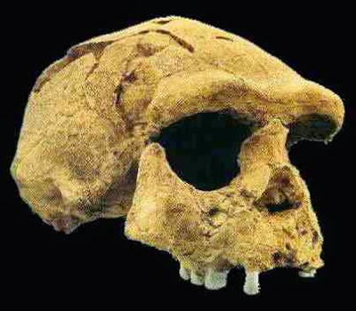
In the 1970s and 1980s a collection of Hominin bones and teeth were
unearthed in the Longtan Cave at Hexian in Anhui Province in eastern China. The
bones of these remains have been extensively studied, and assigned to the
species Homo erectus, though
palaeoanthropologists have differed in their opinions as to whether these
remains were most similar to those from Zhoukoudian in China or Java in
Indonesia. The depositional layer which produced the teeth has been dated to
about 412 000 years ago, and contains traces of fires and fire damaged animal
bones, animal bones with cut marks and tools made from bones, antlers and
teeth, but no stone implements. The fauna and pollen assemblage at this layer
imply a subtropical climate.
In a paper published in the journal PLoS One on 31 December 2014, Song
Xing of the Key Laboratory of Vertebrate Evolution and Human Origins at the
Institute of Vertebrate Paleontology and Paleoanthropology of the Chinese
Academy of Sciences, María Martinón-Torres and José María Bermúdez de Castro of
the National Research Center on Human Evolution, Yingqi Zhang, also of the Key
Laboratory of Vertebrate Evolution and Human Origins, Xiaoxiao Fan of the Hexian
Museum of Anhui Province, Longting Zheng of the Anhui Museum, Wanbo Huang of
the Chongqing Three Gorges Institute of Paleoanthropology at the China Three
Gorges Museum and Wu Liu, again of the Key Laboratory of Vertebrate Evolution
and Human Origins, describe the results of a study of the teeth of the Longtan
Cave Hominins, in which they were compared to a range of other teeth from
ancient and modern populations of Australopithecus
and Homo from Africa, Europe and
China.
The first tooth described is a right upper third premolar (formerly
described as a right upper fourth premolar). The tooth is in generally good
condition, although it has some cracks in its enamel on the crown surfaces.
There is a large fragment alveolar bone attached to the root. The cusps of the
tooth have been flattened by wear, making the cusp pattern unclear. The crown
is oval and slightly asymmetrical, the cusps are joined by a crest that
transverses the central groove. The tooth has one lingual root and two buccal
roots, all of which are quite robust; the mesiobuccal, distobuccal, and lingual
roots are 19.26, 17.88, and 18.96 mm long. The pulp horns are blunt and low,
with a total pulp cavity volume of 82.67 mm3.
Right upper third premolar (PA832) (o: occlusal, B or
b: buccal, M or m: mesial, L or l: lingual, d: distal). 3D reconstructions of
the dentine surface (I–III) and the pulp cavity (IV) obtained from micro-CT scanning.
The arrow points to the transverse crest. Solid lines indicate the bifurcation
of the essential crest. Dotted lines indicate the mesial (I) and distal
accessory ridges (II) and the well-demarcated central ridge onthe buccal
surface (III). Sagittal sectional plane of PA832 (V). Cross-section of PA832
(VI) at the levelindicated by the red line. (I, II, III, IV, V, and VI) are not
scaled. Xing et al. (2014).
The second tooth described is a left upper third premolar, with a
complete crown but a broken root. The cusps of this tooth have been flattened
exposing the dentine, and there are wear facets on the mesial and distal
surfaces of the crown. The exposed dentine of the buccal cusp shows a pronounced
mesial vertical furrow and a weaker distal furrow running towards the apex of
the cusp. This tooth does not have a crest transversing the central groove.
Left upper third premolar (HXUP3) (o: occlusal, B or
b: buccal, m: mesial, l: lingual, D or d: distal). The occlusal (I) and buccal
(II) views of the dentine surface reconstructed from micro-CT scanning. Solid
lines indicate the bifurcation of the essential crest. Dotted lines indicate
mesial and distal accessory ridges. The arrow points to the vertical groove on
the buccal surface. (I and II) are not scaled. Xing et al. (2014).
Transverse grooves crossing the central grooves of upper third
premolars are common in Australopithecus
and early African Homo, but become
increasingly rare in later members of the genus Homo. However it is relatively more common in Early Pleistocene
teeth from East Asia and Middle Pleistocene teeth from Europe. The strong
medial groove seen in the dentine of the buccal cusp of the left premolar is
typical of Early East Asian Hominins, and seen in some Middle Pleistocene
specimens as well, including those from Zhoukoudian. This groove is completely
absent from European Middle Pleistocene specimens, Neanderthals and all modern
Humans. Such grooves are also seen in Homo
ergaster specimens from the Late Pliocene and Early Pleistocene of East
Africa, though it is less well developed in these. The asymmetrical oval
outline of the crowns of these teeth is also seen in Australopithecus and early African Homo including Homo ergaster,
as well as in the Early Pleistocene remains from the Sangiran Dome in Java, and
the middle Pleistocene remains from Zhoukoudian and Chaoxian in China; this
differs from the more symmetrical crowns seen in modern Homo sapiens, which is also seen in a Middle Pleistocene upper
third premolar from in South China.
The triple root of the right tooth is more unusual; this is
otherwise only known from Australopithecus
and early Homo in Africa and in Early
Pleistocene specimens from Java. The robust roots of the tooth are also shared
with the Java specimens, with narrow, tapering roots typical of African and
European Homo specimens.
From left to right, 3D reconstruction of the upper
right premolar from Hexian from micro-CT scanning, Zhoukoudian PA67,
Zhoukoudian 19, and Sangiran S7–35 (MB: mesiobuccal, B: buccal). Xing et al. (2014).
The upper right premolar is also exceptionally large, being wider
than all specimens of Homo ergaster,
Early Pleistocene Hominins from Europe or the specimens from Chaoxian or Panxian
Dadong, and towards the upper end of the size range encountered in Early and
Middle Pleistocene Hominins from East Asia, Middle Pleistocene Hominins from
Europe and Neanderthals. It is longer than almost all known Hominin third
premolars, except some Australopithecusand
early Homo specimens from East
Africa.
The third tooth described is an upper left first premolar. This is
generally well preserved, although the buccal roots are broken off. The tooth
is approximately square in outline. The upper surface is heavily worn, with
large, oval-shaped wear facets, which have largely obliterated the upper
surface of the tooth, though the presence of the four main cusps can still be
detected by the grooves in the exposed dentine. This dentine surface is heavily
crenulated, with crests and ridges on all cusps; the hypocone is large. The lingual
root is very robust, divergent and flattened lengthways. It is 14.72 mm in
length.
Left upper first molar (o: occlusal, B or b: buccal,
m: mesial, l: lingual, D or d: distal). Sagittal sectional plane of PA836 (I).
The occlusal (II) and lingual (III) views of dentine surface reconstructed from
micro-CT scanning. Dotted lines point to the expression of mesial accessory
ridges (II) and a Carabelli’s trait (III). (I, II, and III) are not scaled.
Xing et al. (2014).
The hypocone of the first premolar has tended to decrease throughout
the history of the Hominins, with large hypocones common in Australopithecus, and most Homo specimens from the Early and Middle
Pleistocene, including those from East Asia, although the specimens from Zhoukoudian,
which are close to the Longtan Cave Hominins both geographically and
stratigraphically, have smaller hypocones. The outline of the tooth is similar
to that of other East Asian Pleistocene specimens, and different from the more
rhomboidal outlines seen in teeth from Pleistocene specimens from Europe and
Africa, and from the more square teeth of modern Homo sapiens. The robust divergent root of the tooth is also
typical of Early and Middle Pleistocene specimens from East Asia, and differs
from those of Early Pleistocene Europeans, which diverge little if at all.
However in modern Humans this trait is quite variable, with the Longtan tooth
falling within the range of variation.
This tooth is also large, being wider than most known first molars
of Middle Pleistocene Hominins from Europe or East Asia, Neanderthals or early
modern Humans, though its width is typical for Homo ergaster and Early Pleistocene Hominins from East Asia. Its
length is also typical for Early and Middle Pleistocene East Asian Hominins,
Middle Pleistocene European Hominins, Neanderthals and early modern Humans,
though it is longer than specimens from any known Homo ergaster and Early Pleistocene European Hominins.
The fourth tooth described is a left upper second molar. The roots
of this tooth are completely missing and the tooth is broken along its cervical
line. The cusps have been worn down and flattened, but not enough to expose the
dentine. The outline of the tooth is trapezoidal, with the distal portion being
narrower. There are four main cusps and a smaller fifth cusp visible,
transversed by a series of grooves, the hypocone is medium-sized. Xing et al. calculate that the average enamel
thickness of the tooth is 1.30 mm, but would have been 1.51 mm prior to wear.
Left upper second molar (o: occlusal, B or b: buccal,
m: mesial, l: lingual, D or d: distal). The occlusal (I) and lingual (II) views
of dentine surface reconstructed from micro-CT scanning. Dotted lines point to
the mesial accessory ridges (I) of the occlusal surface and to the expression
of a Carabelli’s cusp on the lingual face of the crown (II). (I and II) are not
scaled. Xing et al. (2014).
The fifth tooth is a right upper second molar, also missing its
roots, and partially broken along its cervical line. Again this tooth had been
worn down and flattened, but not enough to expose the dentine. This tooth is
also trapezoidal, with four main cusps, though there is no sign of a fifth
cusp.
Right upper second molar (o: occlusal, B or b: buccal,
M or m: mesial, l: lingual, d: distal. I and II: The occlusal (I) and lingual
(II) views of dentine surface reconstructed from micro-CT scanning). Dotted
lines point to the mesial accessory ridges of the occlusal surface and to the
expression of a Carabelli’s cusp on the lingual surface of the crown. (I and II)
are not scaled. Xing et al. (2014).
The hypercones of these teeth are typical for Early and Middle
Pleistocene Hominins from East Asia, being smaller than those of Homo ergaster and other early Homo specimens from East Africa, but
larger than those of Middle Pleistocene European Hominins, Neanderthals and
Modern Humans. The Trapezoidal shape of the teeth is typical for Australopithecus, early Homo and Early and Middle Pleistocene
Hominins from East Asia, but unlike the rhomboidal or triangular shape seen in
Middle Pleistocene European Hominins, Neanderthals and Modern Humans.
The teeth also have an average enamel thickness similar to those of
East Asian specimens of Homo erectus
and Middle Pleistocene Hominins from North Africa, and larger than that of most
Middle Pleistocene European Hominins from Europe and Neanderthals, though
within the range of modern Humans.
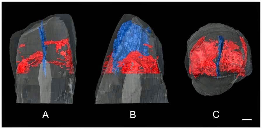
The mesial sectional plane of the Hexian upper left
second molar (the red and grey areas indicate the reconstructed enamel and
dentine, respectively, and green areas show how the occlusal wear was virtually
restored). Xing et al. (2014).
Again these are large teeth, with the width exceeding all previously
recorded Middle Pleistocen East Asian Hominins, and all but the largest Early
and Middle European Hominins, Neanderthals or modern Humans, though the width
does fall within the range of Homo ergaster
and Early Pleistocene Hominins from East Asia. The length of the teeth is at
the upper limit of, or outside the range of, the values seen in Middle
Pleistocene East Asian Hominins, Early or Middle Pleistocene European Hominins,
Neanderthals and modern Humans.
The sixth and seventh teeth described are a lower left second and
third molar from the same individual, still attached by a fragment of alveolar
bone (these were previously reported as the lower left first and second
molars).
The second molar has some damage to the crown and root, and is
severely worn down, preventing analysis of the morphology of the upper surface.
The tooth has two roots, flattened slightly sideways and 15.07 and 16.84 mm
long, respectively. The mesial root has two separate root canals, the distal
root a single canal that splits in two close to the tip.
The third molar is much better prepared, with only minor damage to
the enamel on the upper surface, although it is heavily worn, with a complex
surface to the dentine surface that shows five main and one (or possibly two)
minor cusps and numerous small ridges and crenulations. There are two roots
which bifurcate (split in two) on their third portions and diverge (move apart)
at their tips. These roots are 14.52 and 15.56 mm long. The pulp cavity has
five horns, one for each of the main cusps, and three root canals, two mesial
and one lingual.
Left lower second and third molars (PA834-1 and
PA834-2) (o: occlusal, B or b: buccal, M: mesial, l: lingual, d: distal). The
occlusal (I) and lingual (II) views of dentine surface reconstructed from micro-CT
scanning of the third molar. The occlusal (III) and distobuccal (IV) views of
the pulp cavity of the third molar reconstructed from micro-CT scanning.
Sagittal sectional planes of the second (V) and third (VII) molars.
Crosssection of the second (VI) and third (VIII) molars at the level indicated
by the red lines. Dotted lines indicate theCusp 6 and 7 on the occlusal
surfaces and the protostylid on the buccal surfaces of the crown. (I, II, III,
IV, V,VI, VII, and VIII) are not scaled. Xing et al. (2014).
The eighth tooth described is also a lower left second molar. The
root of this tooth is missing, but the crown is well preserved, if heavily
worn, exposing patches of dentine. The crown of the tooth is an asymmetric oval
in shape, and the tooth has five main cusps and two lesser cusps. The metaconis
and hypoconid are in contact hyperconulid is large and displaces buccally, the
sixth cusp is damaged, but appears medium sized, the seventh cusp is also
medium sized.
Left lower second molar (o: occlusal, B or b: buccal,
M or m: mesial, l: lingual, d: distal. I and II: The occlusal (I) and buccal
(II) views of the dentine surface reconstructed from micro-CT scanning. Dotted
lines indicate the Cusp 6 and 7 on the occlusal surfaces and the protostylid on
the buccal surfaces of the crown. I and II are not scaled. Xing et al. (2014).
The ninth tooth described is another lower second left molar. This
tooth retains its roots, and is also worn on its upper surface, leaving patches
of exposed dentine. This tooth is also oval in outline and slightly asymmetric,
the distal portion being slightly narrower than the mesial portion. There are
five main cusps arranged in a ‘Y’ shape. There are two roots, both very robust,
which bifurcate towards their tips. The mesial and distal roots are 15.96 mm
and 16.00 mm long, respectively.
Left lower second molar (o: occlusal, B or b: buccal,
M or m: mesial, l: lingual, d: distal). Xing et al. (2014).
The surfaces of the lower second molars
from the Longtan Cave have an unusually complex surface, with seven cusps and
numerous crenulations and ridges. This more typical of earlier Hominins, with
complex arrangements being seen in Australopithecus
and Early Pleistocene Hominins from Africa, Europe and Asia, but absent in
Middle Pleistocene Hominins from Europe, as well as Neanderthals and Modern
Humans. However this has been previously recorded in Middle Pleistocene East
Asian Hominins. An oval shape to the upper surface of this tooth is otherwise
only recorded in Early Pleistocene Hominins from Africa, and these have molars
that are more symmetrical and elongate.
The presence of a seventh cusp has previously been recorded in Hominins
from the Middle Pleistocene of Europe, and in Neanderthals. A ‘Y’ shaped
arrangement of cusps seen in these teeth is the ancestral state in Hominins,
but is highly unusual in Middle Pleistocene populations. The structure of the
roots of these teeth is typical of Early Pleistocene Asian Hominins.
These teeth are also large, with a width falling within the range of
Homo ergaster and Early Pleistocene
Hominins from East Asia, but larger than otherwise seen in Middle Pleistocene
East Asian Hominins or Early Pleistocene European Hominins, and towards the
upper end of the range seen in Middle Pleistocene European Hominins and
Neanderthals. Lengthwise these teeth are larger than any known Middle
Pleistocene East Asian Hominin, Neanderthal or modern Human, and falls within
the upper part of the range of Homo ergaster
and Early Pleistocene Hominins from East Asia.
Buccal views of a sample of lower second molars from
the East Asian mid-Middle Pleistoceneperiod (From left to right: Hexian
PA834-1, Hexian PA838, Zhoukoudian PA70). Xing et al. (2014).
The lower third molar has a well-developed, buccally displaced hypoconulid,
which is typical of Australopithecus
and Hominins from the Early and Middle Pleistocen of Africa and Asia, differing
from the reduced, centrally placed hypoconulids seen in Pleistocene European
Hominins and modern Humans. The sixth cusp is also typical of Australopithecus and Hominins from the
Early and Middle Pleistocen of Africa and Asia but not Pleistocene European
Hominins and modern Humans. The root arrangement of this tooth also resembles
Early Pleistocene East Asian specimens, with divergent roots, whereas those
from the Middle Pleistocene Zhoukoudian Locality tend to converge.
Again this is a large tooth, with a width within the ranges of Homo ergaster and Early Pleistocene
Hominins from East Asia but larger than any known Middle Pleistocene East Asian
Hominins, Early or Middle Pleistocene European Hominins, Neandethals or modern
Humans, and a length exceeding everything except Australopithecus and some early Homo
specimens.
The final tooth described is a right central upper incisor. This has
a small fracture on the root tip, but is otherwise in good condition. The tooth
is shovel-shaped, with the front surface relatively flat, while the back is
convex at the base. The root is robust, short and flattened.
High quality replica of the right upper central
incisor (PA835) (o: occlusal, La or la: labial, M orm: mesial, li: lingual, d:
distal). Xing et al. (2014).
The shovel shape seen in the incisor is seen in Australopithecus and some early Homo
specimens, though the more pronounced shape seen in this tooth is more typical
of Pleistocene Hominins from Eurasia, and in particular Neanderthals. This is
another very large tooth, exceeding all but a few Australopithecus and early Homo
specimens in size.
These teeth are generally ‘primitive’ compared to other middle
Pleistocene East Asian ‘Homo erectus’,
more closely resembling Early Pleistocene members of the group, but showing a
mixture of features unlike any other Hominins. Xing et al. suggest this may be indicative of an unrealized level of
diversity among East Asian Hominins, though they do not propose naming any new
taxa on the fragmentary remains available at the current time.
See also…
 Human remains from the Middle Pleistocene of Normandy. Early and Middle Pleistocene Human remains are extremely rare in
northern Europe, having to date been...
 Trying to find Peking Man. Peking Man is a collective name given to a group of hominid fossils
found at Zhoukoudian, near Beijing, between 1929 and 1937. This
comprised remains from at least 15 individuals, assigned to the species Homo erectus,
with an estimated age between 300 000 and 780 000 years old. In 1937
the invading Japanese army was drawing close to Zhoukoudian and the
excavation...
Follow Sciency Thoughts on Facebook.


