Showing posts with label Blastozoans. Show all posts

Tuesday, 16 June 2020

Durhamicystis americana: A new species of Rhipidocystid Blastozoan from the Late Ordovician of Maryland.

Rhipidocystids  are a small group of Palaeozoic Blastozoan Echinoderms, exclusively found in the Ordovician, which had a flattened theca, long brachioles, and reduced stems. The family Rhipidocystidae was erected by Otto Jaekel in 1901 to accommodate the genus Rhipidocystis, then considered a member of the Carpoidea because it bore a flattened theca. A second genus of Rhipidocystids, Batherocystis was described in 1950, with the genus Petalocystites being erected in 1973 and Mandalacystis in 1987; this being one of the better-known taxa within the family, including complete specimens that preserve the stem, brachioles, and the oral area. Some specimens of Rhipidocystis baltica preserved the oral area and show a pentaradial pattern with a BC-DE flattening direction. In 1989 the genus Neorhipidocystis was created to accomodate some material originally described as Rhipidocystis, which included bilateral forms with only the two shared ambulacra. Ambulacral homologies in flattened forms with two ambulacra remain unresolved, and this results in part, from a lack of information from the oral area, which is critical for the understanding of homologies in early pentaradial Echinoderms.

In a paper published in the journal Acta Palaeontologica Polonoca on 5 June 2020, Samuel Zamora of the Instituto Geológico y Minero de España, James Sprinkle of the Department of Geological Sciences at the University of Texas, and Colin Sumrall of the Department of Earth and Planetary Sciences at the University of Tennessee, knoxville, describe a new species of Rhipidocystid Blastozoan from the Late Ordovician of Maryland, based upon a historic collection  of one of the best-preserved Rhipidocystids known.

Zamora et al. describe four specimens from a historic collection housed at the Smithsonian Institution (National Museum of Natural History), Washington DC, USA. These specimens are fully articulated in a dark grey highly lithified limestone. The specimens were apparently hand prepared by the original collectors. They are preserved in calcite that appears to be softer than the surrounding matrix.

Information on the original labels indicates that the material was 'brought in by amateurs' and shown to John Wyatt Durham of the University of California, Berkeley, who was visiting the Smithsonian, on June 6, 1966. This small collection of Rhipidocystid Echinoderms was collected from the 'Edinburg Formation', in western Maryland. Specimens were collected in an old quarry, west of Conococheague Road, east of Wilson, Maryland. The first record of these specimens being deposited at the Smithsonian is from June 24, 1969, indicating that at least 'two specimens of Rhipidocystis sp. and one specimen of disarticulated Rhipidocystid plates' were received by Thomas Phelan, who was Curator Porter Kier’s Assistant at that time, who placed them in the Springer Echinoderm Collection. Since then, nothing has been done with these specimens, with the exception of James Sprinkle who, while visiting Durham at the Smithsonian in August, 1967, made a drawing of one of the Rhipidocystid brachioles to use in his dissertation. While reviewing the collections at the National Museum of Natural History in 2017, Samuel Zamora rediscovered these specimens in a museum drawer, but there is no historical record about what happened with these fossils between 1966 and 2017.

The original label indicates that specimens derive from what was formerly called the 'Edinburg Formation', and were collected from the Schemeompf’s old Quarry, located at the top of a hill, west of Conococheague Creek, about 3.4 km north and 0.8 km east of Wilson, Washington County, Maryland. Coordinates recorded on the label place the quarry at 77º between 49’ + 50’’ W; 39º approximate 41’ N. Based on Google Earth, there is now a mostly filled-in quarry north of Cresspond Road, North Wilson, northwest of Hagerstown, Maryland, that almost certainly is the Schemeompf’s old Quarry.

The Edinburg Formation is now only used in the Shenandoah Valley in western and northern Virginia. The equivalent unit in western Maryland (Hagerstown Valley) and southern Pennsylvania is now called the Chambersburg Formation. The Chambersburg Formation belongs to the Amorphognathus tvaerensis Conodont Zone, in the lower and middle Turinian (Laurentian) Stage, belonging to the lower Mohawkian Series, Sandbian (Upper Ordovician). The Chambersburg Formation in Maryland is about 150 m thick and was deposited in a shallow outer ramp with a small amount of deep ramp deposits. The nodular-bedded outer-ramp facies of the lower Chambersburg Formation contains Echinosphaerites beds, showing that the Chambersburg Formation is fossiliferous and favorable for Echinoderm preservation.

The new species is named Durhamicystis americana, where 'Durhamicystis' honours the palaeontological specialist on Echinoids and Cambrian Echinoderms, John Wyatt Durham, and 'americana' refers to to North America, the continent where the type species was found.

Durhamicystis americana is a Rhipidocystid with large theca composed of ten marginal plates and orals closing the marginal framework. Three apertures are recognised that allow orientation of the material. The peristome appears in the central anterior part, bounded by seven oral plates. Two elongate ambulacra correspond with the BC and DE rays. An anal pyramid covering the periproctal opening appears in the left anterior side of the marginal ring. This is a large aperture of about 2 mm that is covered by a domed anal pyramid composed of about eight?, thin, subtriangular plates. A single small pore (roughly 300 μm) within the plate O1 probably corresponds to the gonopore. The suture between O1 and O7 has a slit corresponding with the hydropore.

Rhipidocystid Echinoderm Durhamicystis americana from the Upper Ordovician Chambersburg Formation of Maryland, USA. (A) Holotype USNM 642510 showing the theca, proximal stem, and brachioles; general view of the complete specimen in ventral view (A₁), detail of oral area and proximal part of the brachioles (A₂), specimen in lateral view showing the periproct surrounded by three plates (A₃). (B) Paratype USNM 642513; general view of the complete specimen in ventral view (B₁), note central plates in thecal interior, and single columnal attached to basals; detail of the anal pyramid on left edge (B₂). Specimens whitened with ammonium chloride sublimate. Zamora et al. (2020).

Nine U-shaped marginal plates contribute to the marginal framework on the 'ventral' surface closing it, and an extra small plate with a vertical suture lies above the periproct on the 'dorsal surface'; thus, most of the marginal framework is singly plated. This framework is closed posteriorly with two large marginal plates (M1, M2), usually considered as basals. Those plates are very large and abut with large sutures closing the marginal frame. They are both trapezoidal in shape and articulate anteriorly with plates M3 and M4, and posteriorly form the stem facet. They both have large, thin projections on the ventral side that contribute to the central part of the theca. Marginal plates M3–M5, and M6 are longer than wide and also have large, thin projections contributing to the central theca. Marginals M4 and M6 are the longest marginals. Marginal plate M7 articulates posteriorly with M5, forms the posterior edge of the periproct, and is overlain by M9 and M10 separated by a vertical suture at the left anterior corner of the theca, followed anteriorly by oral plates. M8–M10 contribute to the peristomial opening; M8 contributes to the marginal framework in both dorsal and ventral aspects, while M10 and M9 are only visible in dorsal and ventral views, respectively. M9 also articulates anteriorly with an ambulacral flooring plate.

Rhipidocystid Echinoderm Durhamicystis americana from the Upper Ordovician Chambersburg Formation of Maryland, USA. (A) Paratype USNM 642511a, b; general view showing two nearly complete superimposed specimens (A₁); detail of the oral area, proximal brachioles attached to marginal, oral, and floor plates, and three apertures (periproct, hydropore slit, and tiny gonopore) (A₂); specimen in left lateral view showing the thin central plates from both integuments (A₃); specimen in right lateral view showing the position of the periproct (A₄); detail of facets between brachiolar plates and cryptic vertical sutures indicated by arrows (A₅); detail showing thin internal projections of weathered, U-shaped, marginal plates (A₆). Specimens whitened with ammonium chloride sublimate. Zamora et al. (2020).

Ventral central plates are large, polygonal, and very thin in cross section. They are probably four in number and form a tessellate pavement that covers the ventral surface of the theca. Dorsal central plates are more disrupted and only visible in one specimen; they are apparently smaller than ventral centrals, polygonal, and also very thin in section. They form a tessellate integument that covers the dorsal surface and connects with marginal plates and anterior oral plates.

Rhipidocystid Echinoderm Durhamicystis americana from the Upper Ordovician Chambersburg Formation of Maryland, USA. (A) USNM 642510. (B) USNM 642511a, b. Photographs of specimens submerged in water to increase contrast of plates and plate boundaries (A₁, B₁). Camera lucida drawings indicating plate arrangements (A₂, B₂). Abbreviations: FP, flooring plate (green); G, gonopore; H, hydropore; M1–M9, marginal plates (yellow); O1–O7, oral plates (red); Pe, periproct. Central plates colored in blue, stem in purple, and brachioles in brown. Zamora et al. (2020).

At least seven oral plates are recognized in the anterior part of the theca. Four of them are large plates that articulate aborally with marginal plates M8–M10 and have facets for feeding appendages. O1 and O6 are on the dorsal side of the oral area and have an indented aboral margin to accommodate a small pentagonal O7 plate. O3 and O4 are large, relatively thick, subpentagonal elements that appear on the anterior side of the oral area. O2? and O5? are questionably recognized (aborally from the central orals) at the ends of the ambulacra (where branches BC and DE would normally split ontogenetically). They are smaller elements bearing facets for feeding appendages and are visible on both sides of the theca.

Flooring plates appear aborally from the large central oral plates and also have facets for feeding appendages They are difficult to distinguish in size and morphology from oral plates O2? and O5?, except that they do not wrap around the ends of the ambulacra.

Brachioles range from 22 and 26 in number, are biserial, long, and slender. Brachiolar plates appear to be adjacent (not alternating), and have cryptic sutures. They attach either in groups of two (rarely three) on both oral plates and flooring plates. Proximally, the three or four lowest segments of the feeding appendages are wider than long and have deep facets composed of a transverse prominent ridge and three fossa, a large posterior one and two small anterior ones. These create wedgeshaped spaces for ligament attachment, and indicate some mobility. Higher segments are longer than high, tightly sutured, and have a thin, vertical suture visible on the aboral side accentuated by weathering. The adjacent brachiolars here may be partly fused, but still show different crystallographic orientations.

The stem is incomplete in all specimens. Proximally, there is a single large, tapering, subtriangular columnal with a distally positioned facet for one or more additional columnals. Only one? columnal is present in the best preserved specimens. Some disarticulated material preserved in slab USNM 642514 suggests the presence of a normal stem with cylindrical columnals showing crenulae with symplexy articulations in the columnal facets, but this columnal material is not closely associated with any of the complete rhipidocystid specimens.

Understanding the orientation of Echinoderms that have undergone ambulacral reduction is sometimes problematic and Rhipidocystids are a clear example. This is exacerbated because flattening in Eocrinoids is polyplyletic, and any attempt to recognise possible orientation requires recognition of oral plates, ambulacra, and in some cases thecal openings. Many clades of Echinoderms have independently developed flattened theca in response to living in close proximity to the substrate or as adaptations to high energy environments with oscillatory currents. Flattening in Echinoderms is recognized to occur in three planes: lateral where the sides of the theca area are compressed along the BC-DE axis, anterior-posterior where the sides of the theca are compressed along the A-CD axis, and dorsal-ventral where the theca is compressed along the polar axis.

Thecae showing lateral flattening have the BC and DE sides of the theca compressed parallel to the polar axis of the theca. This results in a lens-shaped to discoidal theca that is flattened parallel to the A-CD and polar axes, and perpendicular to the BC-DE axis. This occurs only in Rhipidocystis. Anterior-posterior flattening occurs where flattening is along the A-CD axis. This results in a lens-shaped to discoidal theca that is flattened parallel to the BC-DE and polar axes and perpendicular to the A-CD axis. This is seen in Pleurocystitid Glyptocystitoids, the Eocrinids Lingulocystis, Cardiocystites, and Haimacystis, and many Rhipidocystids such as Neorhipidocystis, Petalocystites, and Durhamicystis. Dorsal-ventral flattening occurs where flattening is along to the polar axis of the theca. This is seen in many Echinoderms such as Asteroids, some Echinoids, Edrioasteroids, and Cyclocystoids.

As with other rhipidocystids, Durhamicystis has a flattened theca and large U-shaped marginal plates. The position of inferred oral plates and openings allows comparison with other pentaradial Echinoderms, suggesting that ambulacra correspond to the shared BC and DE rays.

Rhipidocystids have been argued to be related to Paracrinoids based on similar peristomial plate arrangement, three distinctive basal plates, position of main thecal apertures (gonopore, hydropore, and periproct), and proximal stem morphology. Most highly flattened Rhipidocystids with U-shaped marginals, and most lens-shaped paracrinoids with 2–3 ambulacra, appear to be flattened into the anterior-posterior plane (through the A-CD), although there is considerable variability in where the periproct is located and what ambulacra are retained (usually BC and DE). At least three taxa, Petalocystites, Neorhipidocystis, and Durhamicystis agree with this configuration, and thecal plates can be homologized across the three taxa. Rhipidocystis, that gives the name to the family Rhipidocystidae, is apparently flattened in the opposite direction (see discussion above). If confirmed this probably responds to different flattening planes in Rhipidocystidae, and thus the family, as presently described, should be reconsidered.

Rhipidocystids include some of the most enigmatic Echinoderm taxa from the lower Palaeozoic. Their fattened body and aberrant morphology, with little morphological information from the oral area, limited in part the understanding of homologies across different taxa. The discovery and description of Durhamicystis americana from the Upper Ordovician Chambersburg Formation in western Maryland, preserving both thecal sides and the oral area, and major apertures including peristome, periproct, gonopore, and hydropore, allows orientation of the flattening this species and proper comparison across all Rhipidocystids. This questioned previous hypothesis about homology within Rhipidocystids, and suggests that two different groups, one including Rhipidocystis and the other comprising Petalocystites, Neorhipidocystis, and Durhamicystis, show flattening in opposite directions.

See also...

https://sciencythoughts.blogspot.com/2020/06/enodicalix-inornatus-blastozoan.htmlhttps://sciencythoughts.blogspot.com/2020/06/the-fulu-biota-new-fossil-lagerstatte.html
https://sciencythoughts.blogspot.com/2020/03/athenacrinus-broweri-new-species-of.htmlhttps://sciencythoughts.blogspot.com/2019/08/hanusia-sp-thoralicystis-sp-soft-tissue.html
https://sciencythoughts.blogspot.com/2015/12/iocrinus-africanus-new-species-of.htmlhttps://sciencythoughts.blogspot.com/2012/09/a-new-species-of-crinoid-from-southern.html


Follow Sciency Thoughts on Facebook.

Saturday, 13 June 2020

Enodicalix inornatus: A Blastozoan Echiniderm from the Middle Ordovician of the Montes de Toledo, Spain.

The Ordovician Echinoderm fauna of Spain includes some diverse, but sometimes poorly known Diploporite Blastozoans. In several cases these were first described and named on the basis of inadequate type material, such as partial internal moulds, often with no indication of the key characters that are today considered essential to determine their taxonomic affinities. Recent discoveries have improved palaeontologists' knowledge of the morphology of these species, which they are attempting to re-describe to modern standards, and where necessary introduce new names. This requires reviewing the evidence that relates the new material that preserves key characters to the originally described type material. In turn, this should help establish with greater certainty the essential characters of Diploporite families, such as the Aristocystitidae, and hence clarify their evolutionary relationships. 

In a paper published in the journal Geologica Acta on 16 February 2020, Christopher Paul of the School of Earth Sciences at the University of Bristol, and Juan Carlos Gutiérrez-Marco of the Instituto de Geociencias and Departamento de Geodinámica, Estratigrafía y Paleontología, at the Universidad Complutense de Madrid, redescribe one such species, ‘Calixinornatus and shows that it cannot be accommodated within any existing aristocystitid genus, including Calix.

In 1958 Bermudo Meléndez described Calix inornatus as a new species of Diploporite Blastozoan from the Ordovician of the Montes de Toledo, Spain. The species was based on a unique type specimen that preserved only the internal mould of the aboral part of the theca. Thus, the external surface was unknown as were the details of the oral area, which is nowadays considered critical in determining the affinities of blastozoans. Meléndez chose the trivial name inornatus because the internal surface of the theca was basically smooth, except for the fillings of the canals of the diplopores. Most species of Calix have circlets of spiny plates aborally, so the absence of any trace of spines was thought to be significant. In fact, matching internal and external moulds show that sometimes there is no trace of the spiny ornament on the inner surface of the theca. Later Jean Chauvel and Bermudo Meléndez described and illustrated some additional specimens under the names Calix inornatus and Calix toledensis, but these did not add significantly to knowledge of the species. It is worth pointing out that the aboral theca appears to have been more robust in Calix than the oral part, at least to judge by the frequency of preservation. Calix sedgwicki, the type species of Calix, and ‘Calixinornatus are the only two species of Calix in which details of the oral area are known.

Another specimen of ‘Calixinornatus was described in 1999 from the type locality of the species. This speciem preserved the external surface of the adoral part of the theca.  Based upon a review of the literature known to that date, the authors of that study described the morphology thoroughly and presented a revised diagnosis. This revision showed that ‘Calixinornatus was characterized by four ambulacra, each of which terminated in a pair of equal ambulacral facets developed on individual circum-oral plates and with obvious ambulacral orifices. The mouth is covered by relatively large cover plates, as in other Aristocystitid Diploporites. The hydropore is an irregular elongate structure set in a broadly oval tubercle with the adoral margin parallel to the posterior edge of the peristome. The gonopore is circular and set in a chimney-like tubercle apparently within a single thecal plate and the periproct is widely separated from the mouth, heptagonal with narrow ledges for the anal cover plates. The specimen also shows that the trivial name inornatus was most appropriate. The external surface is remarkably smooth, but with traces of small elongate oval diplopores that lack peripore rims.

Diagrammatic representations of the oral areas of Calix inornatus (A), Calix (B), and Phlyctocystis (C). In Enodicalix each circum-oral plate (CO1), (CO2), (CO4), (CO5) bears a pair of rounded facets (F). Food passed into the theca through small holes adjacent to the facets, the ambulacral orifices (black). The mouth was surrounded by eight plates, four central peri-orals (PO1), (PO3), (PO4), (PO6) and the four distal circum-orals. The posterior pair of peri-orals share the hydropore (H), which is oval and parallel to the posterior margin of the mouth. The mouth is covered by immovable cover plates, crudely arranged in two series anterior and posterior. In Calix (B) the same oral plate arrangement occurs, but the four circum-orals each bear four facets for a cluster of brachioles (erect, biserial food-gathering structures). The hydropore (H) is crudely trilobed and extends onto the plates below (PO1) and (PO6). In Phlyctocystis (C) the four ambulacra are proportionately narrower and distally they divide into separate branches that terminate in a single facet, but in the upper left ambulacrum each facet appears to be developed on three plates, not a single circum-oral. The hydropore in this diagram is to the right. This is really exceptional in Aristocystitid, indeed Diploporite, Blastozoans and suggests the diagram was drawn from an external mould, not a latex cast. If so, it is reversed with respect to left and right. For this reason, the ambulacra are not identified. (B)-(E) ambulacra (B)-(E). Paul & Gutiérrez-Marco (2020).

Other aristocystitid genera have four ambulacra, but of these Lepidocalix, Glaphocystis, and Sinocystis all have a single facet in each ambulacrum. Calix sensu stricto has four facets in each ambulacrum that are arranged in an arc and were added in a clockwise direction during growth. Phlyctocystis has four ambulacra that each divide once to give eight ambulacral facets, but the eight branches are widely separated and from the only specimen that preserves the oral area two of the facets are developed on three thecal plates. In contrast the paired facets in ‘Calixinornatus are side by side on a single circumoral plate and flush with the surface. Finally, Triamara and Binocalix have paired ambulacral facets on single circum-oral plates, but in both genera the facets are raised up in oral prominences and Triamara has only three ambulacra. The  number of ambulacra in Binocalix is unknown, but it is also characterised by unusual diplopores that are polygonal and have a central tubercle. They closely resemble those of the Sphaeronitid subgenus Sphaeronites (Peritaphros), but those of Calix gutierrezi are also similar.

Therefore, ‘Calix inornatus cannot easily be accommodated in any of the existing genera of Aristocystitids. Paul and Gutiérrez-Marco therefore consider it useful to establish a new generic name based on the Spanish material, which also has significance for the family level systematics of Diploporite Echinoderms.

Based on present evidence, the geographic and stratigraphic range of ‘Calix inornatus is restricted to a single locality, to the south of Las Ventas con Peña Aguilera, in the Montes de Toledo area, central Spain, in the lower part of the Navas de Estena Formation, in dark mudstones of early Oretanian age (early middle Darriwilian in terms of the global scale). From this classical locality for Spanish Ordovician fossils, in the right bank of the Acebrón stream valley, all the currently known speciemsn have been recovered, including the holotype, and the topotype with the oral area, and an additional topotype which consists of a fairly complete internal mould of an uncrushed theca.

Latex casts were made from the critical external mould (MGM-2000-O) that preserves the details of the oral area. Latex casts and natural internal moulds were whitened with magnesium oxide or white fingerprint powder for photography. Stereophotographs were produced using a see-saw, which rotates specimens through approximately 12º.

The species is placed within the Family Aristocystitidae, which is diagnosed by directly attached diploporites with elongate oral area surrounded by at least eight plates (four central peri-orals and four distal circum-orals), and covered by two series of larger outer and smaller central cover plates; with 2-5 ambulacra bearing 1-5 brachioles each; with a large hydropore shared by plates PO1 and PO6 and often a spout-like gonopore usually within a single plate near the periproct; with thecal plates densely covered with diplopores that are frequently sealed externally by a thin epistereom and sometimes extended into spine-like projections.

It is given the new generic name Enodicalix, meaning 'a cup without knots' in reference to its relationship with the genus Calix (which means 'cup'), giving it the full name Enodicalix inornatus

Enodicalix is diagnosed as an Aristocystitid genus with smooth thecal surface, four ambulacra, each with an equal pair of rounded facets developed on a single circum-oral plate, large oval hydropore close to ambulacrum D, circular gonopore within a circular tubercle, heptagonal periproct, very little evidence of diplopores on the external surface.

(A) Internal mould of specimen MGM-2001-O, showing the overall shape of the theca. (B) Reverse side of MGM-2001-O, showing small area that preserves thecal surface (top). (C) Enlargement of figure (B) to right showing weak traces of diplopores on the external surface. (D) Enlargement of latex cast of MGM-2000-O, showing weak traces of diplopores on external surface. Scale bars (A), (B) 10mm, (C), (D) 5mm. Paul & Gutiérrez-Marco (2020).

The holotype of Enodicalix inornatus is the unique internal mould of the aboral part of a theca, described by Meléndez in 1958 and now in the Department of Geodynamics, Stratigraphy and Palaeontology, Universidad Complutense, Madrid. The only additional material accepted by Paul and Gutiérrez-Marco is another internal mould (MGM-2001-O), and the matching internal and external moulds of (MGM-2000-O), on which the generic diagnosis is based.

(A) Detail of oral surface of latex cast of MGM-2000-O to show ambulacra, hydropore and plate sutures. Scale bar 5mm. (B) Traces of oral frame plates (dashed lines) and cover plates  (solid lines) superimposed over the oral surface. (C) Interpretation of ambulacra and sutures of oral frame plates and cover plates (shaded). Abreviations: AO, Ambulacral Orifices (black); B-E, ambulacra B-E; CO1-CO5, Circum-Oral plates bearing ambulacral Facets (F), H, Hydropore; PO1-PO6, Peri-Oral plates. 1-6, possible primary oral cover plates. Paul & Gutiérrez-Marco (2020).

In MGM-2000-O the theca is moderately large, approximately 30mm in diameter and up to 30 mm of the adoral theca is preserved. The internal mould reaches over 100 mm and is incomplete. The oral surface is regularly rounded with the more or less rectangular peristome at the top. The four ambulacra extend about 13 mm from left to right, by a maximum of 8.5 mm from front to back, as measured to the outside of the rounded brachiole facets. The oral groove reaches 8.3 mm left to right, by 3.5 mm front to back and is covered by cover plates. Each ambulacrum bears a pair of equal facets, reaching 2.0 mm across by about 1.5 mm radially and divided by a median radial ridge. At the adoral end of the ridge is an Ambulacral Orifice 0.25 to 0.3 mm across In ambulacrum E the median ridges of the pair of facets are at 90º to each other.

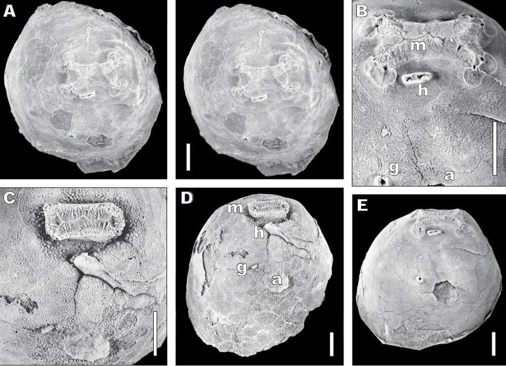
Photographs of Enodicalix. (A) Stereophotos of latex cast of MGM-2000-O, showing oral region and principal thecal orifices. (B) Detail of the thecal orifices in the latex cast (a) periproct, (g) circular gonopore, (h) oval hydropore, (m) mouth with ambulacral cover plates. (C) Detail of the oral area of the natural internal mould showing internal structures associated with the thecal orifices. (D) Oblique view of internal mould to show impression of mouth (m), stone canal leading to hydropore (h), possible gonoduct, septum associated with the gonopore (g) and cast of the periproct (a). (E) Oblique general view of the latex cast showing separation of peristome and periproct. Scale bars 5mm. (A) Whitened with fingerprint powder, (B)-(E) with magnesium oxide. Paul & Gutiérrez-Marco (2020).

The plating around the mouth appears to conform with the usual Aristocystitid pattern, with four more central Peri-Oral Plates, two on the anterior side of the mouth and two on the posterior side which share the hydropore, plus at each end a pair of more distal Circum-Oral plates which bear the ambulacral Facets. Sutures between most of these plates can be seen on the internal mould.

The Hydropore is an irregular slit set within an oval tubercle with a distinct rim 2.5 mm long by 0.9 mm wide. The tubercle is parallel to the posterior margin of the peristome and separated from it by 1.3 mm. The gonopore is a rounded pore 1.1 by 0.7 mm across and set within a raised tubercle, also with a distinct rim 1.2 mm across. The gonopore lies below the D ambulacrum and separated from it by 7.6 mm. It is also 2.3 mm from the nearest edge of the periproct.

The periproct was probably seven-sided, about 4.2 mm across and none of the anal plates remains. The adoral side is 2.3 mm long.

The external thecal surface is entirely smooth. It shows only the faintest traces of either diplopores or plate sutures. The original presence of numerous diplopores is confirmed by the fill of the perpendicular canals on the internal mould.

The internal moulds reveal the overall shape of the theca and the presence of dense diplopores on all plates as well as the prominent rectangular ‘donjon’ around the mouth, the stone canal leading obliquely up towards the hydropore, the rather inconspicuous gonoduct and the fill of the periproct. There is also a trace of an internal mesentery connecting the hydropore and gonopore. Plate sutures and the fill of the perpendicular canals of the diplopores are much more obvious on the internal surface of the theca. The oral ‘donjon’ shows the central suture between the cover plates, which clearly divides towards either end, reflecting the four ambulacra, but there is no evidence of a fifth ambulacrum internally or externally.

Although plate sutures are not easy to detect, the plate arrangement around and over the mouth can be deduced. It shows the usual Aristocystitid pattern of eight plates surrounding the mouth; four central peri-oral plates, two (PO3, PO4) on the anterior side and two (PO1, PO6) on the posterior side which share the hydropore. At either end of the mouth are two pairs of circum-oral plates: CO1 bears the facets for ambulacrum D, CO2 for ambulacrum E, CO4 for ambulacrum B and CO5 for ambulacrum C. The oral cover plates are quite complex, but appear to show six large plates that extend from the outer edge of the peristome to the central suture, one in each inter-ambulacrum and two in the posterior interambulacrum.

The nomenclature is not as secure as desirable. Bermudo Meléndez based his species Calix inornatus on a unique type specimen that preserved only the aboral part of the theca and no trace of the external surface. He explicitly stated that he chose the trivial name ‘inornatus’ because the internal surface of the mould was smooth, apart from the casts of the perpendicular canals of the diplopores. By this Paul and Gutiérrez-Marco presume Meléndez was drawing attention to the fact that most other species that have been attributed to the genus Calix bear obvious spine-like plates, whereas his did not. Unfortunately, that does not help recognising Meléndez’s species, firstly because species of Calix with the spine-like plates often have smooth internal surfaces. In addition, other genera of Aristocystitids, such as Oretanocalix, also have smooth external surfaces. Thus, the unique specimen which exhibits the characters here attributed to the new genus, Enodicalix, can only tentatively be associated with the type specimen of ‘Calix' inornatus, its supposed type species. 

Re-examination of the critical specimen of ‘Calix' inornatus shows that it has four ambulacra, each of which terminates with a pair of equal ambulacral facets, side by side on, and flush with the surface of, single circum-oral plates. The oral area is elongate, covered by two series of larger outer and smaller central cover plates, within which small ambulacral orifices are developed adjacent to each facet. Posterior peri-oral plates PO1 and PO6 share an elongate hydropore set within an oval tubercle. The circular gonopore is developed within a single plate near the heptagonal periproct. These characteristics are typical of Aristocystitid Diploporites, but no currently described genus in this family has these precise characters. Paul and Gutiérrez-Marco therefore erect the new genus Enodicalix for ‘Calix' inornatus, which is currently the only known species in the genus.

See also...

https://sciencythoughts.blogspot.com/2020/03/athenacrinus-broweri-new-species-of.htmlhttps://sciencythoughts.blogspot.com/2019/10/ophiopsila-xmasilluminans.html
https://sciencythoughts.blogspot.com/2019/08/asterodiscides-fourmanoiri-starfish.htmlhttps://sciencythoughts.blogspot.com/2019/08/ophiacantha-scissionis-fissiparous-six.html
https://sciencythoughts.blogspot.com/2019/08/hanusia-sp-thoralicystis-sp-soft-tissue.htmlhttps://sciencythoughts.blogspot.com/2019/07/sollasina-cthulhu-new-species-of.html
Follow Sciency Thoughts on Facebook.