Showing posts with label Tunicates. Show all posts

Monday, 10 April 2023

Trying to identify an invasive colonial Sea Squirt from the Gulf of California.

Invasive species are one of the greatest challenges faced by conservationists today, often rapidly taking over ecosystems where they have no natural enemies and displacing endangered or commercially important local species in the process. The first step in controlling an invasive species is identifying it, which enables appropriate control systems to be introduced. However, this is often difficult and complicated, as invasive species are not necessarily well understood, or even known, in their own ecosystems. This is particularly true in marine environments, where our taxonomic understanding of many groups is poor, and increasingly plagued by a lack of skills as few young scientists are attracted into the field. This is despite the problem of invasive species being particularly acute in marine environments, where international shipping traffic has made it easy for many benthic 'fouling' organisms to rapidly establish global distributions.

Ascidians, or Sea Squirts, are the most abundant class of the Subphylum Tunicata and are distributed along shorelines worldwide. They are sessile marine invertebrates and are widely used as a model organism for developmental and evolutionary studies. Ascidians exhibit multiple morphological characteristics, from small colonial to colorful and large solitary forms. They are divided into three major well-accepted orders, namely, Phlebobranchia, Aplousobranchia, and Stolidobranchia, based on the branchial sac morphology of the adults. However, the class Ascidiacea is paraphyletic (i.e. not everything thought to be descended from the last common ancestor of the group is considered to be an Ascidian) with the  Phlebobranchia and Aplousobranchia showing a close relationship with Thaliaceae (Pyrosomes, Salps, and Doliolids), a non-Ascidian Tunicate class, whereas the Stolidobranchia remains a distinct and monophyletic group. Over the course of several decades, the Ascidiacea have been shown to be an important class of ecological species because of their invasive potential along with their ability to adapt to new environments. Transportation of Ascidians attached to ship hulls as fouling material and within the ballast water of ships has enabled them to invade many new territories. This phenomenon has major impacts on local marine biodiversity as well as aquaculture industries. Therefore, the Ascidiacea are now considered as important model species for the study of non-indigenous species worldwide.

Sea Squirts are particularly problematic from a taxonomic point of view, with a simple body plan which provides few diagnostic features, and very few scientists specializing in their taxonomy. To make matters worse, the group appears to be rife with cryptic species (i.e. species which are physically identical or nearly identical, but biologically distinct), so that many identified species are likely to be clusters of similar-looking species, often with different ecological constraints. Many species of Sea Squirt have proven to be adept at colonizing new environments, where they are often identified as new species, so that particularly successful invasive species often have several different names, further adding to the taxonomic confusion within the group. A combination of careful physical examination and genetic analysis can hopefully unravel some of these problems, particularly in the identifying of pseudo-indigenous species, i.e. invasive species thought to be native to their new habitat because their origin is unknown.

Eleven invasive species of Sea Squirt had been identified from Mexican waters by 2014, five of them from the Gulf of California. In 2015, a colonial Sea Squirt previously unknown to the region was identified in Ensenada de La Paz, which spread rapidly, causing a mass mortality event in Pen Shells, Atrina maura, a Bivalve species commercially important locally, whose large shells provided an ideal substrate for the invaders. The Pen Shell fishery was already suffering from over-harvesting, with a moratorium on their collection introduced in 2013 to allow the population to recover, something which appears to have been significantly hampered by the arrival of the Ascidians (no significant variation in other environmental variables was detected which coincided with the die-back). A subsequent biofouling experiment carried out in Bahía de La Paz found that the Sea Squirts, tentatively identified as Distaplia cf. stylifera, were the most abundant macro-organisms settling on a series of silicone resin coated metal panels placed in the water for two months. Furthermore, the Sea Squirt colonies provided a substrate upon which 28 species of epibiont Polychaete Worms were able to settle, further adding to the biofouling. Subsequent studies of the Sea Squirts, which have continued to suppress Shellfish populations, and become a serious biofouling problem in the area, have used the name Distaplia stylifera, although this is only an assumption, based upon a rough similarity to descriptions and the fact that it is a common invasive species. No formal taxonomic identification of the invader has been attempted to date, something which may be hampering efforts to find an effective control.

In a paper published in the journal ZooKeys on 5 April 2023, Betzabé Moreno-Dávila and Leonardo Huato-Soberanis of the Programa de Ecología Pesquera at the Centro de Investigaciones Biológicas del Noroeste, Jaime Gómez-Gutiérrez of the Departamento de Plancton y Ecología Marina at the Instituto Politécnico Nacional, Carolina Galván-Tirado of the Consejo Nacional de Ciencia y Tecnología, Carlos Sánchez of the Departamento de Ciencias Marinas y Costeras at the Universidad Autónoma de Baja CaliforniaTeresa Alcoverro of the Department of Marine Ecology at the Centre for Advanced Studies of BlanesEduardo Balart, also of the Programa de Ecología Pesquera at the Centro de Investigaciones Biológicas del Noroeste, and Xavier Turon, also of the Department of Marine Ecology at the Centre for Advanced Studies of Blanes, present the results of a study of the invasive Sea Squirt from Ensenada de La Paz, which examines the Mexican species' morphology and compares it to previous descriptions from other parts of the world, as well as attempting a genetic analysis.

Bahía de La Paz is located on the southeastern tip of the Baja California Peninsula. The Ensenada de La Paz is separated from Bahía de La Paz by a 12 km long sandbar known as El Mogote. The Ensenada de La Paz has an area of about 45 km² and a maximum depth of 10 km and a bottom covered by patches of sand and mud-silt. The Ensenada de La Paz and the Bahía de La Paz are connected by a shallow channel about 1 km wide and 4 km long, upon which are seven yacht docks associated with the city of La Paz, while at the entrance lies the Pichilingue commercial harbour.

(A) Area of study in Ensenada de La Paz located in the southern part of Bahía La Paz, Baja California Sur, Mexico. (B) Both bodies of water are located on the southwest coast of the Gulf of California, Mexico. (C) Sampling sites and potential sources of dispersal of Tunicates (circles). Moreno-Dávila et al. (2023).

Colonies representing three different colour-morphs of the invasive Sea Squirt (white, orange, and purple) were collected from four sites within the Ensenada de La Paz, for a total of twelve colonies, which were preserved for morphological analysis as well as having their DNA extracted for genetic analysis. The colonies were found growing at depths of 0.5-3 m, growing on the shells of Sea Pens, a PVC pipe, buoys, a rope, and wooden docks.

'Distaplia stylifera' from Ensenada de La Paz. (A) Live orange colony. (B) Live purple colony. (C), (D) Typical mushroom-shaped colonies. (E) Zooid. Scales bars: 10 mm (A), (B); 2.5 mm (C), (D); 1 mm (E). Moreno-Dávila et al. (2023).

The colonies were predominantly orange, and approximately mushroom-shaped, with mottled white markings around the common cloacal-aperture, with purple and white colonies also present within the population. They reach a maximum of about 2 cm, with the head having a diameter of up to 2.5 cm. The colonies are more heavily pigmented around their tips than at the base, with pigment only remaining in this area after preservation. The tunics of the colonies are firm, and the stalks sometimes branch, so that two or more colonies share a common base; some colonies lack stalks altogether, forming cushion-shaped masses that spread over the substrate. The head of each colony is shared by one or more zooid systems, each sharing a common cloacal-aperture, which is surrounded by a single or double ring of zooids.

The zooids are up to 5 mm in length, excluding the gonadal sacs. Each zooid is divided into a thorax and abdomen. Two sacs are attached to this structure, a smaller one, containing the gonads, is attached to the right posterior side of the abdomen. The second sac contains embryos and developing larvae, and is often longer than the zooid. This second sac is attached to the posterior part of the pharynx by a thin peduncle.

'Distaplia stylifera'. (A) Zooid (thorax and abdomen). (B) Thorax. (C) Dissected thorax. (D), (E) Stomach. (F) Gonads. (G) Larvae. (H) Enlargement of one larva showing two pigmented spots. Scale bars: 10 mm (A); 0.5 mm (B), (C), (F), (G); 0.25 mm (D), (E). All images except (F) correspond to stained zooids. Moreno-Dávila et al. (2023).

The oral siphon on the thorax is smooth-rimmed or has six slight lobulations, and a large atrial opening which exposes most of the branchial sac. A  wide flap-like lid with smooth or lobed margins called the atrial languet lies on top of the atrial opening. This languet is crossed by several transverse muscular bands. Each side of the thorax has about 30 bands of longitudinal muscle. About 14 simple oral tentacles are also present on the thorax. The pharynx has four stigmata rows clearly divided by parastigmatic vessels, with the first two rows typically having 18-19 stigmata, and the posterior two rows having 15-16. Three simple dorsal languets are found between these rows, slightly displaced to the left.

'Distaplia stylifera' (A) Zooid (thorax and abdomen). (B) Abdomen. (C) Larva. Abbreviatures: a. anus; am. ampullae; ap. adhesive papillae; oc. ocellus; o. oocyte; pv. parastigmatic vessels; pg. pyloric gland vesicle; sc. statocyte; sg. stigmata; st. stomach; t. testes. Scales bars: 1 cm (A); 0.5 mm (B), (C). Moreno-Dávila et al. (2023).

Within the abdomen lies an elongated and curved stomach, the wall of which has more than 20 fine longitudinal plications (folds), which can be seen on the inner and outer surfaces when the specimens are sectioned, although they are sometimes interrupted or divided. A short post-stomach connects to an enlarged mid-intestine at the bottom of the gut-loop. The distal intestine runs to the anterior, and ends in a bilobed anus at the base of the atrial aperture. A pyloric (mucus) gland between the stomach and the intestine and continues anteriorly forming sinuous tubules over the intestine in front of the stomach.

The gonads are held within a pedunculated sac, with one or two oocytes at the bottom, above which lie a cluster of five or six elongated or wedge-shaped testes. A  common sperm duct arises posteriorly from the cluster of testes, but turns anteriorly at its very beginning, without overlapping the oocytes.

All of the colonies examined had larvae incubating in long sacs that reach posteriorly deeper than the zooids themselves in the colonies. These sacs typically contained one or two well developed larvae, about 1.3 mm in length, plus three embryos. When fully developed the larvae reach about 1.5 mm, and possess three adhesive papillae, two dorsal and one ventral, with a globular ampulla each in the stalks. The four rows of stigmata have developed by this stage, with an incipient abdomen folded under the branchial sac. A sensory vesicle contains two pigmented spots, with a larger one above and a smaller one below, though these can be hard to differentiate, as they are close together and the larvae are not transparent. 

Distaplia stylifera was originally described in the Red Sea, and subsequently has been recorded from several areas of the Indo-Pacific, Australia, the Philippines, and the South China Sea, as well as. under the possible synonym Distaplia mikropnoa, from Palau. There have been reports from the Mediterranean, although these are considered dubious. It has also been reported in the Western Atlantic, from the coast of North Carolina south as far as Jamaica, and at several locations in the Caribbean, and possible further south around Sao Paulo in Brazil. It has not, however, previously been reported from the Eastern Pacific.

Sites of previous records of Distaplia stylifera: (1) Red Sea, 1874 (type locality). Indo-Pacific; (2)-(9). Mediterranean, (10). Western Atlantic Ocean, (11)-(18). Eastern Pacific Ocean, (19) present study. The type locality in the Red Sea and the record of the present study are indicated with stars. Moreno-Dávila et al. (2023).

Despite successful amplification of DNA apparently collected from the Ascidian colonies, all of this proved, upon analysis, to be closer to that of Algae, Bacteria, or Fungi, than to any Animal, despite great care being taken to avoid any contamination. For this reason the taxonomic comparison had to be made entirely upon physical examination of the specimens, and comparison to other members of the genus Distaplia.

The taxonomy of the genus Distaplia is mainly based on characters such as colony shape, arrangement of zooids in systems, presence or not of gonadal sac, stigmata per row, stomach shape and external surface, and muscle arrangement. Based upon these criteria, the Ensenada de La Paz specimens would appear to be entirely consistent with Distaplia stylifera. However, Moreno-Dávila et al. note that the original description of the species was made in 1874, and like a lot of descriptions from this period, was a lot less detailed than descriptions made by modern taxonomists. Furthermore, that description was made upon a specimen that was probably a juvenile, lacking larvae and having under-developed gonads. 

Descriptions of populations of modern populations of 'Distaplia stylifera' show some variation, making it possible that the global population is in fact a cluster of closely related species. Notably, the specimens collected at Ensenada de La Paz all had parastigmatic vessels, something also reported in almost all specimens collected from Australia, as well as specimens collected from the Caribbean. An absence of such vessels has been noted in specimens from Madagascar, and it has been suggested that the presence or absence of these vessels might be a feature which can be used to differentiate between Distaplia stylifera and Distaplia mikropnoa. However, a number of other features can be used to differentiate Distaplia mikropnoa, which include a long double rows of zooids converging to the terminal common cloacal apertures, a long post-pyloric part of the gut loop, and a lack of a gastric reservoir. Furthermore, in Distaplia mikropnoa the course of the gastro-intestinal ducts that does not cross from the stomach to the ascending limb of the gut loop but extends down the descending loop. Based upon this, Patricia Kott concluded that the two species are both valid, but that they have widely confused within the literature. Notably, she concluded that the Palau population belonged to Distaplia stylifera rather than Distaplia mikropnoa.

Oval follicles have been suggested as another feature which could be used to differentiate the two species, with specimens which have up to 15 oval follicles assigned to Distaplia mikropnoa, while those having only five or six, which would include the Ensenada de La Paz population, belong to Distaplia stylifera

The sperm duct is another feature apparently variable in populations assigned to Distaplia stylifera, with some populations having a sperm duct running posteriorly and making one or several loops over the oocytes before turning anteriorly, while in other populations, including the Ensenada de La Paz population, it is straight. Furthermore, in some populations, including Ensenada de La Paz, the gonadal sac is attached via a peduncle, while in others it is almost flush with the abdomen, separated by a wide neck. 

Moreno-Dávila et al. suggest that this wide range of variable features indicate that 'Distaplia stylifera' is probably a cluster of closely related species, often mistaken with the closely related Distaplia mikropnoa. This probably cannot be resolved without a more detailed taxonomic study, using both morphological and genetic methods to analyse populations from different regions of the world. Nevertheless, the reporting of a member of the species cluster from the Eastern Pacific represents a significant range-expansion for a group previously known from tropical regions of the Western Atlantic, Indian Ocean, and Western Pacific. 

Moreno-Dávila et al. were unable to extract DNA from their specimens of 'Distaplia stylifera', and note that no records exist within the GenBank and BOLD public databases. The co-generic Distaplia bermudensis is present in these databases, and shows a 14–20% genetic variance between different populations and morphotypes, indicating either a remarkable level of genetic variation within a single species, or that Distaplia bermudensis is also a species cluster. 

This taxonomic uncertainty makes it difficult to assess to what extent the different populations of 'Distaplia stylifera' represent introductions or local species, although Moreno-Dávila et al. note that the populations from North Carolina and the Caribbean appear to strongly favour lagoons and artificial structures, which is behaviour typical of invasive fouling organisms, and that this population appears to be expanding southwards to the coast of Brazil, where again it has been found only on artificial substrates. 

Despite this uncertainty, Moreno-Dávila et al. believe that the Ensenada de La Paz population does represent a high-impact invasive species, and given the absence of any other known populations of 'Distaplia stylifera' in the Eastern Pacific, and the behavioural similarities of this species to the Western Atlantic populations, one which has probably arrived via the Panama Canal. probably crossing the canal on a large ship, then secondarily transferring to a smaller vessel capable of entering the harbours around La Paz. 

See also...

Follow Sciency Thoughts on Facebook.

Follow Sciency Thoughts on Twitter.


Wednesday, 1 July 2020

Biomineralising Tunicates from the Late Triassic of Italy.

Biologically controlled biomineralisation is a fundamental process in the evolution of the animal kingdom. Though it was sporadically achieved already in the latest Precambrian, the so-called 'Cambrian Explosion' around 540 million years ago, marks the general advent of Animals with hard parts in the fossil record. It is well known, that this event was not an instantaneous process, but that it was accomplished in a time span of about 20–25 million years. The acquisition and evolution of hard skeletons within the great majority of phyla and classes are the basic prerequisites for the reconstruction of animal form and function through time. Once hard parts were created in order to protect and stabilize soft tissues and organs, this evolutionary advantage indeed developed a great variety of morphologies, but in spite of this diversity and modification, solid skeletons were generally maintained as such until extinction of the equivalent groups.

In a paper published in the Journal of Paleontology on 24 January 2020, Jobst Wendt of the Fachbereich Geowissenschaften der Universität Tübingen, describes and decrypts an unusual case of biomineralisation in fossils of the rather poorly known class Ascidiacea (Sea Squirts) of the subphylum Tunicata.

The reduction of a hard skeleton into a mere softbodied stage seems to have been achieved in some Coleoid Cephalopods and Opisthobranch Sea Slugs, but this accordance is based on notional palaeontological evidence. An alleged late acquisition of a hard skeleton inherited from soft-bodied ancestors is suspected in Bryozoans and Scleractinian Corals, but in contrast to Ascidians, once these groups had acquired calcified skeletons, they maintained this protective and stabilizing advantage until Recent. Thus, the very late acquirement of a solid calcareous skeleton among Tunicates and its later loss in favor of an almost soft-bodied stage is a very rare phenomenon in the geological record.

From their form and mode of life, Ascidiacea (Sea Squirts) appear as rather simple-structured organisms that superficially and functionally are evocative of Porifera. But the presence of a tubular dorsal nerve cord and a notochord in their larval stage and pharyngeal clefts allocates them amuch higher position within the Animal Kingdom as a subphylum of the Chordata. Ascidians are an artificial, polyphyletic group that comprises three orders (Aplousobranchiata, Stolidobranchiata, Phlebobranchiata) with a total of about 2940 living species. They generally lack hard parts, thus minimizing the possibility of preservation. Only a few genera of the orders Aplousobranchiata and Stolidobranchiata segregate tiny isolated spicules, embedded in the mantle (tunica) or other organs, but their origin remains unknown. Tunica is the Latin word for mantle. Therefore, many zoologists regard both terms as synonymous. Others, however, distinguish an inner layer (tunic), in which the spicules are secreted, from an outer one (mantle) In order to guard against misunderstandings, Wendt uses both terms as synonyms because, with regard to fossils, the differential application of both terms would appear arbitrary.

Spicules of cpossible Tunicates have been found in rocks as old as lower Liassic. Generally, ascidian spicules consist of aragonite, more rarely of vaterite or other minerals. Unlike some siliceous sponge spicules, they are never fused together and thus could not form solid skeletons. The discovery of complete soft-bodied ascidians (lacking spicules, however) in the lower Cambrian of China has revealed that ascidians existed already at an early stage of Metazoan evolution, but this finding does not allow to reconstruct their relationships to Mesozoic to recent representatives of this class. A new evolutionary branch among Ascidians is the recent discovery of calcareous Tunicate exoskeletons in the Upper Triassic and their ancestors in the Permian. 

Wendt suggests that these very unusual skeletons, which are composed of irregular aragonitic plates, can be assigned to no phylum other than the Tunicates. This systematic position of these previously unknown organisms to the class Ascidiacea has been the fundamental clue for the systematic attribution of the new discoveries described by Wendt. The fact, however, that they are endoskeletons, opens an unusual insight into a new aspect of the evolution of this poorly known class which has no counterpart among their living representatives. Tunicate exoskeletons have long been known from Permian deposits in east Asia and Europe, but until recently they were erroneously attributed to Rugose Corals. These skeletons are composed of a varying number (2 to about 35) of irregular plates that consist of acicular aragonite. This very unusual construction and mineralogy were crucial for their systematic attribution, though similar living representatives of this class are unknown. Comparably organized endoskeletons now add a new aspect to the fossil record of this largely ignored subphylum of the Chordata.

Endoskeletons are widespread in the Animal Kingdom and display a great variety of shape, function, and mineralogical composition. They are typical for the major phyla of Deuterostomia (Echinodermata and Vertebrata, including Conodontophorida) and many Protista (Foraminifera and Radiolaria), which exhibit a stunning variety of shapes and geometries. Endoskeletal spicules (sclerites) of different mineralogical composition evolved independently among several phyla and classes (Porifera, Octocorallia, Vermes, Holothuroidea, Tunicata). Apart from the calcareous octocoral Tubipora musica, only some Porifera only some Porifera (Hexactinellida and Lithistida), produced siliceous spicules, which are fused or articulated into compound endoskeletons forming cubic or irregular meshworks, thus giving more stability to the soft body. Among other phyla, compact solid calcareous endoskeletons occur only among the Mollusca. Composite calcareous endoskeletons consisting of numerous plates with flexible boundaries are characteristic for Echinodermata and for the newly discovered fossil Tunicata.

Wendt describes two incomplete specimens from the the Cassian Formation (lower Carnian) of the Dolomites (northern Italy). Extensive searches for additional, either biostratigraphically older, contemporaneous, or younger specimens from European and North American reef specialists and collections were unsuccessful. The two specimens were discovered among far over a million of skeletal remains from the the Cassian Formation. The number of taxa and the diversity of skeletal remains collected from the Cassian Formation during almost two centuries are indeed impressive and assign this rock unit a singular rank among the Fossil-Lagerstätten in the geological record. As of 2019, 1429 species have been described so far from this formation. A special feature of this unrivaled fauna is their often excellent state of preservation, exemplified by diagenetically almost unaltered aragonitic microstructures, which are among the oldest in earth history. In this state of perfection and considering its age, the Cassian Fauna can even be regarded as unique.

Fossil record of Tunicates. Black dot, soft-bodied; black triangles, calcareous exoskeletons; black square, calcareous endoskeletons; asterisks, spicules. Wendt (2020).

The fossils are placed in a new order and family of Ascidians, named the Cassianosomidae and Cassianomorpha respectively; both names refer to the Cassian Formation. Both are placed in the new genus Toscanisoma, which is named in honour of Maria Luigia Toscani, who collected the fossils upon which the study is based. Each of the two fossils is described as a separate species within the new genus.

The first species described is named Toscanisoma multipartitum, where 'multipartitum' means 'consisting of several parts'. This is a sessile colonial species consisting of seven branching tubes (zooids) composed of irregular plates that are joined by straight or curved boundaries on the outer and zigzag ones on the inner side. The only complete zooid of the colony is closed at the top by six plates that are smaller than the remaining ones. The other zooids are incomplete, but were probably slightly larger. Spicules have not been observed.

Toscanisoma multipartitum holotype (GPIT/TU 82). (1) Lateral view (drawing); (2) opposite side; (3) view from top; (4) base. Asterisks mark complete zooids, arrows indicate zigzag sutures on inner surfaces. Scale bar 10 mm. Wendt (2020).

The only specimen of Toscanisoma multipartitum available is not complete, lacking the basal portion, which probably served as a holdfast. The seven zooids, which bud from the flattened base, have different shapes ranging from almost circular to elongate or flattened in cross section. Only the smallest zooid is complete, 25 mm long and consists of about 10 plates with very faint and barely recognisable outer boundaries. The inner plate boundaries of the incomplete zooids show zigzag sutures similar to contemporaneous representatives of the order Khmeriamorpha. The zooids fit closely together at the base and are separated by small open interspaces higher up.

The second new species is named Toscanisoma triplicatum, meaning 'triplicate'. as the only known specimen consists of three zooids. This species is distinguished from Toscanisoma multipartitum by the presence of spicules, the different kind of budding, in which the separation into three individual zooids starts at a distance of about 2 cm above the base. Thus, in their early growth stage, the individual zooids share the outer walls of the adjacent ones, but higher up than in Toscanisoma multipartitum.

Toscanisoma triplicatum holotype (GPIT/TU 83). (3) Lateral view; (4) view from base; (5) view from top; 1, 2, 3 mark cavities of three incomplete zooids. Scale bar 15 mm. Wendt (2020).

Because of the fragileness of the specimen, the inner side of the basal cup and the individual zooids could not be sufficiently prepared, but it is assumed that the inner plate boundaries show zigzag sutures similar to Toscanisoma multipartitum. Due to the incomplete state of preservation and the lack of comparable material, speculations about the final growth form and the number of zooids are premature.

The morphological and mineralogical features described by Wendt raise the pivotal question of the systematic attribution and the functional morphology of these enigmatic remains. Solid skeletons (whether exo- or endo-) composed of irregular plates that consist of acicular aragonite crystals are unknown in the Animal Kingdom, with one exception: Permian/Triassic Ascidian Tunicates with a compound calcareous skeleton, which appeared in the early(?) Permian and became extinct during the Late Triassic. These remains reveal a certain relationship to some living Ascidians (e.g. Chelyosoma and Forbesella), although these lack any hard parts. But they are partially composed of irregular soft plates that can be moved by muscles. The hinge-like sutures on the inner plate surfaces of the newly described fossil endoskeletons also suggest a certain flexibility of the compound skeleton, which is indispensable for their here proposed assignment to Ascidian Tunicates. The latter are filter-feeders in which a steady flow of seawater moves through an atrial and a branchial siphon. If we assign a similar function to the fossil counterparts described by Wendt, it must be required that one or two of the top plates could be opened by muscles during the life-time of the organism because this is the case in the genera Chelyosoma and Forbesella. Unfortunately, the upper part of the examined specimens generally is not preserved. But one zooid of Toscanisoma multipartitum is complete and exhibits a mosaic of tiny plates, which probably could be opened by muscles to allowaccess for the protruding siphons.

At a first glance, the presence of a solid calcareous endoskeleton in Cassianomorpha might be surprising. However, one should bear in mind that, apart from Arthropoda (which have a totally different kind of growth), all invertebrate classes with a calcareous exoskeleton show well-developed growth lines reflecting an intermittent growth at the edge of the skeleton-secreting tissue. This is not the case in the Cassianomorpha in which the skeleton is formed within the mantle (tunica) starting more or less simultaneously at several nucleation points. Faced with these observations, it is less surprising that the extinct Cassianomorpha developed an endoskeleton, as did the other main Deuterostome phyla or classes (Echinodermata, Vertebrata, Jurassic to recent Ascidiacea), than the fact that the other contemporaneous fossil Ascidian order Khmeriamorpha strangely developed an exoskeleton.

By mid-Cambrian times, representatives of almost all Metazoan phyla and subphyla had reached a high degree of biomineralisation in such away to enable a reliable reconstruction of their evolution. Why is this not the case with Tunicates, which must have existed contemporaneously? It is possible to speculate about a change in seawater chemistry or a possible global perturbation of the carbon cycle near the Carboniferous/Permian boundary, but such speculations about the appearance of mineralised (aragonitic) tunicate skeletons at this interval appear rather theoretical, because the time-span of an 'aragonitic ocean' (Mississippian to Middle Jurassic) is not consistent with the existence of aragonitic Ascician skeletons.

A possible answer to this fundamental question is that compound pre-Permian tunicate skeletons might have existed in earlier Paleozoic times, but they have not yet been discovered or recognized as such. It can also not be totally dismissed that they are hidden among the great number of previously described fossil Problematica or those of incertae sedis. The discovery of rare spicules embedded in the solid skeleton clearly points to a new and successful attempt of Tunicate biomineralisation in the Late Triassic, which persisted until recent. However, post-Triassic survivors of Tunicates with compound calcareous, either endo- or exoskeletons, are unknown from the fossil record. Wendt speculates that post-Triassic Ascidians developed other protective strategies that served as a defensive function (e.g., secretion of indigestible chemicals), which, of course, cannot be detected in fossil remains. Not taking into account these theoretical considerations, the total loss of a compound calcareous skeleton in Ascidians during the Late Triassic in favor of a much less stable one consisting of isolated spicules only remains an unresolved question.

See also...

https://sciencythoughts.blogspot.com/2020/06/investigating-genetic-diversity-of-sea.htmlhttps://sciencythoughts.blogspot.com/2020/06/spirobranchus-spp-christmas-tree-worms.html
https://sciencythoughts.blogspot.com/2020/02/searching-for-suspended-and-salp.htmlhttps://sciencythoughts.blogspot.com/2019/01/mercury-and-selenium-levels-in.html
Follow Sciency Thoughts on Facebook.

Tuesday, 30 June 2020

Investigating the genetic diversity of Sea Squirts in Rongcheng Bay, Shandong, China.

Ascidians, or Sea Squirts, are the most abundant class of the subphylum Tunicata and are distributed along shorelines worldwide. They are sessile marine invertebrates and are widely used as a model organism for developmental and evolutionary studies. Ascidians exhibit multiple morphological characteristics, from small colonial to colorful and large solitary forms. They are divided into three major well-accepted orders, namely, Phlebobranchia, Aplousobranchia, and Stolidobranchia, based on the branchial sac morphology of the adults. However, the class Ascidiacea is paraphyletic (i.e. not everything thought to be decended from the last common ancester of the group is considered to be am Ascidian) with the  Phlebobranchia and Aplousobranchia showing a close relationship with Thaliaceae (Pyrosomes, Salps, and Doliolids), a non-Ascidian Tunicate class, whereas the Stolidobranchia remains a distinct and monophyletic group. Over the course of several decades, the Ascidiacea have been shown to be an important class of ecological species because of their invasive potential along with their ability to adapt to new environments. Transportation of Ascidians attached to ship hulls as fouling material and within the ballast water of ships has enabled them to invade new territories. This phenomenon has major impacts on local marine biodiversity as well as aquaculture industries. Therefore, the Ascidiacea were recently considered as important model species for the study of nonindigenous species worldwide.

In a paper published in the journal Ecology and Evolution on 10 March 2020, Punit Bhattachan and Runyu Qiao of the Key Laboratory of Marine Genetics and Breeding at the Ocean University of China, and Bo Dong, also of the Key Laboratory of Marine Genetics and Breeding at the Ocean University of China, and of the Laboratory for Marine Biology and Biotechnology at the Qingdao National Laboratory for Marine Science and Technology, and the Institute of Evolution and Marine Biodiversity at the Ocean University of China, present the results of a comparative analysis of three Ascidian species from northeast of China, with samples from elsewhere in the world, using the cox1 gene sequence as a genetic marker to distinguish native from invasive ascidian populations.

Bhattachan et al. collected adults of three Ascidian species, Ciona robusta, Ciona savignyi, and Styela clava, from the Rongcheng Bay area of Shandong Province, which is a part of the Yellow Sea, in northeast China. These were maintained in the laboratory in seawater tanks with aeration and constant illumination, where species were identified morphologically, and internal tissues were collected for DNA extraction and sequencing.

Collection site of the Ascidian samples (black arrow). Bhattachan et al. (2020).

The cox1 sequences from three ascidian populations at different regions of the world were retrieved from the NCBI database to build multiple sequence alignments. Only the unique haplotype datasets were used for the multiple sequence alignments. Neighbor-Joining  and maximum parsimony  methods were employed to construct a phylogenetic tree with 1000 bootstrap estimations in the default setting using MEGA7.0. The barcode region of the cox1 sequence (accession no. HM151268.1) of the Sea Pinapple, Halocynthia roretzi, was used as an out-group.

Multiple sequence alignments of cox1 from the three Ascidian species were performed separately in ClustalW hosted by MEGA7.0 using default settings. Genetic diversity parameters, including haplotype number, haplotype diversity, nucleotide difference, mutation number per sequence, number of segregating sites, and nucleotide diversity, were estimated using DnaSP software.

Relationships among the three Ascidians cox1 haplotypes found globally, including those from China, were determined using a median-joining method in the network software. To infer the population structure and understand the connectivity between native and invasive ascidian populations, we performed molecular variance analysis using cox1 haplotypes from samples available in the database as well as those in Bhattachan et al.'s dataset study using the ARLEQUIN 3.11 software.

Morphological identification of the three Ascidian species. (b) Ciona robusta adult with oral siphon (os), atrial siphon (as), sperm duct (white arrow), oviduct (black arrow), and red colour at the tip of the sperm duct (arrowhead). (c) Ciona savignyi adult with oral siphon (os), atrial siphon (as), sperm duct (white arrow), and oviduct (black arrow). (d) Adult Styela clava with oral siphon (os) and atrial siphon (as). Scale bar represents 1 cm. Bhattachan et al. (2020).

Bhattachan et al. cloned the full length of the cox1 gene from genomic DNA of 50 individuals of the three Ascidian species. Each resulting sequence was subjected to BLASTN, with the results indicating that these sequences belonged to the three respective ascidian species. The open reading frames of the cox1 sequence from three species were variable. Bhattachan et al. identified a deletion polymorphism of cox1 in Ciona savignyi, but not in Ciona robusta and Styela clava. For instance, only a single 1560 and 1543 base pair-length of cox1 sequence was identified in Ciona robusta and Styela clava, respectively, whereas two different lengths of cox1 (1545 and 1548 base pairs) were identified in Ciona savignyi. All these sequences were deposited in the NCBI database.

Bhattachan et al. also retrieved the cox1 barcode sequences from the NCBI database Only the cox1 barcode regions of unique haplotypes were used for multiple sequence alignments and phylogenetic tree construction. The resulting phylogenetic trees allowed us to delineate different haplotypes among all of the samples. In the Ciona robusta tree, Bhattachan et al. found that the haplotypes (H_1 to H_9) from China did not form a single clade in either Neighbor-Joining or maximum parsimony trees, but rather clustered with some haplotypes from individuals originating from Korea and the USA. Similarly, the haplotypes (H_1 to H_16) of Ciona savignyi from China did not cluster in a single clade in either Neighbor-Joining or maximum parsimony trees. Instead, they grouped with other haplotypes from Korea and the USA. In addition, Neighbor-Joining and maximum parsimony trees did not resolve the haplotypes (H_1 to H_14) of Styela clava from China into a single clade either. Conversely, they formed a cluster with some haplotypes from New Zealand and the USA, which were invasive populations.

Bhattachan et al. used the cox1 gene for molecular diversity analysis. Nine haplotypes were identified among 14 Ciona robusta samples, 14 haplotypes among 19 Styela clava samples, and 16 haplotypes among 17 Ciona savignyi samples. The results of the comparative analysis using different genetic diversity parameters also revealed that Ciona savignyi was diverse compared with Ciona robusta and Styela clava. The haplotype diversity was comparatively higher in Ciona savignyi (0.993 + 0.038) than that in Ciona robusta (0.912 + 0.059), and Styela clava (0.947 + 0.038). Similarly, the detected average number of nucleotide difference in Ciona savignyi (20.618) was higher than that in Ciona robusta (8.143) and Styela clava (11.550). Nucleotide diversity and average number of mutations were also relatively higher in Ciona savignyi (0.02630, 0.05061) compared with Ciona robusta (0.01094, 0.01811) and Styela clava (0.01919, 0.03097), respectively.

A Tajima neutrality test produced negative values for all three species, but these values were significant only in the Ciona savignyi population, indicating that there was an excess of low-frequency polymorphisms, and the Ciona savignyi population was expanding. However, in the Ciona robusta/Styela clava populations, the values were not statistically significant, indicating that these two species populations did not deviate from the neutral expectations. Similarly, for Fu and Li's D* statistic, negative values were observed in all three species. The values from Ciona robusta and Ciona savignyi were statistically significant, whereas those from Styela clava were not. The results from these two analytical approaches indicate that the population of Ciona savignyi is undergoing positive selection and expansion.

Bhattachan et al. divided the three Ascidian species populations into native and invasive groups, with populations located within eastern Asian countries-like China, Japan, and Korea being considered as native groups. Since these species are believed to have originated from this region while the rest of the populations from other regions were grouped as invasive populations. Network analysis revealed that there were three haplogroups (1, 2, and 3) in Ciona robusta and Ciona savignyi, respectively. No haplogroups were found for Styela clava. In the Ciona robusta network, Bhattachan et al. found native populations in haplogroup 1, and haplogroup 3 consisted of invasive populations. On the other hand, haplogroup 2 was comprised mainly of native populations, including those from China, but few haplotypes were shared from invasive populations as well. Haplogroups 1 and 2 were connected with haplogroup 3. Similarly, in the Ciona savignyi network, Bhattachan et al. found native populations in haplogroup 1, but haplogroup 2 was entirely composed of only native populations, and haplogroup 3 consisted only of invasive populations. By contrast, there were no haplogroups present in the Styela clava network, and all haplotypes from both native and invasive populations, including those from China, were connected to each other.

Bhattachan et al. also performed a hierarchical analysis of molecular variance using cox1 haplotypes from both native and invasive populations of the three Ascidian species. There was no clear structure between native and invasive populations in Ciona robusta and Ciona savignyi, but these values were not statistically significant. In addition, we recorded a negative value for Styela clava, indicating that there was no population differentiation. By contrast, among populations of Ciona robusta, Ciona savignyi, and Styela clava, there were significant variations, with the highest level of variation appearing in Ciona savignyi. Surprisingly, within these variations, the highest value was recorded for Styela clava (77.37%),  followed by Ciona savignyi (22.77%) and Ciona robusta (21.07%).

Bhattachan et al. identified three Ascidian species from Northeast China using both morphological characteristics and genetic marker analysis. The tunic of Ciona spp. is soft and semi-transparent, whereas that of Styela clava is relatively rough and opaque. Since the tunic is mainly composed of a cellulose-like material resembling that of plants, we assume that tunic composition varies among different species. In addition, Ciona spp. absorb more water, as demonstrated by dry tunic weight, and potentially as a result, this organ became semi-transparent in nature. Furthermore, Ciona robusta is comparatively larger in size than Ciona savignyi. Recently, it was also revealed that the morpho-physiological properties play an essential role in the control of size between these two Ascidians. Hence, Bhattachan et al. use these characters to distinguish between them. It is also interesting to note that there is a red coloration at the tip of the sperm duct in Ciona robusta, which is absent in Ciona savignyi. The evolutionary and functional property of this pigmentation is not yet known. Strikingly, egg morphology also varies among these three species. For instance, long follicle cells are present on the outer covering of Ciona robusta eggs, comparatively shorter follicular cells overlay Ciona savignyi eggs, and no outer follicle cells are present on Styela clava eggs. Generally, the Ascidian egg consists of two layers of follicle cells, with a vitelline coat next to the egg membrane and several test cells between them. These outer follicle cells are vacuolated and elongated and are speculated to provide buoyancy to eggs in seawater. This may help Ascidian eggs disperse by the water current and thereby be transported to distant places. Follicle cells are also the first contact of sperm entry, and it is widely known that they function to prevent self-fertilization via a chemical reaction. Long follicle cells might have enabled a higher dispersal rate of Ciona robusta. This characteristic might also inhibit more self-fertilization in comparison to Ciona savignyi and Styela clava.

The genetic marker cox1 has been widely used for identification and characterization of genetic diversity. On the basis of barcode region of the cox1 gene from these three Ascidian species as well as other available sequences in the databases, Bhattachan et al. constructed the phylogenetic trees to infer their identification, which showed that the Ciona spp. from China was closely related to native populations, mostly from Korea to Japan. This result indicates that the Ciona spp. samples collected here from China are indeed native Ascidians, and these were not introduced from other geographical areas. However, Styela clava formed a clade with invasive populations. Bhattachan et al. also found that some haplotypes from invasive populations formed a cluster with native populations. This result indicates that there was incursion of native and invasive Ascidian populations to different parts of the world. A similar phylogenetic method was used for Ascidian identification in other geographical regions as well.

Ascidians are marine organisms with a relatively high level of genetic diversity, and there exist differences in levels of genetic diversity among the Ascidians themselves. How these differing levels of genetic diversity are maintained remains unknown. Bhattachan et al.'s current analyses confirmed that these Ascidians have a high level of genetic diversity, with Ciona savignyi exhibiting a comparatively high level of genetic diversity at the molecular level. One possible explanation might be that Ciona savignyi has a large effective population size, with differing life-history traits compared to Ciona robusta and Styela clava. Of note, a previous genome-wide study also revealed that Ciona savignyi exhibited the highest level of genetic diversity. Other comparative studies on Ascidians also confirmed that they have different evolutionary rates. This could be another reason causing the different levels of genetic diversity among these three species. In addition, the neutrality tests showed that Ciona robusta and Styela clava are undergoing neutral evolution, and Ciona savignyi is experiencing population expansion and positive selection. This also explains why Ciona savignyi exhibits a higher level of genetic diversity compared with Ciona robusta and Styela clava. Given the widespread distribution of Ascidians, it is possible to exhibit high genetic diversity across populations. This kind of observation is also seen in a wide range of other organisms.

Another important characteristic feature of Ascidians is their invasive potential. Some Ascidian species are dispersed to different geographical or ecological niches because of both anthropogenic and natural causes and are considered as invasive species. Bhattachan et al. compared the global cox1 haplotypes of these three Ascidians to understand their connectivity and population genetic structure. Global haplotypes were divided into native and invasive populations. The network analysis indicated that Ciona spp. formed haplogroups with separate native and invasive populations, although some haplotypes were shared. However, in the network of Styela clava, there was no such haplogroup formation as all of its haplotypes were interconnected, suggesting extensive incursion for this species in different geographical areas. A previous global study of Styela clava also suggested its extensive incursion, in which it was categorized as invasive species. In addition, a regional study of this species indicated the multiple sources of incursions. The results of the hierarchical analysis of molecular variance of the three species of Ascidian were also consistent with the network analysis. Bhattachan et al. found a weak population genetic structure in Ciona spp. and less genetic differentiation in Styela clava populations. An occasional gene flow between native and invasive populations of Ascidians might have occurred previously, most likely via ship transport. Bhattachan et al. clearly show that the Ciona robusta and Styela clava invasive potential is attributed to the neutral genetic diversity, whereas the invasive potential of Ciona savignyi might not be due to neutral evolution, but rather by population expansion and positive selection. Previous work indicated that a neutral force plays a role in the biological invasion and subsequent structuring of a population, but equally natural selection within biological invasion was also well characterised. It is worth noting that our analysis was based on the small sample size, because of the fewer collection sites. Increase of collection sites and sample sizes could be more accurate for the population genetic evaluation, but would not change the conclusion. Bhattachan et al.'s study reveals a global relationship between native and invasive populations and has implications in understanding the invasive potential of these three species. Thus, their work provides approaches useful for risk evaluation and management of invasive species.

See also...

https://sciencythoughts.blogspot.com/2020/06/spirobranchus-spp-christmas-tree-worms.htmlhttps://sciencythoughts.blogspot.com/2020/02/searching-for-suspended-and-salp.html
https://sciencythoughts.blogspot.com/2019/04/hagfish-from-late-cretaceous-hadjula.htmlhttps://sciencythoughts.blogspot.com/2019/01/mercury-and-selenium-levels-in.html
https://sciencythoughts.blogspot.com/2019/01/tarimspira-artemi-new-species-of.htmlhttps://sciencythoughts.blogspot.com/2016/12/ontogeny-in-siphonodellid-conodonts.html
Follow Sciency Thoughts on Facebook.

Saturday, 6 June 2020

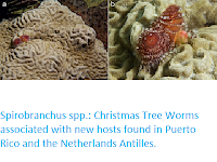
Spirobranchus spp.: Christmas Tree Worms associated with new hosts found in Puerto Rico and the Netherlands Antilles.

Caribbean Christmas Tree Worms, Spirobranchus spp., are considered host generalists in their associations with Anthozoan (Scleractinia) and Hydrozoan (Millepora) Stony Corals. As planktonic larvae, they settle on Coral surfaces and start secreting a calcareous tube to be used as a dwelling. This tube usually becomes overgrown by the host Coral (except for its opening) and may get encapsulated deep inside the Coral skeleton. In this manner, the well-protected Worms grow and survive predation and other hazards, allowing them to live for over four decades. When the host Corals are overgrown by other organisms, such as Octocorals and Sponges, these may act as secondary hosts.

In a paper published in the journal Diversity on 21 March 2020, Bert Hoeksema of the Taxonomy and Systematics Group at the Naturalis Biodiversity Center, the Groningen Institute for Evolutionary Life Sciences at the University of Groningen, and the Institute of Biology Leiden at Leiden University, Jaaziel García-Hernández of the Marine Genomic Biodiversity Laboratory at the University of Puerto Rico - Mayagüez, Godfried van Moorsel of Ecosub and the ANEMOON Foundation, Gabriël Olthof also of the Taxonomy and Systematics Group at the Naturalis Biodiversity Center, and the Institute of Biology Leiden at Leiden University, and Harry ten Hove, again of the Taxonomy and Systematics Group at the Naturalis Biodiversity Center, report two new primary hosts (Scleractinians) and two new secondary hosts (a Zoantharian Soft Coral and an Ascidian colonial Sea Squirt), discovered during recent surveys (2015–2019) in the southern and eastern Caribbean, as well as in the Greater Antilles.

The Coral–Worm associations occurred in shallow subtidal water (less than 4 m depth), with the Knobby Brain Coral, Pseudodiploria clivosa, hosting Spirobranchus giganteus at St. Eustatius in the northern Leeward Islands, and the Golfball Coral, Favia fragum, hosting both Spirobranchus giganteus and Spirobranchus polycerus at Bonaire in the southern Leeward Islands. The secondary host observations, both for Spirobranchus giganteus, involved the Zoantharian Palythoa caribaeorum at Puerto Rico and the Ascidian Trididemnum solidum at Bonaire and Curaçao. Palythoa caribaeorum represents a first record as a secondary host for a species of the order Zoantharia. Until now, the only other Anthozoan secondary hosts were species in the order Alcyonacea (subclass Octorallia), whereas Trididemnum solidum represents an entirely new host phylum, the Chordata. The only other non-Anthozoan secondary hosts known to date are Sponges (Porifera).

A Coral of Pseudodiploria clivosa at 2 m depth, Scubaqua House Reef, St. Eustatius, Eastern Caribbean (2015) hosting Spirobranchus giganteus: (a) overall view and (b) close-up. Hoeksema et al. (2020).

The two new Scleractinian hosts are both typical for shallow subtidal water near the shoreline (less than 4 m depth), where a lack of previous surveys may explain why they have not previously been reported. The new records of secondary hosts are remarkable because these encrusting Animals are known to be aggressive in competition for space with Scleractinians by allelopathy (the production of harmful biochemicals) and can be abundant on shallow reef flats and slopes, where they usually outcompete and kill Scleractinian Corals by overgrowing them. In both cases, the Christmas Tree Worms survive by withstanding this overgrowth and maintain an open space near the tube opening.

Favia fragum hosting Spirobranchus spp. at 3–4 m depth, dive site 'Front Porch', Bonaire, Southern Caribbean (2019). (a)–(c) Spirobranchus giganteus: overall view (a), overgrown tube section indicated by red arrow (b); antler-shaped opercular spines showing dark pink colouration indicated by yellow arrow (c). (d) Spirobranchus polycerus: two individuals, one showing white spines on its operculum (blue arrow). Hoeksema et al. (2020).

Hoeksema et al.'s new host records confirm two Caribbean Christmas tree worms as generalist symbionts capable of infesting a large spectrum of host Corals. They are also strong survivors when their primary hosts become overgrown by more aggressive competitors for space. Previous host records mostly concern  Spirobranchus giganteus, but Hoeksema et al. also report a new host Coral for Spirobranchus polycerus. This worm species occurs in shallow water (less than 4 m depth), whereas Spirobranchus giganteus is commonly found down to 40 m depth. Both Spirobranchus species can easily be distinguished, as Spirobranchus giganteus shows long dark pink opercular spines, whereas those of Spirobranchus polycerus are short and white. Furthermore, Spirobranchus giganteus may be larger than Spirobranchus polycerus and usually shows six to seven (maximum eight) whorls in its branchial spires, whereas Spirobranchus polycerus has two to three (maximum five).

Palythoa caribaeorum acting as a secondary host for Spirobranchus giganteus at 5 m depth, Cayo Media Luna (La Parguera Natural Reserve), Puerto Rico, Greater Antilles (2017): (a) Worm extended and (b) retracted, showing the tube opening surrounded by dead Coral; damage to the Zoantharian host caused by the operculum of the extended Worm indicated by a black arrow. Hoeksema et al. (2020).

Hoeksema et al.'s observations suggest that future surveys may discover other hosts for both Spirobranchus species with the possibility of more host overlap. Whether such host sharing is related to their phylogenetic affinities or to ecological similarities (e.g., overlapping bathymetric distributions) is an open question that merits assessment.

Trididemnum solidum acting as a secondary host for Spirobranchus giganteus in the Southern Caribbean: (a), (b) dive site 'Thousand Steps', Bonaire (2019); (c) Marie Pampoen, Curaçao, 12 m depth (2017); (d), (e) Daaibooi Bay, Curaçao (2017). Extended Worms (a), (d) and the same individuals retracted, showing an open space in front of the Worm tube mouth (b), (e). Hoeksema et al. (2020).

See Also...


Follow Sciency Thoughts on Facebook.

Tuesday, 18 February 2020

Searching for suspended and Salp-ingested microplastic debris in the North Pacific, using epifluorescence microscopy.

Marine debris is a worldwide ocean pollution problem, with plastics found in virtually all aquatic environments. The majority of marine debris analyzed to date has been microplastic, plastic particles less than 5 mm in size. However, findings suggest even smaller plastics (less than 333 μm) are both under-sampled due to the inappropriate mesh size of common sampling nets and far more numerous because plastic particles physically degrade over time into progressively smaller pieces. Such small debris can be consumed by, and deleterious to, suspension-feeding marine organisms, including Salps. Salps are pelagic Tunicates that possess the highest per-individual filtration rates among marine zooplankton, ingesting particles from under 1.0 μm to about 1.0 mm in size. They primarily feed in the upper water column, where microplastics are abundant. Once plastics are ingested by zooplankton, they have the potential to bioaccumulate in the food web into larger organisms, along with adsorbed persistent organic pollutants and harmful chemical additives, with unknown physiological consequences.

In a paper published in the journal Limnology and Oceanography Letters on 27 November 2019, Jennifer Brandon of the Scripps Institution of Oceanography at the University of California San Diego, Alexandra Freibott, also of the Scripps Institution of Oceanography, and of the Pacific Northwest Research Station of the United States Forest Service, and Linsey Sala, again of the Scripps Institution of Oceanography, present the results of a study which aimed to isolate, identify, and quantify microplastics 5–333 μm in size, a subgroup of microplastics which they termed mini-microplastics, from surface seawater samples and salp specimens collected from the North Pacific.

Although many zooplankton species consume microplasticc in a laboratory setting, the ecologically significant question lies in whether they are ingesting such particles in situ. Although the measured abundance of surface seawater microplastics is high, it is 1–3 orders of magnitude below model predictions of plastic inputs. In 2014 a considerable influx of both salp tunics and fecal pellets to about 4000 m depth following a bloom of Salpa spp. in the northeast Pacific. Thus, Salps could be a key link explaining the discrepancy between modeled and measured abundances of buoyant plastics, because fast sinking Salp fecal pellets and carcasses may be a vector moving ingested surface microplastics to the deep sea.

A Salp (plural Salps) is a barrel-shaped, planktic Tunicate. It moves by contracting, thus pumping water through its gelatinous body, one of the most efficient examples of jet propulsion in the Animal Kingdom. The Salp strains the pumped water through its internal feeding filters, feeding on phytoplankton. Wikipedia/Oregon Department of Fish and Wildlife.

Isolating, identifying, and quantifying microplastics 5–333 μm in size is a difficult task in the ocean due to their small size and irregularity. Brandon et al. took advantage of the well-documented autofluorescence of many plastics, and modified an epifluorescence microscopy approach normally used to enumerate planktonic microorganisms, to quantify oceanic mini-microplastics in surface seawater and Salp gut contents. Specifically, they asked: What are the distribution and abundances of these mini-microplastics in surface seawater? Are Salps ingesting mini-microplastics in situ? And, does the size distribution of ingested particles reflect that of available plastic particles?

Surface seawater samples and salp specimens used in this analysis came from the following cruises: SEAPLEX (02–21 August 2009), R/V Falkor (21–30 October 2013), SKrillEx I (26–31 July 2014), and SKrillEx II (11–17 June 2015). Surface seawater samples (1–2 m) were collected in metal buckets, immediately filtered onto 5 μm pore polycarbonate filters, and frozen. Brandon et al. sorted Salps from sodium borate-buffered 1.8% formaldehyde preserved plankton samples. These were collected via a 202 μm mesh bongo net at a tow depth of approximately 200 m or a surface-dwelling 333 μm mesh manta net. They tested for airborne plastic contamination during sample collection on a separate cruise in January 2017 by separately filtering both surface seawater samples and ultra-filtered Milli-Q water.

Maps of sampling locations, for testing of microplastics both in surface seawater and in Salp gut contents via epifluorescence microscopy. Surface seawater samples were taken via bucket tow in the open ocean (A) on the R/V Falkor (yellow, California Current; purple, transition region; mint green, North Pacific Subtropical Gyre; grey, SKrillEx sites), in 2013 and the nearshore (B) on SKrillEx I in July 2014 (blue) and SKrillEx II (green) in June 2015. Salp samples were taken via manta tow in the open ocean (A), on SEAPLEX (red), in August 2009, and in the nearshore environment (C), on SKrillEx I in July 2014. In (C) the bright green dots indicate stations with 10 Salps present, dark green indicate less than ten Salps present, and grey indicate no Salps in the sample. Brandon et al. (2019).

Brandon et al. sorted, measured, and identified Salp species and life history stage from preserved specimens from each sampling location, located in three open ocean regions: North Pacific Subtropical Gyre, California Current, the transition region, and a nearshore region. Life history stage was designated as blastozooid, the sexual chain-forming generation, or oozooid, the asexual solitary generation. Salp guts were dissected; however, any existing mucous nets and gill bars were not analyzed to avoid artifacts of net feeding.

Traditional epifluorescence microscopy techniques add fluorochromes to stain the DNA and proteins of plankton so that identifying features appear under different reflected wavelengths of light. Because Brandon et al.'s target was identification of plastics, not living organisms, they did not add any fluorochromes. Brandon et al. left prepared slides at room temperature for at least 24 hours to diminish chlorophyll a autofluorescence of plankton before visualization. This ensured the most fluorescent particles on microscopy images were likely microplastics, bacteria, or transparent exopolymeric particles. Brandon et al.tested multiple plastic and nonplastic reference materials, such as cotton and wool, under the four light excitation channels of our microscope to determine their autofluorescence. Filtered surface seawater samples and Salp gut contents were prepared for microscopy using an all-glass filtration apparatus.

Brandon et al. created a decision tree to determine if a particle was plastic. Generally, plastics appeared as long, thin fibers or flat fragments with sharp edges. Plastic particles fluoresced uniformly and did not have inner striations, coloration patterns, or features suggestive of biological particles, such as spines, nuclei, or organelles. Not all plastics fluoresce, so this was not used as a diagnostic feature Particles that were invisible under transmitted light but fluoresced under another light channel were determined to be transparent exopolymeric particles. Particles identified as likely diatom frustules, including chain-formers and pennates like Pseudo-nitzschia, were not counted as plastics. When in doubt, particles were not counted as plastic, so Brandon et al.'s estimates are conservative and most likely underestimate total mini-microplastic abundance.

Decision tree for enumerating plastic microdebris on slides, used to determine which particles were plastic and which were biota or other detritus. Brandon et al. (2019).

Particles were categorized as short or long fibers and fragments. The lengths, widths, areas, and fluorescence were recorded for every fragment and short fiber (under 300 μm) in automated images. Long fibers (at least 300 μm) were enumerated in separate, manual visual transects at lesser magnification to eliminate the possibility of double-counting single large fibers that were not visualized in their totality in automated images. Fibers under 200 μm in length were not counted in manual transects; however, there may be some overlap between the short fibers counted in automated images and long fibers counted in manual transects, due to the 200–300 μm overlap. Brandon et al. recorded long fiber color, length, and width with an ocular micrometer.

Brandon et al. analyzed plastic particles in filtered salp gut contents via epifluorescence microscopy. Because Salp gut walls and ingested biogenic material can fluoresce, fluorescence was considered a secondary characteristic of ingested plastic over particle shape and reflectivity under transmitted light. However, fluorescence was checked to visualize inner striations or patterns characteristic of diatom chains. When in doubt, particles were not counted as plastic. The thick gut walls of Salps and ingested biogenic material most likely occluded some plastic, so our data underestimate total plastic ingestion.

To calculate salp ingestion rates of plastic, mini-microplastic counts were divided by gut clearance times for each species identified, which ranged from 2.5 to 6.25 hours.

Brandon et al. found different patterns of fluorescence between plastic and biological materials, and when in doubt, particles were not counted as plastic. Using a controlled test, they determined that the vast majority of mini-microplastic materials in these filtered seawater samples were not from contamination during processing.

Transmitted light and epifluorescence images of microplastics from surface seawater, including a plastic fragment (A), thick and thin short plastic fibers (B), a long fiber and transparent exopolymer particles (C). Column (1) transmitted light; Column (2) Excitation 450-490 nm, Emission greater than 515 nm; Column (3) Excitation 340-380 nm, Emission 435-485 nm; Column (4) Excitation 465-495 nm, Emission 635-685 nm. Brandon et al. (2019).

Brandon et al. detected no significant spatial heterogeneity in seawater plastic concentrations across the Falkor transect (at 12 h intervals) for three open ocean regions: North Pacific Subtropical Gyre, California Current, the transition region. Nearshore samples from SKrillEx I and II were collected at approximate 15 km intervals, and showed no significant spatial heterogeneity possibly due to small sample sizes. Mean open ocean mini-microplastic concentrations compared to nearshore demonstrated significant heterogeneity between regions. Nearshore mini-microplastic concentrations differed from all other regions

Open ocean mini-microplastic concentrations were on the order of 10-100 per litre for short fibers and fragments with lower long fiber concentrations (1-10 per litre). In contrast, the fluorescent long fibers were 3.5–6.5 times more abundant than mean concentrations of nearshore short fibers and fragments on SKrillEx I, and almost eight times more abundant on SKrillEx II.

Almost every open ocean fragment and short fiber was below 333 μm in length and would have been missed by previous studies using larger mesh nets. Long fibers were usually over 333 μm, but thin enough to easily slip through 333 μm mesh. The minimum lengths of fragments and short fibers were between 14 and 50 μm for all locations, approaching the 5-μm pore size of the filters. For long fibers, both surface area and length were significantly different among regions, with significantly shorter fibers in the transition region. Similarly, in the nearshore samples (SKrillEx I and II), every measured fragment and short fiber length was under 333 μm

Individual particle surface area ranged from 0.0 003 to 0.71 mm², compared to earleir studies using a 333 μm net, which detected particles 0.01–565 mm². These earlier studies found plastic particle lengths ranging from 0.34 to 65.7 mm, while Brandon et al found lengths from 0.01 to 16.27 mm (including long fibers). Ultimately, the most pronounced difference between Brandon et al.'s findings and those of earlier studies was not the size range of particles, but rather their concentrations. Mini-microplastics in this study were five orders of magnitude more abundant than the over 333 μm microplastics of earlier studies. However, when concentration was multiplied by surface area Brandon et al. found that the over 333 μm microplastics had significantly higher areal concentrations than the under 333 μm mini-microplastics.

Salps have a very interesting life cycle, known as alternation of generations.  Salps have two different life stages: a solitary asexual stage and a colonial sexual stage.  The solitary stage of Salps (also referred to as the oozooid stage) has a special structure called a stolon.  This stolon develops into chains of the colonial stage.  When a chain of the aggregate stage has grown large enough within the solitary organism the chain will be released.  The chain is the sexual stage of the Salp (also referred to as the blastozooid stage).  In many species, the colonial stage (chain) looks very different from the solitary stage, and in fact when scientists first discovered Salps they often thought that the colonial and solitary stages of the same species were actually different species.  Chains of the colonial stage can extend for several meters in the water, and include hundreds of individuals.  The chains start off as females, and are fertilized by the sperm from older chains.  Once the eggs within a chain are fertilized, an embryo (of the solitary stage) will grow within each individual of the colonial stage.  The colonial stage will then give live birth to the solitary (asexual) stage so that the process can repeat itself.  As the chains matures it will switch from a female to a male. FSU Zooplankton Ecology and Biogeochemistry Lab.

Every single Salp gut analyzed contained plastic. Blastozooids had higher ingestion rates than oozooids. In general, nearshore Salps were larger than open ocean Salps  The California Current Salps were the smallest and had the lowest plastic ingestion rates. Excepting the North Pacific Subtropical Gyre and transition region oozooids, there was no detectable relationship between body length and plastic ingestion rate for dissected Salps. Although Brandon et al found regional differences in mini-microplastic concentrations in the water column, there was no significant effect of region on Salp plastic ingestion rate. Fibers made up 91% of the total ingested particles. The surface area and lengths of fibers and fragments differed significantly between most regions.

Brandon et al. compared the size of ingested mini-microplastics with that of ambient mini-microplastics in surface seawater, both from their data and from earlier studies Most of the net-collected particles from earlier studies fell within the size range of potential Salp food particles. At all sample locations, the average size of particles consumed by Salps was significantly smaller than the size of ambient seawater plastic.

Brandon et al. successfully used epifluorescence microscopy to identify mini-microplastic particles in natural seawater samples and Salp gut contents. This method required careful judgment and expertise to distinguish biotic from plastic materials. Furthermore, autofluorescence of Salp gut walls and biogenic materials made ingested plastic fluorescence only a secondary identification characteristic. This method allowed us to distinguish plastic from nonplastic particles and fluorescent from nonfluorescent plastic, but not to identify specific plastic types. Isolating plastic-type autofluorescence patterns under specific emission wavelengths may permit such differentiation in future work. However, our ultimate goal was to use standard epifluorescence microscopy techniques to differentiate plastics from nonplastic particles in order to obtain accurate bulk measurements of plastics under 333 μm, which the method accomplished.

This study may be one of the first to estimate the abundance of the smallest mini-microplastics in surface seawater, which are consistently under-sampled. Brandon et al. found a mean plastic concentration across all locations of 8277 particles per litre (8 277 000 particles per m³). Their particle concentrations averaged 5–7 orders ofmagnitude higher than previous studies. This highlights the previously unquantified significance ofmini-microplastics inmarine debris counts.

Nearshore samples had higher plastic concentrations than open ocean samples. This agrees with published findings that have recorded similar spikes in plastic concentrations nearshore, close to populated areas, with a decline in plastic moving offshore. The difference in plastic concentrations between SKrillEx I and II may be explained by annual differences in rainfall and watershed input to these nearshore waters.

Many estimates of macro- and microdebris, including modeled debris trajectories agree that the highest concentrations of open ocean marine debris occur in convergence zones of subtropical gyres. However, Brandon et al. did not detect a significant increase in mini-microplastic concentration in the North Pacific Subtropical Gyre and their open ocean samples were not significantly different across regions. Many possible sinks of mini-microplastics could account for this. Plastic below 5 μm in size presumably degrade beyond the detection limit of Brandon et al.'s method. Plastics can also be biofouled and sink out of surface water, or ingested and removed from the water. As plastic accumulates in the North Pacific Subtropical Gyre and breaks down into progressively smaller pieces, Brandon et al.'s data suggest that plastic under 333 μm is removed from the gyre through biofouling, ingestion, or degradation at the same rate it is being supplied. In the nearshore zone, however, mini-microplastics, especially long fibers (in the 200 μm–17 mm range), likely have a higher rate of input than loss. All of these sources and sinks require further research to be better parameterized.

Earlier studies sampled almost no particles smaller than 0.333 mm × 0.333 mm (0.11 mm²), due to the mesh size of the sample collection net, while Brandon et al. detected many particles below that limit (minimum size 0.000 3 mm²). Their results show the majority of plastic concentrations occur between under 333 μm and 0.11 mm². Although the mini-microplastics they measured were more numerically abundant, they did not comprise the majority of the plastic surface area in the water. Organisms that colonize surface substrates in the ocean are more likely to find surface area on micro- and macroplastics rather than mini-microplastics, despite the numerical abundance of mini-microplastics.

This is the first record of Salp ingestion of microplastic in situ. Every Salp dissected had plastic in its gut, regardless of species, life history stage, or region of the ocean sampled. Salp gut clearance times are on the order of 2–7 hours, so Brandon et al. are confident that by only analyzing the gut, they avoided artifacts of net feeding or other contamination. Airborne contamination is a major concern in modern microplastic work, especially when samples are dominated by fibers, as in this study (91% of the Salp-ingested particles). However, our processes of seawater filtration, slide preparation, and salp dissection limited contamination. Compared to filtered control samples, most of the plastics in Brandon et al.'s surface seawater samples were not contamination.

Brandon et al. detected no regional differences in plastic ingestion by Salps, excluding the much lower values of the California Current Salps. This finding is likely attributable to the very small body size of those salps. The California Current Salps had the lowest ingestion rate of any region, whereas for surface seawater, concentrations of mini-microplastics in the nearshore environment were significantly higher than the entire open ocean. Overall, however, both Salp ingestion and surface seawater plastic concentrations had limited regional differences.

Salps are predominantly generalist suspension feeders with ingestion based primarily on particle size, typically from less than 1 μm to 1 mm. All seawater mini-microplastic measured, and almost all the plastic in earlier studies, fall within their possible ingestion range. Yet, the Salps sampled by Brandon et al. ate significantly smaller pieces of plastic than were available in the ambient surface water. This may be explained by the fact that Salps can efficiently collect down to submicron particles and feed throughout a greater area of the water column than the surface, where larger,more buoyant plastic is retained.

Salps are of ecological importance due to several factors: their notoriously rapid growth and opportunistic reproductive rates that can lead to extremely high population densities or 'blooms', higher filtration rates per individual than any other zooplankton grazer, and production of dense fecal pellets that can result in high vertical fluxes of this material to deeper depths. The large fecal pellets of Salps have proven to possess rapid sinking and slow decomposition rates such that they can reach the deep ocean relatively intact, transporting organic carbon and potential microplastics with them. Brandon et al.'s evidence for the widespread and universal consumption of microplastics by Salps leads Brandon et al. to believe that Salps may be an important vector of marine debris transport from the surface ocean to deep-sea communities. The transport of microplastics via Salps may be critical to incorporate into microplastic export calculations as an overlooked output from surface waters.

See also...

https://sciencythoughts.blogspot.com/2019/12/assessing-impact-of-plastic-waste-on.htmlhttps://sciencythoughts.blogspot.com/2019/09/cetacean-sightings-within-great-pacific.html
https://sciencythoughts.blogspot.com/2019/01/mercury-and-selenium-levels-in.htmlhttps://sciencythoughts.blogspot.com/2015/10/microplastics-in-deep-sea-marine.html
https://sciencythoughts.blogspot.com/2014/12/counting-floating-plastics-in-worlds.htmlhttps://sciencythoughts.blogspot.com/2014/05/marine-litter-on-european-seafloor.html
Follow Sciency Thoughts on Facebook.