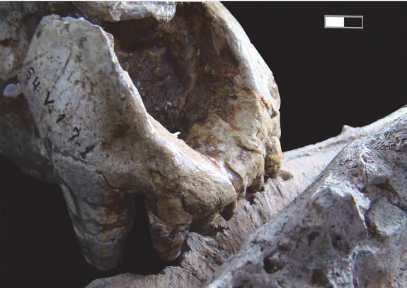
Glyptodonts, Cingulata, are known from the late Eocene to the early Holocene of South, Central, and North America. Their fossil record is represented mainly by disarticulated osteoderms during the Eocene and Oligocene, and the oldest known skull remains are from the early Miocene. Glyptodont skulls are relatively well known from the early Miocene through the earliest Holocene, when Glyptodonts became extinct. The oldest known Glyptodont skulls belong to the Propalaehoplophorinae, from the Santa Cruz Formation (late early Miocene) in Patagonia, Argentina, and to Glyptodontidae incertae sedis from the Chucal Formation (early Miocene) in the Chilean Altiplano.
From the Santa Cruz Formation, several skulls and mandibles have been described and assigned to five traditionally recognised genera (Propalaehoplophorus, Cochlops, Asterostemma, Eucinepeltus, and Metopotoxus), and although we have information about the phylogeny, taxonomic issues, body mass, locomotion, mastication, and feeding habits of these Glyptodonts, our knowledge on the juvenile stages is still poor.
Juvenile specimens of Miocene Propalaehoplophorinae Glyptodonts were mentioned by Florentino Ameghino, William Berryman Scott, and Aden Tauber, but never described in detail. Additional information of Pliocene and Pleistocene juvenile Glyptodonts appeared in several different publications. Other contributions exclusively referred to the description of juvenile specimens of Pliocene and Pleistocene Glyptodonts are mostly based on isolated osteoderms or teeth, with the remarkable exception of a fetal specimen of Glyptodon, described in 2009.
In a paper published in the Journal of Vertebrate Paleontology on 5 August 2020, Laureano González Ruiz of the Laboratorio de Investigaciones en Evolución y Biodiversidad and Centro de Investigaciones Esquel de Montaña y Estepa Patagónica at the Universidad Nacional de La Patagonia San Juan Bosco, and the Concejo Nacional de Investigaciones Científicas y Técnicas, Diego Brandoni of the Laboratorio de Paleontología de Vertebrados at the Centro de Investigación Científica y de Transferencia Tecnológica a la Producción, and the Concejo Nacional de Investigaciones Científicas y Técnicas, Alfredo Zurita of the Centro de Ecología Aplicada del Litoral and Universidad Nacional del Nordeste, and the Concejo Nacional de Investigaciones Científicas y Técnicas, Jeremy Green of the Department of Geology at Kent State University at Tuscarawas, Nelson Novo of the Instituto Patagónico de Geología y Paleontología, and the Concejo Nacional de Investigaciones Científicas y Técnicas, Adan Tauber of the Escuela de Geología at the Universidad Nacional de Córdoba and Museo Provincial de Ciencias Naturales ‘Dr. Arturo Umberto Illía’, and Marcelo Tejedor, also of the Instituto Patagónico de Geología y Paleontología, and the Concejo Nacional de Investigaciones Científicas y Técnicas, describe a newly recovered mandible belonging to a juvenile specimen of a Propalaehoplophorinae Glyptodont, collected by their team in field work carried out during 2015 in Santa Cruz Province, Argentina, and discuss the mandibular and dental characters of Glyptodonts.
The specimen was collected at the cliffs of the estuary of the Gallegos River in the property of Killik Aike Norte farm, Santa Cruz Province (Patagonia, Argentina). The locality of Killik Aike Norte, where the Santa Cruz Formation crops out, has been known since 1845 and is dated in this area from about 18 to 16 million years old (Burdigalian, late early Miocene).
(A) Location map of the fossiliferous locality Killik Aike Norte, with the Santa Cruz Formation indicated; (B) photograph of the profile; (C) generalised geological profile. González Ruiz et al. (2020).
Three fossiliferous levels have been described for Killik Aike Norte, which are placed at the base of the section: two of sandstone (NF1, NF3) and one tufaceous (NF2) interbedded. The most abundant fossiliferous level (NF2) has been dated to ca. 17.0 million years ago, and all the fossil levels (NF1, NF2, NF3) are below a guide level of the Santa Cruz Formation, the white tuff (TB, ‘Toba Blanca’) dated to about 16.89 million years ago.
The new specimen was found in an isolated fragmentary rock, filled and surrounded by a matrix of volcanic ashes, of massive structure, probably coming from the NF2 (about 17.0 million years old) or any of the potentially fossiliferous upper levels of ash, ranging from about 16.4 to about 16.9 million years old. This specimen increases the numerous fossil vertebrates known for Killik Aike Norte, especially the Cingulate association represented by Peltephilus, Stegotherium, Prozaedyus, Stenotatus, Proeutatus, and Eucinepeltus.
The specimen described is MPM PV 17408, a fragment of mandible (left dentary) with most of the horizontal ramus (corpus mandibularis), the mandibular symphysis, mf1–mf7, and the external wall of the alveoli corresponding to the mf8. The mf8 and most of the upper region of the vertical ramus (ramus mandibularis) are not preserved. This is tentatively referred to the genus Eucinepeltus as cf. Eucinepeltus.
The preserved portion measures 73 mm in length. The horizontal ramus is elongate; the posterior half is regularly curved, convex labially, and flat lingually, and the anterior half is ventrolingually curved and anteroventrally expanded, forming the symphyseal area. The vertical ramus is not preserved. In occlusal view, there is an almost complete, short predental zone (‘spout’) of 4.1mm, only slightly everted, followed posteriorly by the tooth row. In lateral view, anteriorly, there is no labial depression (for the descending process of the zygomatic arch) and there are three mental foramina: the anterior-most below the mf2, a second small mental foramina below the mf2–mf3, and the posterior-most below the mf3. Posteriorly, the base of the ascending ramus is preserved, represented by the base of the coronoid process at the anterior face of the mf7. In medial view anteriorly, the mandibular symphysis extends dorsally from the rostral tip of the mandible ahead of the mf1 to the posterior margin of the mf3 ventrally and exhibits a rough surface and a rounded anteroventral lingual edge. The symphysis extends slightly below the ventral margin of the horizontal ramus. The mandibular/dental canal is ventral to mf2, and lingual to the mf3–mf7 because the roots of mf4–mf7 extend ventrally almost to the ventral margin of the mandibular ramus, which is broken lingual to the mf6 where the mandibular/dental canal is present.

MPM PV 17408, cf. Eucinepeltus, left hemimandible in (A) labial, (B) lingual, and (C) occlusal views. Abbreviations: cp, base of the coronoid process; emf, external wall of the mf8 alveoli; mc, mandibular canal; mef, mental foramina; mf, molariform; pz, predental zone; rsvm, rounded symphyseal ventral margin; sa, symphyseal area. Scale bar equals 10 mm. González Ruiz et al. (2020).
There are seven molariforms (mf1–mf7). In occlusal view, mf1–mf3 are oriented obliquely to the longitudinal axis of the tooth row, whereas mf4–mf7 are oriented in the same longitudinal axis. In lateral view, all molariforms are conical, with the base ventral and the lateral faces oblique to the plane of the dental series. The preserved tooth row measures 57.4 mm in length. Molariforms have the long axis anteroposteriorly oriented, increasing in size from mf1 to mf5 and decreasing from mf5 to mf7. The mf1–mf4 are simpler than mf5–mf7, with increasing complexity of the development of lobes and grooves from mf1 to mf5, and mf5–mf7 are subequal in complexity.

MPM PV 17408, cf. Eucinepeltus, left hemimandible, digital transverse sections. (A) At mf2 level (first mental foramina); (B) at mf2 level (second mental foramina); (C) at mf3 level; D, at mf4 level. Abbreviations: bn, bone; mc, mandibular canal; mef, mental foramina; mf, molariform; sed, sediment. Scale bar equals 5 mm. González Ruiz et al. (2020).
The mf1 has a reniform outline, lingually convex and labially concave, with no deep grooves clearly delimiting lobes. The mf2 is more distinctly reniform than mf1 in outline, with two shallow grooves not reaching the tip of the tooth, one lingual anterior and one labial central defining two lobes, thus making an asymmetrical bilobed outline. The mf3 is reniform and sigmoid in outline, similar to mf2 although larger, with a lingual deep anterior groove and a labial deep central groove not reaching the tip of the tooth and defining an anterior lobe and a larger posterior one; the central groove has two inner, shallow and narrow grooves, which do not reach the tip of the tooth and define the incipient middle labial lobe, absent in mf1–mf2, indicating a lingual bilobed outline and an incipient labial trilobed outline. The mf4 is reniform and sigmoid in outline, with a deep anterior groove lingually and a shallow, narrow posterior groove, both reaching the tip of the tooth and defining an incipient middle lobe from the larger posterior lobe, and two deep, narrow grooves labially that reach the tip of the tooth and define anterior and posterior larger lobes and a small middle lobe, indicating a more trilobed outline than mf3 but less than mf5. The mf5–mf7 have the typical trilobed outline, with anterior, middle, and posterior lobes limited by two labial and two lingual deep and wide grooves, one anterior and one posterior in each case, all reaching the tip of the tooth. The lobes are rounded; the anterior and middle lobes are symmetrical, and the posterior lobe is asymmetrical with the labial half larger; the posterior face of the tooth is flat and oblique to the long axis. All molariforms retain the external observable outline from the top to the base, having open roots.

MPM PV 17408, cf. Eucinepeltus, left hemimandible, digital rendering in (A) labial, (B) lingual, (C) occlusal, and (D) ventral views. Bone is transparent. Abbreviation: mf, molariform. Scale bar equals 10 mm. González Ruiz et al. (2020).
The occlusal surfaces of the mf1–mf2 are blunt and lack wear facets, having a central rounded cusp. The mf3 has a central rounded cusp similar to that of mf1–mf2, and a posterolingual, subcircular, small facet oblique to the horizontal plane of the horizontal ramus, occupying a small part of the occlusal surface. The mf4 has one anterior small, oval facet and one posterior large, reniform facet occupying most of the occlusal surface; both facets are perpendicularly oriented, and together produce a beveled occlusal surface. The anterior (smaller) and the posterior (larger) facets are mesioventrally and distoventrally oriented, respectively, oblique to the plane of the horizontal ramus. The mf5–mf7 have a single, trilobed, and almost flat facet oblique distoventrally to the plane of the horizontal ramus, which occupies all the occlusal surface of the tooth.

MPM PV 17408, cf. Eucinepeltus, left lower molariforms, digital renderings in occlusal view. (A) mf1; (B) mf2; (C) mf3; (D) mf4; (E) mf5; (F) mf6; (G) mf7. Abbreviations: alb, anterior lobe; alagr, anterior labial groove; aligr, anterior lingual groove; clagr, central labial groove; cgr, central groove; crc, central rounded cusp; horth, hypermineralized orthodentine; lahalb, labial half anterior lobe; lahmlb, labial half middle lobe; lahplb, labial half posterior lobe; lihalb, lingual half anterior lobe; lihmlb, lingual half middle lobe; lihplb, lingual half posterior lobe; mf, molariform; mlalb, middle labial lobe; mlb, middle lobe; mlilb, middle lingual lobe; orth, orthodentine; ostd, osteodentine; plagr, posterior labial groove; plb, posterior lobe; pligr, posterior lingual groove; wfc, wear facet. Scale bar equals 1 mm. González Ruiz et al. (2020).
The mf1 and mf2 have no attrition facets, and the external layer of orthodentine covers all the molariform; in the posterior facet of mf3 and in the anterior facet of mf4, the external and the middle layers of orthodentine are observable, whereas the internal osteodentine layer is not; in the posterior facet of mf4 and in the facets of mf5–mf7, the external and the middle orthodentine layers and the internal osteodentine layer are observable. In the facets, the external orthodentine and the internal osteodentine (when observable) layers are elevated with respect to the middle orthodentine layer. The external orthodentine layer stands out as a rim in all facet outlines, and the internal osteodentine layer forms a straight, elevated, central line without branching, extending from the center of the anterior lobe to the posterior lobe where it is inclined labially.

MPM PV 17408 (cf. Eucinepeltus), mf1, mf3, mf4, and mf5 of left hemimandible, digital renderings. (A) mf1 in occlusal view; (B) detail of microwear in nonchewing region of mf1; (C) mf3 in occlusal view; (D) detail of the wear facet of mf3; (E) mf4 in occlusal view; (F) detail of the beveled wear facets of mf4; (G) mf5 in occlusal view; (H) detail of the growth lines of mf5. Abbreviations: mf, molariform; Lab, labial; Ling, lingual. Scale bars equal 1 mm. González Ruiz et al. (2020).
In the external layer of orthodentine, all molariforms have transverse, rounded, regularly spaced long-period incremental lines in the dentine (‘Andresen lines’) around the tooth and from the base to the tip. These ‘Andresen lines’ are parallel or subparallel to the horizontal plane of the horizontal ramus, but some of them, especially those of the labial face of the posterior lobe, are oblique; in the mf5, González Ruiz et al. counted 17 per 50 mm.
The predental zone of the mandible is short and slightly everted in MPM PV 17408 and Eucinepeltus, whereas it is long and strongly everted in Propalaehoplophorus.The horizontal ramus has no labial depressions and the ventral margin is regularly curved in MPM PV 17408 and Eucinepeltus, whereas the labial depressions are present and the ventral margin is more convex in Propalaehoplophorus. The hypsodonty indices in MPM PV 17408 (0.338) and in adult Eucinepeltus (0.369) are lower than in adult Propalaehoplophorus (0.470), as a result of a shallow horizontal ramus in the first two.
Glyptodont left hemimandibles in labial view. (A) MPMPV 17408, cf. Eucinepeltus; (B) MACN A 4760, Eucinepeltus; (C) MLP 16-15, Propalaehoplophorus. Scale bars equal 10 mm. González Ruiz et al. (2020).
The mf4 of MPM PV 17408 is more similar to the mf1–mf3 than to the mf5–mf7, especially in the reniform and sigmoid outlines, with a marked labial concavity and lingual convexity; mf4 has also a small labial middle lobe but no lingual middle lobe, producing an incipient labial trilobed outline and a lingual bilobed outline. This morphology is similar to that of adult specimens of Eucinepeltus but differs from Propalaehoplophorus.
Glyptodont left hemimandibles in lingual view. (A) MPM PV 17408, cf. Eucinepeltus; (B) MACN A 4760, Eucinepeltus; (C) MLP 16-15, Propalaehoplophorus. Scale bars equal 10 mm. González Ruiz et al. (2020).
Considering that we do not know the complete morphological variations during Propalaehoplophorinae Glyptodont ontogeny, and that Eucinepeltus has three described species from the Santa Cruz Formation (Eucinepeltus petesatus, Eucinepeltus complicatus, and Eucinepeltus crassus) with unresolved taxonomy, González Ruiz et al. assign the specimen to cf. Eucinepeltus.
Glyptodont left hemimandibles in occlusal view. (A) MPM PV 17408, cf. Eucinepeltus; (B) MACN A 4760, Eucinepeltus; (C) MLP 16-15, Propalaehoplophorus. Scale bars equal 10 mm. González Ruiz et al. (2020).
The preserved portion of the dental series (mf1–mf7) measures 57.4 mm in length, ca. 49% of the same portion in an adult (116.2 mm) of Eucinepeltus. In a complete hemimandible of an unborn specimen of Glyptodon, it represents 27% of an adult. The anteroventral edge of the mandibular symphysis is lingually rounded in juvenileand straight in adult specimens of Eucinepeltus and Propalaehoplophorus; the dorsal margin is less distinctly marked in the juvenile than in the adults, and well developed and straight in the latter; the posteroventral margin of the juvenile extends to the mf3–mf4 boundary, to the middle of the mf4 in adults of Eucinepeltus, and to the mf3–mf4 boundary or the middle of the mf4 in Propalaehoplophorus. The mandibular symphysis is not expanded lingually in the juvenile, being almost a flat rugged surface, indicating that the symphysis was not fused as in all adult Glyptodonts.
In MPM PV 17408, the space at occlusal surfaces between molariforms is wider than in adults, as in juvenile specimens of Glyptotherium. The morphology of the molariforms in lateral view is conical, but rectangular in adults of Propalaehoplophorinae and in other glyptodonts. The main differences in the occlusal surfaces between juvenile and adult specimens occur in the mf1–mf4; in this case, the first two have rounded cusps, the third has a rounded cusp and a small facet, and the fourth has a beveled occlusal surface; all eight molariforms in adults have flat occlusal surfaces, similar to the mf5–mf7 in the juvenile, suggesting a stepwise dental eruption.
The ‘Andresen lines’ observed in the external orthodentine layer of the molariforms were interpreted by Óscar Carranza-Castañeda and David Gillette as probable growth lines in juvenile specimens of the Glyptodont Glyptotherium, but they counted 16 per 100 mm, whereas González Ruiz et al. counted 17 per 50 mm, which could indicate a slow down of growth toward adulthood. The relation of the transverse bands with the relative age of the specimens needs to be explored with more specific techniques and a larger sample of specimens.
Florentino Ameghino described the eruption sequence of anterior lower molariforms in Propalaehoplophorinae Glyptodonts based on an ontogenetic sequence of three mandibles, and he observed, from the youngest to oldest: (1) two germs of incisors, the mf1 not erupted, and the mf2 erupted and unworn; (2) two or three semipartitioned cavities in front of mf1 attributed to probable abortive incisors, and the mf1 not erupted; and (3) three small alveoli of the incisors and the mf1 completely erupted and worn. Ameghino concluded that the dental germs are in an alveolar canal that closes progressively, the incisors never erupt, and anterior-most molariforms erupt successively, with the mf1 the last. Unfortunately, the specimens mentioned by Ameghino were not found in collections; they were published without collection numbers or figures, and only the first one was clearly assigned by Ameghino to Asterostemma; in addition, the presence of alveoli of atrophied incisors in Glyptodonts has not yet been corroborated. Previous studies found no evidence of the complete eruption sequence in Glyptotherium, but they have identified a relative late eruption of the Mf8 in one specimen, in which the occlusal surface is somewhat shorter than the alveolar portion and the Mf8 is smaller than Mf7, and in a second specimen they found an apparent later eruption of mf2 and mf3; unfortunately, there are no records of juvenile specimens with the mf8.
Although the preserved molariforms appear to be erupted in the new specimen MPM PV 17408, the alveolar dorsal margin of the horizontal ramus is not fully preserved. This implies the possibility that mf1–mf3 are not fully erupted, especially mf1–mf2, which are unworn. This new specimen supports the view of Florentino Ameghino, who observed that the mf1 is the last tooth to erupt, or at least the last to be functional. This condition of MPM PV 17408, as well as that observed in Glyptotherium by later authors, indicates an eruption progression from the center (mf5–mf7) to the anterior region (mf1–mf3), and also probably to the posterior region of the tooth row (mf8); unfortunately, the mf8 was not preserved in the new specimen. This also agrees in part with a 2011 study in that the eruption of the permanent dentition generally occurs from posterior to anterior in the Armadillo Dasypus. Finally, beyond fragmentary segments of the mandibular/dental canal, González Ruiz et al. could not find evidence of internal structures (i.e., other mandibular canals), probably because of the low quality of preservation of MPM PV 17408, that may indicate the existence of teeth ahead of the mf1 as those found in other Xenarthrans, nor alveoli or dental germs of atrophied incisors as proposed by Ameghino, nor evidence of tooth replacement in any molariform.
Attrition is produced by tooth-to-tooth contact, which tends to form macrowear facets. In MPM PV 17408, attrition is absent in mf1–mf2 but is evident in mf3 due to a small facet, as well as in mf4, which has two facets, one small and one large, forming a beveled occlusal surface; attrition is also seen in mf5–mf7, each with a complete and almost flat facet occupying all the molariform surface. All facets are at oblique angles with respect to the horizontal plane of the horizontal ramus, from ca. 20° in mf4 reducing progressively to ca. 5° in mf7, whereas all occlusal surfaces are subparallel to the horizontal plane of the horizontal ramus in adults. The different degrees of development of the occlusal surfaces allow us to infer that mf5–mf7 are functional first, because they have all occlusal surfaces occupied by attrition facets like in adult specimens, and also because they erupted first; then, the incorporation of molariforms in the masticatory process progresses anteriorly. This progression probably goes also posteriorly, as indicated by the presence of Mf8 with less development of the occlusal surfaces than the preceding Mf7, and so forth, as was indicated for juvenile specimens of Glyptotherium.
The beveled mf4 of this specimen is unique in Glyptodonts because all adult Glyptodonts do not have beveled teeth; the flat occlusal surface is a synapomorphy of Glyptodontia. Beveled molariforms are present in Armadillos due to the occlusion of the lower tooth between its immediate upper homologue and its more anterior neighbor, as reported for Dasypus. Beveled teeth are present in the oldest known Armadillos from the Eocene (Utaetus, Lumbreratherium, and Astegotherium), and in other Armadillos, both extinct (e.g. Prozaedyus, Proeutatus, and Eutatus) and extant (e.g. Dasypus and Euphractus). In this sense, the temporary presence of this character in the juvenile specimen reported by González Ruiz et al. could be a reflection of this beveled occlusal surface or the result of the masticatory movements.
Original microwear usually occurs on occlusal surfaces and does not extend to nonchewing regions of a molar or molariform. The incidence of indistinguishable microwear on both chewing and non-chewing regions of a single tooth has long been cautioned in the interpretation of the originality of microwear features tooth. The microscopic patterns of MPM PV 17408 are visible on occlusal surfaces and extend to all nonocclusal regions of the teeth in this particular mandibular series. The similarity is obvious under both low-magnification light microscopy and scanning electron microscopy. Therefore, González Ruiz et al. cannot reject the hypothesis that the visible scratches and pits in this specimen are a result of random, non-food-related causes. As such, they cannot objectively analyse the ecological potential of microwear on this specimen.
Eucinepeltus has been interpreted, on the basis of the hypsodonty index and the relative muzzle width index, as highly selective feeder in relatively closed habitats, living within the flora of the Santa Cruz Formation (late early Miocene) on the Atlantic coast represented by a mixture of open, semiarid temperate forests and humid warm-temperate forests. Unfortunately, the relative muzzle width could not be calculated for cf. Eucinepeltus because the skull was not preserved, so the niche requirements could not be inferred, although at least the hypsodonty indice is similar to that of an adult Eucinepeltus.
González Ruiz et al. have described in detail one of the few and oldest known juvenile specimen of a Miocene (Burdigalian Age) Glyptodont from Patagonia (Argentina).
The assignment as cf. Eucinepeltus is based on (1) mandibular predental zone not much everted, almost straight and subparallel to the molariform series; (2) horizontal ramus with no labial depressions, and with the ventral margin regularly curved; and (3) mf4 more similar to mf1–mf3 than to mf5–mf7.
The preserved portion of the dental series (mf1–mf7) represents about 49% of the same portion in an adult of Eucinepeltus, and the hypsodonty indice is similar between the new specimen (0.338) and an adult Eucinepeltus (0.369). The main juvenile characters were found at the symphysis (not expanded lingually, rounded anteroventral edge, dorsal margin less distinctly marked, and posteroventral margin extends to the mf3–mf4 boundary) and at the molariforms (conical in lateral view, mf1–mf2 with rounded cusps, mf3 with a rounded cusp and a small facet, and mf4 with a beveled occlusal surface).
In cf. Eucinepeltus, the mf1 is the last erupted tooth, or at least the last to be functional, and the eruption progresses from the centre (mf5–mf7) to the anterior region (mf1–mf3) of the tooth row. Attrition is absent in mf1–mf2 and is evident from mf3 to mf7. The beveled mf4 of this specimen is unique because adult Glyptodonts do not have beveled teeth.
See also...

Online courses in Palaeontology.
Follow Sciency Thoughts on Facebook.
Follow Sciency Thoughts on Twitter.