
Mennerotodus is an extinct Odontaspidid (Sand) Shark that was originally named based on Middle Eocene (Bartonian) teeth from Kazakhstan. The teeth were distinguished by their overall shape and the development of denticulation at the base of the main cusp. Although the genus Mennerotodus was named in 1989, no type specimens were designated, nor were any specimens illustrated in the original publication, so the generic name was therefore a nomen nudum. The original author used the name again soon thereafter, but the genus was not formally recognized until teeth were described and figured in 1994. Three species of Mennerotodus were originally erected, but only the type species, Mennerotodus glueckmani, is currently recognised. Within this species three subspecies, Mennerotodus glueckmani glueckmani, Mennerotodus glueckmani usunbassi, and Mennerotodus glueckmani boktensis, have been named. There was an intent to utilise the subspecies as biostratigraphic tools, as the type stratum for Mennerotodus glueckmani boktensis is the Middle Eocene (Lutetian) Amankisilit Formation, Mennerotodus glueckmani glueckmani is from the lower Shorym Formation (Bartonian), and Mennerotodus glueckmani usunbassi was in the upper Shorym Formation (Bartonian). The current reported temporal distribution for the genus is Late Paleocene (Thanetian) to late Eocene (Priabonian). Since the original description, teeth atributed to Mennerotodusi have been found in Thanetian deposits of the Paris Basin, and the Middle Eocene (Bartonian) of southern Alabama.
In a paper published in the journal Fossil Record on 22 July 2020, David Cicimurri of the South Carolina State Museum, Jun Ebersole of the McWane Science Center, and George Martin of Auburn, Alabama, describe two new species of Mennerotodus from Palaeogene strata of the southeastern United States.
The older of the two species was recovered from the Lower Palaeocene (Danian Stage) Pine Barren Member of the Clayton Formation of southeastern Alabama. The other species derives from the Middle Eocene (Bartonian–Priabonian) Clinchfield Formation in central Georgia. Cicimurri et al. also provide additional morphological features that allow for differentiation of Mennerotodus from similarly shaped teeth of other, coeval genera. Heterodonty within the two new species is discussed, and artificial dentitions of each species are presented. In addition, they comment on the paleobiogeographic and stratigraphic distribution of Mennerotodus in North America.

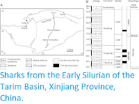
Generalised location and surface stratigraphy of site ALn-13, Lowndes County, Alabama, USA. (a) Geographic maps showing the location of site ALn-13 in country, state, and county contexts. (b) Danian surface stratigraphy in Alabama, USA. Shaded areas on the stratigraphic chart represent unconformities. Cicimurri et al. (2020).
The Danian type specimens described by Cicimurri et al. were all collected by George Martin at site ALn-13 in Lowndes County, Alabama, USA. This sample of teeth was obtained by surface collecting specimens that were eroding directly from a roadside outcrop consisting of numerous beds of the Pine Barren Member of the Clayton Formation. In Butler, Lowndes, and Wilcox counties of Alabama, the Clayton Formation is divided into two members, including the Pine Barren Member and overlying McBryde Limestone Member. The Pine Barren Member is the lowermost Palaeocene unit exposed in Alabama, and it is separated from the underlying Upper Cretaceous (Maastrichtian) Prairie Bluff Chalk by a Type 1 unconformity. In central Alabama the Pine Barren Member can attain a thickness of up to 45 m, 8 vertical meters of which are exposed at site ALn-13. The thick section of the Pine Barren Member in this area spans across parts of calcareous nannoplankton zones NP1 to NP3.
The Clayton Formation is believed to represent shallow marine and marginal marine settings. However, at site ALn-13 the Pine Barren Member exposures are thought to have been deposited in an estuarine setting during a high-stand systems tract. The Pine Barren Member exposures at site ALn-13 has been subdivided into 30 informal units, with beds 14–30 consisting largely of alternating sandy micritic limestones or marlstones and induratedfine- to medium-grained sandy mudstones. Although all of the exposed beds at site ALn-13 are fossiliferous, the Danian teeth reported by Cicimurri et al. were concentrated within the lower two-thirds of the section, within beds 14 to 18.
Previous studies have eported the occurrence of reworked Maastrichtian microfossils within the Pine Barren section at site ALn-13, and extremely rare Cretaceous macrovertebrate fossils were collected from the outcrop by George Martin. These macrofossils include teeth from a Pycnodont Fish, Anomoeodus sp., and the Sharks Squalicorax kaup and Serratolamna serrata. Despite the presence of these reworked specimens, the Mennerotodus teeth collected from this locality all appear to be Danian in origin, as this genus is abundant in the Pine Barren section but absent from any of the Cretaceous units in the state.
The Bartonian type specimens described by Cicimurri et al. were obtained from the Clinchfield Formation, the basal lithostratigraphic unit within the middle to late Eocene Barnwell Group. The fossils were collected from within a defunct kaolinite surface mine, locally known as the Hardie Mine, in Wilkinson County, Georgia, USA. At this site, the Riggins Mill Member of the Clinchfield Formation was exposed, and this formation was disconformably underlain by unfossiliferous middle Eocene kaolinite and overlain by orange–red, cross-bedded sand of the Priabonian-aged Dry Branch Formation. The Clinchfield Formation represents estuarine deposits, and the unit accumulated during calcareous nannofossil zone NP17.

Generalized location and surface stratigraphy of the Hardie Mine
site,Wilkinson County, Georgia, USA. (a) Geographic maps showing the
location of the Hardie Mine site in country, state, and county contexts.
(b) Middle to late Eocene lithostratigraphic units formerly exposed in
the mine. Shaded areas on the stratigraphic chart represent
unconformities. Cicimurri et al. (2020).
The majority of the specimens in Cicimurri et al.'s sample was recovered from spoil piles of the Clinchfield Formation. At the Hardie Mine the apparent mining procedure was to remove Dry Branch Formation deposits from the site and then strip away the Clinchfield Formation and place it in piles throughout the mine. Close examination of in situ kaolinite, Clinchfield Formation, and superjacent Dry Branch Formation deposits showed that the upper and lower confining units were unfossiliferous, and Cicimurri et al confidently say that the specimens they describe herein were all derived from the Clinchfield Formation.
During their study Cicimurri et al examined a number of Mennerotodus specimens from several different states and lithostratigraphic units. The type specimens for the Danian taxon are all housed at the McWane Science Center in Birmingham, Alabama, USA, and the Bartonian type specimens are housed at the South Carolina State Museum in Columbia, USA. Unfortunately, the Hardie Mine has been reclaimed and is no longer available to collectors, but additional fossils from this mine are housed at the South Carolina State Museum; the Georgia College and State University in Milledgeville, USA; and the Mississippi Museum of Natural Science in Jackson, USA. Mennerotodus teeth from the Gosport Sand in Alabama are part of the Alabama Museum of Natural History collections in Tuscaloosa, USA, and additional specimens from the Clayton Formation in Alabama are housed at Mississippi Museum of Natural Science, McWane Science Center, and South Carolina State Museum. A sample of teeth from middle to late Eocene deposits in St Francis County, Arkansas, were examined in the collections of Mississippi Museum of Natural Science and the United States National Museum of Natural History in Washington, DC; Danian specimens from Arkansas were examined in the Mississippi Museum of Natural Science collections.
The jaws of several extant Selachian taxa housed at the South Carolina State Museum, McWane Science Center, and United States National Museum of Natural History were also closely examined as part of Cicimurri et al.'s study. They found that the Mennerotodus teeth discussed are remarkably similar to those of the extant Sand Tiger Shark, Carcharias taurus. Two sets of Carcharias taurus jaws from the South Carolina State Museum collection (SC200.120.6 and SC86.62.2) and an unnumbered jaw from the Gordon Hubbell Collection in Florida were used as models for reconstructing the artificial dentitions for the new Mennerotodus species. Previous studies have commented on the utility of using dentition of extant Sharks to elucidate the morphological variations among fossil teeth, and Carcharias taurus has previously been used as the basis for reconstructions of dentitions of extinct species like Araloselachus cuspidatus, Araloselachus vorax, and even Striatolamia.
There are inter- and intrageneric variations in tooth counts within the Odontaspididae, and Cicimurri et al. concede that the artificial Mennerotodus denti tion as they reconstructed them may not have matched Carcharias taurus exactly. Although the number of intermediate, lateral, and posterior files may vary within Carcharias taurus, the number of anterior files does not, and Cicimurri et al. presume the same held true within the Mennerotodus dentition. Cicimurri et al.'s reconstructions are based on isolated teeth and rely heavily on comparisons with extant Carcharias taurus, and these hypotheses can best be tested only through the discovery and analysis of tooth-bearing jaws of the extinct species. To create the artificial dentitions, in particular to account for ontogenetic size differences between the teeth, the fossil teeth were scaled to reflect the size range of teeth within Carcharias taurus dentition. The photographs of certain teeth were reversed when necessary to help complete the artificial dentitions (i.e., a tooth from the left jaw to represent a file from the right jaw).
Elasmobranch tooth terminology has varied within the literature, and terms like symphyseal, parasymphyseal, medial, alternate, anterior, intermediate, lateral, and posterior have been used to identify the location of a tooth within the jaws of Galeomorph Sharks. The terms 'symphyseal', 'medial' or 'median' and 'alternate', and 'parasymphyseal' have all been used to identify teeth occurring directly on or immediately adjacent to the jaw symphysis. However, an alternative has not been adopted and medial or median has generally only been used to identify the very wide, centrally located teeth within Batoid dentitions like those of the Myliobatidae. An alternative would be restricting the use of symphyseal to only those teeth occurring at the midline of the jaw, where the right and left halves articulate, as seen, for example, in the upper jaw of extant Galeocerdo cuvier and Squalus acanthias. Such a restriction in terminology would therefore result in a tooth file immediately adjacent to the symphysis being considered a parasymphyseal or anterior position. Extant lamniform sharks like Carcharodon carcharias and Isurus paucus, lack teeth directly on the symphysis of the upper and lower jaws, and the first tooth file is considered an anterior tooth. We follow the convention that, within the Meckel’s cartilages of Lamniform Sharks, the tooth file immediately adjacent to the symphysis is the first anterior position. This file occurs within a mesial hollow along with other anterior teeth, all of which are distinctly separated from the elongated furrow that contains the lateral and posterior tooth files. On the palatoquadrates of Lamniform Sharks, a cartilage bar separates the mesial and distal hollows, and teeth occurring just on the mesial side of the bar are identified as occupying an 'intermediate' position. These teeth are much reduced in size compared to the preceding anterior teeth and succeeding lateral teeth, and this phenomenon occurs in Carcharias taurus dentition (SC86.62.2 and SC2006.120.6). In contrast to an obvious morphological difference between the anterior and lateral files of Lamniform dentition, the transition from lateral to posterior files can be more gradational and the term 'lateroposterior' has been applied to tooth files occurring distally to the intermediate file of the upper dentition and the last anterior file in the lower dentition. For the purposes of their report, Cicimurri et al. recognise anterior, intermediate, lateral, and posterior tooth files when identifying isolated teeth.
Mennerotodus teeth can be divided into anterior, intermediate, lateral, and posterior tooth groups. The upper dentition consists of three anterior tooth files, whereas the lower dentition includes four anterior files, and in general the anterior teeth have a tall and narrow triangular main cusp, with a highly convex lingual face and a virtually flat labial face. The enameloid on the main cusp is generally smooth, but faint longitudinal ridges have been observed on the lower half of the crown on a small number of teeth. The main cusp is sinuous in profile view, and the teeth have a single pair of small and conical lateral cusplets. The cutting edges on the main cusp are incomplete and do not extend to the base of the crown. On some teeth one or more denticles are present between the cutting edge and the lateral cusplets, and at times this denticle is expressed as a small ridge that is separate from the main cutting edge. The root lobes are thin with rounded extremities, and a conspicuous nutritive groove is located on a robust lingual root protuberance. The interlobe area on the anterior teeth is generally U-shaped. The first lower anterior tooth is conspicuously smaller in size than the other anterior teeth, and it has an extremely narrow main cusp and elongated distal root lobe. The main cusp on the third upper anterior tooth has a slight mesial bend and an extended mesial root lobe.
The intermediate teeth have a much lower main cusp than the anterior teeth and are smaller in overall size. The height of the root is greater than the height of the crown, and the teeth are labiolingually compressed. The main cusp has a slight distal bend, and a single pair of lateral cusplets are extremely tall in comparison to the overall height of the main cusp.
The lateral teeth are similar to the anterior teeth but have a shorter and more broadly triangular main cusp. The main cusp has a profile that is flat to only slightly sinuous, and this sinuosity is not to the degree seen on anterior teeth. The lateral teeth have one pair or very rarely two pairs of lateral cusplets, and these cusplets are larger and more triangular than those on the anterior teeth. When a second pair of cusplets occurs, they are generally diminutive and united to the outer edge of the larger, more medial cusplet. Denticles occur at the base of the main-cusp cutting edge (medial to the lateral cusplets) on about 30% of the teeth. These denticles are larger than those observed on the anterior teeth and can occur in greater numbers. Although the crown faces are generally smooth, fine vertical ridges are developed on some teeth. The root lobes are short and divergent and have rounded extremities, and the interlobe area can be U-shaped or V-shaped. A conspicuous nutritive groove occurs on a robust lingual root boss. The main cusp is distally inclined on the upper lateral teeth but more erect in the lower lateral files.
The posterior teeth are similar to the lateral teeth but can be differentiated by their having a labiolingual width that is nearly equal to the overall height of the tooth. The main cusp is extremely low and triangular, and the teeth have a single pair of wide and triangular lateral cusplets. The teeth have a shallow and V-shaped interlobe area, and the upper posterior teeth have a more distally inclined main cusp than those in the lower posterior positions.
The first new species described is named Mennerotodus mackayi, in honour of John Mackay, initial President and CEO of the McWane Science Center, for his distinguished career in informal education. The description of the species is based upon speciemens from the Lower Danian (Paleocene) Pine Barren Member of the Clayton Formation, approximately 10m above lower contact with the late Maastrichtian (Upper Cretaceous) Prairie Bluff Chalk Formation, zones NP1 to NP3, at the ALn-13 site in Lowndes County, Alabama. Due to the sensitive nature of the site, the precise location is not provided, but detailed locality information is on file at the McWane Science Center and available to qualified researchers.
The first upper anterior teeth, not exceeding 25mm in total height, are slightly asymmetrical in labial view. The main cusp is very narrow, is slightly distally curving, and has a weakly sigmoidal profile. Mesial and distal cutting edges are sharp, smooth, and subparallel but never reach the base of the main cusp. There may be a minuscule tubercle or very short ridge near the base of the main cusp, well separated from the main cutting edge, on one or both sides of the crown. A single pair of diminutive triangular cusplets is located at the crown foot. When present, the short ridge is connected to the lateral cusplet. The labial face of the main cusp is smooth and flat except for where basal convexity occurs at each side of the labial crown foot. The lingual face is very convex and generally smooth, although faint longitudinal ridges on the lower half were occasionally observed. The root is bilobate and has a large lingual boss that is bisected by an elongate and deep nutritive groove. A conspicuous dental and (often deeply impressed) occurs at the lingual crown foot. Root lobes are rather short and may be cylindrical or mesiodistally compressed, and the mesial lobe is slightly longer than the distal lobe.

Mennerotodus mackayi upper-dentition hypodigm, Paleocene (Danian) Pine Barren Member of the Clayton Formation, site ALn-13, Lowndes County, Alabama, USA. (a)–(e) MSC 42411, first upper right anterior tooth (paratype). (a) Basal view. (b) Labial view. (c) Lingual view. (d) Mesial view. (e) Close-up of distal cusplet. (f)–(j) MSC 42408, second upper right anterior tooth (paratype). (f) Basal view. (g) Labial view. (h) Lingual view. (i) Mesial view. (j) Close-up of distal cusplet and denticle. (k)–(o) MSC 42413, third upper left anterior tooth (paratype). (k) Basal view. (l) Labial view. (m) Lingual view. (n) Mesial view. (o) Close-up of distal cusplet. (p)–(t) MSC 42412, upper lateral tooth (holotype). (p) Basal view. (q) Labial view. (r) Lingual view. (s) Mesial view. (t) Close-up of distal cusplet and denticle. Labial at top in (a), (f), (k), and (p). Scale bars are 5 mm. Cicimurri et al. (2020).
The largest complete tooth from the second upper anterior tooth file measures 20mm in total height, which is smaller than the largest first upper anterior tooth available to Cicimurri et al.. Larger second upper anterior teeth are present in the sample, but they are incomplete and represented fragmentary specimens. The main cusp is tall and narrow, sharply tapering, weakly sigmoidal in profile, and more distally inclined than the main cusp of the first anterior tooth. Cutting edges are subparallel, and although they do not reach the cusp base, the edges extend further basally than on teeth from the first anterior file. The labial face is smooth and flat to very weakly convex, but the lingual face is very convex and may bear faint vertical ridges on the lower half. The main cusp is flanked by a single pair of small cusplets that are triangular, are sharply pointed, and have distinct cutting edges. A short cutting edge or small denticle, well separated from the main cutting edge, may occur on the medial side of one or both lateral cusplets. The lingual dental band is conspicuous and may be impressed, and the robust lingual root boss bears a thin nutritive groove. The root is bilobate, and although the lobes are of nearly the same length, the mesial lobe is thin and pointed basally, whereas the distal lobe is wide and rounded basally.
The largest specimens of third upper anterior teeth do not measure more than 20mm in total height. The main cusp is broad-based but sharply tapering, is strongly distally inclined but with weak mesial curvature, and has only a very weak sigmoidal profile. The labial face is flat and smooth, but the lingual face is moderately convex and usually smooth. The smooth cutting edge is continuous across the entire main cusp. A single pair of broadly triangular cusplets flanks the main cusp, and these cusplets bear sharp cutting edges. A small denticle may be located medially to the lateral cusplet, on one or both sides of the main cusp. The lingual dental band is conspicuous and may be impressed. The root is bilobate with highly divergent lobes, with the mesial lobe being much more elongated than the distal one. The lingual boss is thin and shelflike, bisected by a thin nutritive groove.
No intermediate teeth have been identified in the available sample.
Upper lateral teeth can be differentiated from the anterior teeth in that the main cusp is labiolingually thinner, flat to only weakly sigmoidal in profile, and distally inclined, and the base is broader. The smooth cutting edge is continuous across the entire main cusp, reaching the crown foot. The mesial and distal cutting edges may be straight, but more often the main cusp appears distally curving because the mesial edge is slightly convex and the distal edge straight to concave. One or two tiny denticles may occur on one or both sides of the crown foot, adjacent to lateral cusplets. A single pair of broadly triangular, tall, sharply pointed cusplets flanks the main cusp. The lingual dental band is impressed. The bilobate root bears a small lingual boss that is bisected by a deep nutritive groove. Root lobes are rather short, wide, and divergent. Within the first few lateral files, the mesial root lobe is more elongated and thinner than the distal lobe, but other lateral teeth have more equidimensional root lobes. Total tooth height exceeds root width.Within our sample of upper lateral teeth, it appears that the crown decreases in size but becomes more strongly distally inclined towards the commissure.
No upper posterior teeth have been identified in the sample.
Teeth from the first lower anterior tooth file are not known to exceed 15mm in total height. The main cusp is very narrow and sharply inclined mesially, may be straight to distally curving, and has a strong lingual curve in profile but is not sigmoidal. The labial face is smooth and nearly flat, whereas the lingual face is very convex and smooth. Cutting edges are smooth, sharp, and subparallel and extend to the crown foot. A single pair of lateral cusplets flanks the main cusp, and in labial view the mesial cusplet appears to be located higher on the tooth than the distal cusplet. The cusplets are small, triangular, sharply pointed, and divergent. The root is laterally compressed, weakly bilobate (mesial lobe is shorter and narrower than the distal lobe), and the large lingual boss is bisected by an elongate nutritive groove. Crown height slightly exceeds root height.

Mennerotodus mackayi lower-dentition hypodigm, Paleocene (Danian) Pine Barren Member of the Clayton Formation, site ALn-13, Lowndes County, Alabama, USA. (a)–(d) MSC 42407, first lower left anterior tooth (paratype). (a) Distal view. (b) Lingual view. (c) Labial view. (d) Basal view. (e) Close-up of distal cusplet. (f)–(i) MSC 42405, second lower right anterior tooth (paratype). (f) Distal view. (g) Lingual view. (h) Labial view. (i) Basal view. (j) Close-up of mesial cusplet. (k)–(n) MSC 42410, third lower right anterior tooth (paratype). (k) Mesial view. (l) Lingual view. (m) Labial view. (n) Basal view. (o) Close-up of mesial denticle and cusplet. (p)–(s) MSC 42406, fourth lower left anterior tooth (paratype; reversed). (p) Mesial view. (q) Lingual view. (r) Labial view. (s) Basal view. (t) Close-up of mesial denticle and cusplet. (u)–(x) MSC 42409, lower right lateral tooth (paratype). (u) Mesial view. (v) Lingual view. (w) Labial view. (x) Basal view. (y) Close-up of mesial denticle. (z)–(ad) MSC 42632, lower right posterior tooth. (z) Close-up of distal cusplet. (aa) Lingual view. (ab) Labial view. (ac) Mesial view. (ad) Basal view. Labial at bottom in (d), (i), (n), (s), (x), and (ad). Scale bars are 5 mm. Cicimurri et al. (2020).
Second lower anterior teeth are the largest in the available sample, reaching 24.4mm in total height. The main cusp is tall, narrow, and erect and has a sigmoidal profile. The labial face is smooth and flat, whereas the lingual face is very convex and may bear fine longitudinal ridges on the lower half. The cutting edges are smooth and appear biconvex due to medial curvature, and the edges end well before the cusp base. A small node or short ridge may occur near the crown base, well separated from the main cutting edge, on one or both sides of the main cusp. A single pair of very small, triangular lateral cusplets flanks the main cusp. When present, the short basal ridge is closely connected with the lateral cusplet. The root is bilobate with a large lingual boss that is bisected by a deep nutritive groove, and the thin dental band is impressed. Root lobes are elongated but of equal length, although the distal lobe may be slightly wider. Root height is roughly one-third (30 %) of the total tooth height.
The third lower anterior teeth are very similar to those of the second lower anterior file. They can be distinguished by their less convex cutting edges and root with a more elongated and narrower mesial lobe compared to the distal lobe.
In the fourth lower anterior teeth the main cusp is erect and slightly curved distally and has a sigmoidal profile. The cutting edges are smooth and convex apically but otherwise subparallel, nearly reaching the crown foot. The labial face is weakly convex and smooth, whereas the lingual face is very convex and only occasionally bears faint longitudinal ridges. A very convex ridge or small denticle, well separated from the main cutting edge, may occur on one or both sides of the cusp base. A single pair of broadly triangular cusplets flanks the main cusp. The lingual dental band is impressed. The lingual boss is indistinct, bisected by a thin nutritive groove. The bilobate root has a very elongated and narrow mesial lobe and very short, rounded distal lobe. Root height represents 30% (one-third) of the total tooth height. Teeth from this file are somewhat similar to the third upper anterior tooth, but they differ in that the main cusp is rather erect and has an obvious distal curvature. In contrast, the third upper anterior tooth has a highly distally inclined cusp that exhibits distinctive mesial curvature.
In general, the main cusp in lower lateral teeth is broadbased but sharply tapering, vertical to very slightly distally inclined, and flat to weakly sigmoidal in profile. The labial face is smooth and flat to very weakly convex, but the lingual face is convex and may bear faint vertical ridges on the lower half. The cutting edges are smooth and sharp and extend to the very base of the main cusp. The mesial and distal cutting edges are usually straight, but some teeth exhibit a mesial edge that is somewhat concave. The base of the cutting edge may be smooth and continuous or punctuated by one (rarely two) rounded-to-pointed denticle (or denticles). The main cusp is generally flanked by a single pair of tall, triangular, sharply pointed lateral cusplets, but an inconspicuous second cusplet may occur on one or both sides of the crown. The dental band is thin and impressed. The bilobate root bears a conspicuous boss that is bisected by a deep nutritive groove. The lobes are divergent, roughly of equal length, and separated by a V-shaped interlobe area, and their ends may be rounded or pointed, Root width is approximately two-thirds of the total tooth height.
Teeth within the first few lateral files have a slightly wider mesial lobe compared to the distal lobe. Other lateral teeth have more equidimensional root lobes and are difficult to place into a specific file. Within our sample of lower lateral teeth, it appears that the crown decreases in size and becomes slightly distally inclined towards the commissure. Lower lateral teeth are distinguished from upper lateral teeth in having erect main cusps as opposed to conspicuously distally inclined ones, and root lobes are less robust.
The lower posterior teeth are similar to the lower lateral teeth, but they are significantly smaller in size and have a much shorter main cusp. The total root width is greater than the overall tooth height, the latter of which does not exceed 7 mm. No denticulations are present on any of the lower posterior teeth observed.
The second new species described is named Mennerotodus parmleyi, in honour of Dennis Parmley, retired faculty member at Georgia College and State University, in recognition of his numerous contributions to our knowledge of the middle Eocene vertebrate faunas of central Georgia. The description is based upon specimens from the Riggins Mill Member of the Clinchfield Formation, middle Eocene (Bartonian Stage), calcareous nannofossil zone NP17, collected from Hardie Mine near Gordon in Wilkinson County, Georgia.
The first upper anterior teeth are small and do not exceed 21 mm in total height, are slightly asymmetrical in labial view. The main cusp is very narrow, is slightly distally inclined, and has a sigmoidal profile. Mesial and distal cutting edges are sharp, smooth, and subparallel but never reach the base of the main cusp. There may be a minuscule tubercle or very short and sharp ridge at the very base of the main cusp, well separated from the main cutting edge. A single pair of short, conical cusplets is located at the crown foot. The labial face of the main cusp is smooth, is flat apically but weakly convex on its lower half, and in distal view appears to have a slight twist. In contrast, the lingual face is very convex and may bear faint longitudinal ridges on the lower half. The root is bilobate and has a large lingual boss that is bisected by an elongate and deep nutritive groove. The lingual dental band at the crown foot is conspicuous and sometimes deeply impressed. Root lobes are rather short and may be cylindrical or mesiodistally compressed. The distal lobe is more elongated and more obviously angled away from the nutritive groove.

Mennerotodus parmleyi upper-dentition hypodigm, Eocene (Bartonian) Clinchfield Formation, Hardie Mine, Wilkinson County, Georgia, USA. (a)–(e) SC2013.44.117, first upper left anterior tooth (paratype). (a) Basal view. (b) Labial view. (c) Lingual view. (d) Mesial view. (e) Close-up of distal cusplet. (f)–(j) SC2013.44.119, second upper left anterior tooth (paratype). (f) Basal view. (g) Labial view. (h) Lingual view. (i) Mesial view. (j) Close-up of mesial cusplet. (k)–(o) SC2013.44.122, third upper left anterior tooth (paratype). (k) Basal view. (l) Labial view. (m) Lingual view. (n) Mesial view. (o) Close-up of mesial cusplet. (p)–(t) SC2013.44.120, upper left intermediate tooth (paratype). (p) Close-up of mesial cusplet. (q) Labial view. (r) Lingual view. (s) Mesial view. (t) Basal view. (u)–(y) SC2004.34.175, upper left lateral tooth (holotype). (u) Close-up of mesial denticle and cusplets. (v) Labial view. (w) Lingual view. (x) Mesial view. (y) Basal view. Labial at top in (a), (f), (k), (t), and (y). Scale bars are 5 mm. Cicimurri et al. (2020).
The largest second upper anterior tooth measures 33 mm in total height. The main cusp is tall and narrow, more triangular in appearance than the main cusp of the first anterior tooth, and slightly distally inclined. Cutting edges are biconvex apically but otherwise subparallel, and they do not reach the cusp base. The labial face is smooth and flat to very weakly convex, but the lingual face is very convex and may bear fine vertical ridges on the lower half. The main cusp is flanked by a single pair of cusplets, although a second diminutive cusplet was occasionally observed on the mesial side. Cusplets are conical to triangular, sharply pointed, and lingually curved. Conical cusplets lack cutting edges, but more triangular cusplets exhibit complete cutting edges. The lingual boss bears a thin nutritive groove, and the dental band may be impressed. Although root lobes are of nearly the same length, the mesial lobe is labiolingually thick, mesiodistally thin, and pointed basally, whereas the distal lobe is labiolingually thin, mesiodistally wide, and rounded basally.
The largest third upper anterior tooth measures 32 mm in total height. Teeth from this position differ from those of the other anterior positions in having a main cusp that is distally directed, often mesially curving, and only weakly sigmoidal in profile. In addition, root lobes are asymmetrically developed, with the mesial lobe being much more elongated than the distal one, as well as sharply divergent from the nutritive groove. The labial face of the main cusp is smooth and very nearly flat, whereas the lingual face is convex and may bear very fine and discontinuous vertical ridges on the lower half. The cutting edges are smooth and sharp and extend to the crown foot. The base of the cutting edge may be continuous or denticulated. Lateral cusplets are small but broad, more labiolingually compressed, and with a more conspicuous cutting edge than those of the first two anterior files. The lingual dental band is conspicuous and may be impressed, and although the nutritive groove is elongated, the boss is less robust than is seen on the other two anterior files.
A single left intermediate tooth is represented. It measures nearly 8mm in total height and 4mm in width. The crown consists of a rather short and narrow main cusp that is straight and flat in profile (not sigmoid) and slightly distally inclined. There is a single pair of lateral cusplets, with the distal cusplet being larger. The labial face of the main cusp is flat, whereas the lingual face is very convex, and the cutting edge is continuous from the apex to the lateral cusplets. The root is bilobate with short (the distal lobe is longer), divergent lobes having rounded ends. A large lingual boss is bisected by an elongated nutritive groove.
Upper lateral teeth can be differentiated from the anterior teeth in that the main cusp is labiolingually thinner, flat, and distally inclined, and the base is broader. Root lobes are shorter but wider, and they are more strongly divergent. The first few upper lateral tooth files are identified by their more elongated, narrower, and basally pointed mesial lobe, compared to the short, wide, rounded distal lobe. Other lateral teeth have more equidimensional root lobes and are difficult to place into a specific file. Within Cicimurri et al.'s sample of upper lateral teeth, it appears that the crown decreases in size but becomes more strongly distally inclined towards the commissure.
The main cusp of lateral teeth is broad-based but sharply tapering, distally inclined, and straight in profile view. The labial face is smooth and flat to very weakly convex, but the lingual face is convex (although not as strongly as anterior teeth) and may bear fine vertical ridges on the lower half. The cutting edges are smooth and sharp and extend to the very base of the main cusp. The mesial and distal cutting edges may be straight, but more commonly the upper part of the main cusp appears distally curving because the mesial edge is convex and the distal edge straight to concave. The base of the cutting edge may be continuous and sharp or sometimes punctuated by one or more rounded-to-pointed denticles. The main cusp is usually flanked by a single pair of low, broadly triangular lateral cusplets, but occasionally a poorly developed second pair was observed. The lingual face of each cusplet is more convex than the labial face, and the cutting edge is complete. The interlobe area is V-shaped, and many teeth have a labiobasal depression at the crown base. The lingual dental band is impressed, but the root boss is less distinctive than on anterior teeth. Root width is nearly equal to total tooth height.
No upper posterior teeth have been identified in the sample.
Teeth from the first lower anterior tooth file are not known to exceed 13mmin total height. The main cusp is very narrow and may be straight to weakly curved distally, and it is inclined towards the symphysis. The labial face is weakly convex and smooth, whereas the lingual face is very convex. Cutting edges are smooth, sharp, and subparallel and do not reach the crown foot. A minuscule tubercle or very short and sharp edge, well separated from the main cutting edge, may occur. A single pair of lateral cusplets flanks the main cusp, and in labial view the mesial cusplet appears to be located higher on the tooth than the distal cusplet. Cusplets are small, conical, sharply pointed, and lingually curving. The root is laterally compressed and bilobate with a much shorter mesial lobe, and the large lingual boss is bisected by a nutritive groove. Root height is equal to crown height.

Mennerotodus parmleyi, lower-dentition hypodigm, Eocene (Bartonian) Clinchfield Formation, Hardie Mine, Wilkinson County, Georgia, USA. (a)–(e) SC2013.44.128, first lower right anterior tooth (paratype). (a) Close-up of distal cusplet. (b) Basal view. (c) Labial view. (d) Lingual view. (e) Distal view. (f)–(j) SC2013.44.130, second lower left anterior tooth (paratype). (f) Close-up of mesial cusplet. (g) Basal view. (h) Labial view. i) Lingual view. (j) Mesial view. (k)–(o) SC2013.44.132, lower right anterior tooth (paratype). (k) Closeup of distal cusplet. (l) Basal view. (m) Labial view. (n) Lingual view. (o) Mesial view. (p)–(t) SC2004.34.182, fourth lower left anterior tooth (paratype). (p) Close-up of mesial cusplet. (q) Basal view. (r) Labial view. (s) Lingual view. (t) Mesial view. (u)–(y) SC2013.44.157, lower left lateral tooth (paratype). (u) Close-up of mesial cusplet. (v) Basal view. (w) Labial view. (x) Lingual view. (y) Distal view. (z)–(ad) SC2004.34.181, lower right posterior tooth (paratype). (z) Close-up of distal cusplet. (aa) Basal view. (ab) Lingual view. (ac) Labial view. (ad) Mesial view. Labial at bottom in (b), (g), (l), (q), (v), and (aa). Scale bars are 5 mm. Cicimurri et al. (2020).
Second lower anterior teeth are symmetrical and reach at least 32 mm in total height. The main cusp is tall, narrow, and erect and has a sigmoidal profile. The labial face is smooth and flat apically but may be more convex near the base, whereas the lingual face is very convex and may bear fine vertical ridges on the lower half. The cutting edges are subparallel and appear biconvex due to medial curvature near their base, and the edges end well before the cusp base. A single pair of conical, sharply pointed, and lingually curved lateral cusplets is present. The root is bilobate with a large lingual boss that is bisected by a deep nutritive groove, and the dental band is wide and impressed. Root lobes are elongated and of equal length, although the mesial lobe may be slightly wider. Root height is roughly one-third (30 %) of the total tooth height.
The third lower anterior teeth are essentially the same as those of the second anterior file. However, they can be distinguished by their more divergent root lobes and an elongated and narrower mesial root lobe compared to the distal lobe.
Teeth in the fourth lower anterior file are morphologically similar to those in the third upper anterior file, but they differ in that the main cusp is distally inclined but not mesially curving and both root lobes are slightly more elongated. Cutting edges very nearly reach the crown foot. A single pair of lateral cusplets flanks the main cusp, and these cusplets bear conspicuous cutting edges and are broader than those on the more proximal anterior teeth. The distal root lobe is shorter, wider, and more rounded than the mesial lobe, which is elongated and pointed at the end. The root lobes of teeth from this file are more divergent than on the third lower anterior file.
Lower lateral teeth can be differentiated from the anterior teeth in that the crown is shorter, labiolingually thinner, and rather flat and cutting edges extend to the crown foot. Root lobes are shorter but wider, and they are more widely separated. The root lobes in the first few lower lateral files have a slightly shorter and wider mesial lobe compared to the distal lobe, but other lateral teeth have more equidimensional root lobes and are difficult to place into a specific file. Within our sample of lower lateral teeth, it appears that the crown decreases in size and becomes slightly distally inclined towards the commissure. Lower lateral teeth are distinguished from upper lateral teeth by having erect main cusps as opposed to conspicuously distally inclined ones, and root lobes are shorter, lower, and more pointed.
In general, the main cusp is broad-based but sharply tapering, vertical to slightly distally inclined, and virtually flat with little to no lingual curvature. The labial face is smooth and flat to very weakly convex, but the lingual face is convex (although not as strongly as on anterior teeth) and may bear fine vertical ridges on the lower half. The cutting edges are smooth and sharp and extend to the very base of the main cusp. The mesial and distal cutting edges are usually straight, but some teeth exhibit a mesial edge that is convex on its upper part. The base of the cutting edge may be smooth and continuous or punctuated by one or more rounded-topointed denticles. The main cusp is flanked by a single pair of low, broadly triangular lateral cusplets, but a poorly developed second pair on one or both sides sometimes occurs. The lingual face of the cusplet is more convex than the labial face, and the cutting edge is complete from the mesial to distal side. An elongated and deep lingual nutritive groove divides the root into low, roughly equidimensional lobes with rounded or pointed ends. The lingual dental band is impressed, but the root boss is less distinctive than on anterior teeth. Root width nearly equals total tooth height.
A single lower posterior tooth measures 5 mm in height and 5.5 mm in width. The crown is very low, broadly triangular, bluntly pointed, and slightly distally directed. There is a single pair of rather large but low, broad, and blunt lateral cusplets. The labial crown face is flat and bears heavy basal vertical wrinkling. The root is bilobate and bisected by a lingual nutritive groove. Lobes are short, wide, and basally pointed, separated by a V-shaped interlobe area. Posterior teeth having a very low, convex crown that is poorly differentiated from the lateral cusplets, like those occurring near the jaw commissure of extant Carcharias taurus, are unknown for Mennerotodus parmleyi.
The morphologies of the various Mennerotodus teeth described by Cicimurri et al. compare closely to those of extant Carcharias taurus, and the dentitions of both of the new fossil species are based on this extant taxon. As is the case with Carcharias taurus, the artificial dentitions we reconstructed for Mennerotodus mackayi and Mennerotodus parmleyi exhibit disjunct monognathic and dignathic heterodonty. With respect to monognathic heterodonty, the upper dentition of both new species was differentiated into anterior (three files), intermediate (at least one file), and lateral and posterior files. The lower dentition includes anterior (four files), lateral, and posterior files. No intermediate teeth were identified in the Mennerotodus mackayi sample, and posterior teeth were uncommon for both new species. Cicimurri et al. believe that this paucity is related to a collecting bias, as the morphologies would be difficult to see in the field without the aid of magnification. The number of intermediate tooth files occurring in Carcharias taurus varies from zero to four, and Cicimurri et al. assume that this was true for Mennerotodus parmleyi (and presumably Mennerotodus mackayi, for which no intermediate teeth are currently known).

Lingual view of right dentitions of Carcharias taurus, Mennerotodus mackayi, and Mennerotodus parmleyi. (a)–(b) C. taurus, unnumbered specimen from Gordon Hubbell Collection, natural tooth set. (a) Upper dentition. (b) Lower dentition. (c)–(d) Mennerotodus mackayi, artificial tooth set. (c) Mennerotodus mackayi upper dentition, from left to right: MSC 42411, paratype; MSC 42408, paratype; MSC 42413, paratype (reversed); MSC 42495; MSC 42718; MSC 42412, holotype; MSC 42421; MSC 42494; MSC 42497; MSC 42416 (reversed). (d) Mennerotodus mackayi lower dentition, from left to right: MSC 42407, paratype; MSC 42405, paratype (reversed); MSC 42410, paratype; MSC 42406, paratype (reversed); MSC 42719 (reversed); MSC 42409, paratype; MSC 42500; MSC 42498; MSC 42501; MSC 42632. (e)–(f) Mennerotodus parmleyi, artificial tooth set. (e) Mennerotodus parmleyi upper dentition, from left to right: SC2013.44.117, paratype; SC2013.44.119, paratype (reversed); SC2013.44.122, paratype (reversed); SC2013.44.120, paratype (reversed); SC2004.34.175, holotype (reversed); SC2004.34.178; SC2004.34.179; SC2004.34.177 (reversed); SC2013.44.1123 (reversed); SC2013.44.153; SC2004.34.38. (f) Mennerotodus parmleyi lower dentition, from left to right: SC2013.44.128, paratype; SC2013.44.130, paratype; SC2013.44.132, paratype; SC2004.34.182, paratype; SC2013.44.157, paratype; SC2004.34.176 (reversed); SC2013.44.154; SC2013.44.155; SC2013.44.156; SC2004.34.181, paratype. Scale bars are 5 mm. Cicimurri et al. (2020).
For both new species of Mennerotodus, anterior teeth differ slightly from each other in overall size, and the crown of the third upper anterior tooth is often mesially curved. Anterior tooth root lobes become more widely separated within each file, moving distally from the symphysis, with the mesial lobe also being narrower and more elongated, and within lateral positions, crowns become shorter and more distally inclined towards the jaw hinge, particularly in the upper jaw. Root lobes are shorter but wider than observed on anterior teeth and more widely separated. Cicimurri et al. could not confidently assign lateral teeth to a specific file beyond the first lateral file because crown size reduction and inclination towards the commissure appear to have been gradual. The number of lateral tooth files varies in extant Carcharias taurus, and Cicimurri et al. presume that a similar number of files (from five to seven, excluding posterior files) was present in Mennerotodus dentitions. On SC86.62.2 and SC2000.20.6, overall tooth size is sharply reduced, and crowns are very sharply distally inclined after the seventh upper lateral file, and a similar tooth size reduction was observed after the fifth or sixth lower lateral file. Cicimurri et al. consider these smaller teeth as being part of posterior files. The diminutive posterior teeth in our samples for each of the new species are attributed to the lower jaw because they have vertical or only slightly distally inclined crowns.
Dignathic heterodonty is evident in the anterior files of both new species, as the upper teeth are more sigmoidal (there is a stronger S-shaped curvature) than on lower teeth. The root lobes of upper anterior teeth are slightly wider and shorter than those of the lower jaw, and the interlobe area of upper anterior teeth is more rounded than seen on lower anterior files. In the lateral files, the main cusp of upper teeth is broader and distally inclined, whereas those of lower laterals are comparatively narrower and more erect. The root lobes of upper lateral teeth have a more rectangular appearance and are more pointed than those of lower teeth. In general, the root lobes of lower teeth are more elongated than on teeth of the upper jaw. Very few lateral teeth of either new species exhibit two pairs of lateral cusplets, but within Mennerotodus parmleyi Cicimurri et al. observed that most of those with two pairs are from the lower dentition. They presume that upper posterior teeth were conspicuously distally inclined, based on Cicimurri et al.'s observations of extant Carcharias taurus dentitions (SC86.62.2 and SC2000.20.6), as opposed to rather erect as seen on SC2004.34.181.
Interestingly, Cicimurri et al. found that the presence of lingual ornamentation and/or denticulation on the teeth of Mennerotodus mackayi and Mennerotodus parmleyi is not dependent on tooth size or jaw position, as these features can occur on anterior and lateral teeth of all sizes. The denticulation Cicimurri et al. observed is variable and best developed on lateral teeth. On both upper and lower lateral teeth of Mennerotodus parmleyi, the denticulation can be expressed as one or more tiny rounded-to-pointed projections located at the very base of the cutting edge of the main cusp and sometimes on the medial side of the lateral cusplet. The denticulation on MMennerotodus mackayi is much more subtle and generally expressed as a single sharp denticle or an elongated convex cutting edge. At the base of the main cusp of anterior teeth of both new species, a small tubercle may be seen or, more commonly, a short and sharp edge that is clearly separated from the main cutting edge may occur. Cicimurri et al. consider these structures as analogous to the denticles occurring on lateral teeth, and they included anterior teeth with tubercles and/or short ridges in their count of teeth possessing denticles. In all tooth positions, the feature is usually more prominent on the mesial side of the crown.

Variation in denticle and cusplet morphology on Mennerotodus teeth. (a)–(f) Mennerotodus parmleyi. (a) SC 2013.44.125, upper right lateral tooth in labial view. (b) SC 2004.34.19, upper left lateral tooth in labial view (reversed). (c) SC 2013.44.78, fourth lower left anterior tooth in labial view. (d) SC 2004.34.185, upper right lateral tooth in labial view. (e) SC 2013.44.158, upper left lateral tooth in labial view. (f) SC2013.44.151, second lower right anterior tooth in mesial view. (g)–(l) Mennerotodus mackayi, teeth in labial view. (g) MSC 42408, second upper right anterior tooth (paratype; reversed). (h) MSC 42412, upper left lateral tooth (holotype; reversed). (i) MSC 42410, third lower right anterior tooth (paratype; reversed). (j) MSC 42405, second lower right anterior tooth (paratype; reversed). (k) MSC 42406, fourth lower left anterior tooth (paratype). (l) MSC 42409, lower right lateral tooth (paratype; reversed). Cicimurri et al. (2020).
Only 30% of the teeth in both the Mennerotodus mackayi and Mennerotodus parmleyi samples exhibit denticulation, and when present these features are usually difficult to see without the aid of magnification. The presence of lingual ornamentation is also a challenge to discern with the naked eye, and if present, it ranges from barely perceptible to moderately well developed. Those specimens in our samples that lack denticles were identified as Mennerotodus because they are otherwise morphologically identical to teeth possessing denticles. Similarly, teeth bearing denticles may be smooth or bear lingual crown ornamentation. Within both species, the development of denticles does not appear to be related to ontogeny, as this feature may be present or absent on small and large teeth from the same tooth file.

Ontogenetic variation in Mennerotodus. (a)–(l) Mennerotodus parmleyi teeth. (a)–(f) Upper lateral teeth in labial view. (a)–(c) SC2004.34.175, left tooth, holotype, (reversed). (a) Close-up of mesial denticle and cusplets. (b) Whole tooth. (c) Close-up of distal cusplet. (d)–(f) SC2004.34.23, right tooth. (d) Close-up of mesial denticulations and cusplets. (e) Whole tooth. (f) Distal denticulations and cusplets. (g)–(l) Third upper anterior teeth in labial view. (g)–(h) SC2004.34.157, right tooth (reversed). (g) Close-up of distal denticle and cusplet. (h) Whole tooth. (i)–(j) SC2004.34.184, left tooth. (i) Close-up of distal denticle and cusplet. (j) Whole tooth. (k)–(l) SC2013.44.122, left tooth, paratype. (k) Close-up of distal cusplet. (l) Whole tooth. (m)–(t) Mennerotodus mackayi (m)–(p) Second lower anterior teeth in labial view. (m)–(n) MSC 42634, right tooth: (m) Whole tooth. (n) Close-up of mesial denticle and cusplet. (o)–(p) MSC 42633, right tooth. (o) Whole tooth. (p) Close-up of mesial denticle. (q)–(t) Third upper anterior teeth in labial view. (q)–(r) MSC 42635, left tooth (reversed). (q) Whole tooth. (r) Close-up of mesial cusplet. (s)–(t) MSC 42636, right tooth. (s) Whole tooth. (t) Close-up of mesial denticle and cusplet. Scale bars are 5 mm. Cicimurri et al. (2020).
Cicimurri et al. found that Mennerotodus mackayi and Mennerotodus parmleyi are not restricted to their respective type localities or type strata. A small sample of teeth made available by the Mississippi Museum of Natural Science shows that Mennerotodus mackayi occurs in the Clayton Formation of Hot Spring County, Arkansas, USA, and Cicimurri et al. believe that the teeth previously identified as Carcharias cf. whitei and Carcharias sp. are actually Mennerotodus mackayi. This new species was also found stratigraphically higher within the Clayton Formation and in slightly younger deposits of the Porters Creek Formation in Alabama. These occurrences demonstrate that Mennerotodus mackayi is present within multiple Danian units in Alabama (zones NP1 to NP4) and in the Clayton Formation of Arkansas.
A previous study led by Jun Ebersole was the first to recognise Mennerotodus within an Elasmobranch paleofauna of the USA. Their material was derived from the middle Eocene (Bartonian, lower part of Zone NP17) Gosport Sand, which appears to be time equivalent to the Clinchfield Formation of Georgia. Comparison of the Alabama specimens to teeth of Mennerotodus parmleyi from Georgia revealed that the material is conspecific.
As part of their study Cicimurri et al. directly examined a sample of 34 Odontaspidid teeth reported and originally identified as Odontaspis hopei in 1984. These specimens were collected from strata exposed along Crow Creek in St Francis County, Arkansas, but the fossiliferous unit has not been specifically identified. However, the Crow Creek beds have been correlated to the Jacksonian (late Eocene) Moodys Branch Formation (upper part of NP17) and the Yazoo Clay (Priabonian). Based on their inspection of the Odontaspis hopei specimens, Cicimurri et al. conclude that they are also conspecific with Mennerotodus parmleyi. This occurrence is therefore the youngest temporal record of Mennerotodus parmleyi, but a more precise age remains to be determined. A detailed analysis of known Moodys Branch Formation Elasmobranch faunas from the Gulf Coastal Plain has yet to be undertaken.
Mennerotodus is not yet known to occur in the Elasmobranch palaeofaunas from the Yazoo Clay (NP18–NP21) of Alabama or the Parkers Ferry (NP19–NP20) and Harleyville (NP21) formations of South Carolina. It is possible that Odontaspidid teeth from the Priabonian Dry Branch Formation (NP19–NP20) of South Carolina represent Mennerotodus parmleyi, but the material is imperfectly preserved. Cicimurri et al. did not directly examine the specimens, and no description or illustration makes the presence of denticulation clear, but teeth that were identified as Odontaspis acutissima are quite similar to Mennerotodus parmleyi as shown in Cicimurri et al.'s reconstructed dentition. In Cicimurri et al.'s oppinion these teeth include a second upper right anterior tooth, an upper right lateral tooth, a third lower left anterior tooth, and a first lower left anterior tooth. Similarly, specimens identified as Odontaspis cuspidata from the Twiggs Clay facies of the Dry Branch Formation of Georgia appear to represent various upper and lower anterior teeth of Mennerotodus parmleyi.
Two new species of Mennerotodus are described from Palaeogene deposits of the Atlantic and Gulf coastal plains. One species, Mennerotodus mackayi, occurs in the lower Paleocene (Danian) Clayton and Porters Creek formations of Alabama and Arkansas. The type material for the second species, Mennerotodus parmleyi, is from the late middle Eocene (Bartonian) Riggins Mill Member of the Clinchfield Formation of central Georgia. This taxon also occurs in the roughly contemporaneous Gosport Sand of Alabama and potentially in the slightly younger Moodys Branch Formation in St Francis County, Arkansas. The available data indicate that Mennerotodus parmleyi is restricted to the southeastern US Atlantic and Gulf coastal provinces, during the time represented by Zone NP17. It is interesting to note that Mennerotodus parmleyi appears to be absent from the approximately time-equivalent Tupelo Bay Formation of South Carolina. Mennerotodus mackayi is thus far only known from Alabama and Arkansas, but it may also occur at other sites within the Mississippi Embayment where the Clayton Formation is exposed (i.e. Mississippi, southern Illinois). Temporally equivalent palaeofaunas in the Atlantic Coastal Plain, like those reported from the Hornerstown Formation of New Jersey, or the Brightseat Formation of Maryland, need to be re-evaluated for the presence of Mennerotodus. The Danian occurrence of Mennerotodus mackayi, within 10m of the Cretaceous-Palaeocene boundary, represents the oldest record of Mennerotodus and could indicate a North American origin for the genus.
Mennerotodus dentition appears to have been very similar to that of extant Carcharias taurus, but subtle morphological differences can be used to differentiate isolated teeth of the two genera. Based on our analysis of the two new Mennerotodus species, the presence of denticulation at the base of the main cusp, a feature previously considered as a characteristic of the genus, was documented on only 30% of the samples. Denticles on Mennerotodus mackayi are particularly difficult to see without the aid of magnification. These facts, combined with the overall close similarity to teeth of other genera, has contributed to the lack of recognition of Mennerotodus in Elasmobranch palaeofaunas. Cicimurri et al. believe that Mennerotodus is more widespread, both temporally and geographically, than is currently known, and future records are likely to be documented.
See also...


Follow Sciency Thoughts on
Facebook.