Showing posts with label Ryukyu Islands. Show all posts

Monday, 28 December 2020

Eruption on Mount Otake, Japan.

The Japan Meteorological Agency has reported an eruption on Mount Otake, a 979 m high stratovolcano (cone shaped volcano made up of layers of lava and ash, in the Tokara Islands, part of the Ryukyu Archipelago, on slightly before 2.50 am local time on Monday 28 December 2020. The eruption is reported to have thrown rocks up to 1.5 km from the volcano's crater, with a small ash column rising to about 300 m above the volcano's summit. There are no reports of any damage or injuries associated with the eruption, although people have been warned not to approach it to closely.

 
Eruption on Mount Otake, Japan, on 28 December 2020. Japan Meteorological Agency/Kyodo News.

The Tokara Islands lie at the northeast end of the Ryukyu Island Arc, which sits on top of the boundary between the Eurasian and Philippine Plates. The Philippine Plate is being subducted beneath the Eurasian Plate, in the Ryukyo Trench, to the Southeast of the Islands. As it is drawn into the interior of the Earth, the tectonic plate is partially melted by the heat of the Earth's interior, and liquid magma rises up through the overlying Eurasian Plate to form the volcanoes of the Ryukyu Islands and Kyūshū.

 
The movement of the Pacific and Philippine Plates beneath Japan. Laurent Jolivet/Institut des Sciences de la Terre d'Orléans/Sciences de la Terre et de l'Environnement.

See also...


Follow Sciency Thoughts on Facebook.

Follow Sciency Thoughts on Twitter.


Wednesday, 3 June 2020

Cyaegharctus kitamurai: A new species of Marine Tardigrade from a submarine cave in the Ryukyu Archipelago, Japan.

Tardigrades, or Water Bears, are a distinctive group of small (usually less than 1 mm) invertebrates related to Arthropods, Nematodes and Velvet Worms. They have a simple segmented body with four pairs of limbs, and are remarkably resilient to environmental stress, being able to withstand extremely high and low temperatures, complete desiccation and even exposure to vacuum. Marine Tardigrades, specifically Arthrotardigrades, exhibit remarkable morphological diversity. The Styraconyxidae is one of the Arthrotardigrade families and it is currently comprised of 38 species and subspecies placed withingten genera: Angursa (eight species), Bathyechiniscus (one species), Lepoarctus (one species), Paratanarctus (one species), Pleocola (one species), Raiarctus (five species), Rhomboarctus (three species), Styraconyx (15 species and subspecies), Tetrakentron (one species), and Tholoarctus (three species and subspecies). In addition to these ten genera, an undescribed genus related to Styraconyx and Tetrakentron has been reported from a submarine cave in Japan, although no formal description of that species has ever been made and two voucher micrographs of a specimen used for a molecular phylogenetic study have been published.

In a paper published in the journal Zoosystematics and Evolution on 23 March 2020, Shinta Fujimoto of the Research Center for Marine Biology at Tohoku University, and Naoto Jimi of the Japanese National Institute of Polar Research formerly describe this submarine cave-dwelling Tardigrade.


Specimens were collected from Daidokutsu, a submarine cave off Iejima Island in the Okinawa Islands, part of the Ryukyu Archipelago, Japan, by Koshin Yasumura and Shinta Fujimoto in 2013 and 2019. For extraction of meiofauna, the cave sediment samples were stirred with tap water and the supernatants were concentrated using a 30 μm opening mesh net to separate coarse sediment and to wash away seawater. Subsequently, the meiofauna and fine sediment were separated using LUDOX HS-40 colloidal silica and a 32 μm opening mesh net. The type material was sorted under a stereomicroscope and fixed in 2–4% buffered formaldehyde. Specimens for light microscopy were mounted in distilled water for brief observation and mounted in glycerol. Differential interference contrast microscopy was conducted using an Olympus BX53 and phase contrast microscopy was conducted using an Olympus BX41. One specimen for scanning electron microscopy was post-fixed in 2% OsO₄ for 2 hours, dehydrated through a series of ethanol and acetone washes, critical point dried, osmium coated, and observed using a JEOL JSM-7001F Schottky Emission Scanning Electron Microscope. Type material was deposited in the Zoological Collection of Kyoto University. Adobe Illustrator CS6 and Photoshop CS6 were used to prepare figures and to obtain morphometric data.

The species is placed in a new genus, named Cyaegharctus, which is a combination of 'Cyäegha-', a deity of darkness and caves in the writings of Eddy Bertin, and '-arctus', a Latinised Greek word meaning 'bear', commonly used as a suffix when naming Tardigrades, and given the specific name kitamurai, in honour of Akihisa Kitamura of Shizuoka University, who has been studying Daidokutsu Cave and its Bivalve assemblage. The species is described from four specimens, three adult females and a juvenile, all collected from Daidokutsu Cave.

The holotype of Cyaegharctus kitamurai (when describing a new species one specimen is designated the holotype; all future specimens determined to belong to the same species as this holotype therefore bellong to the species) is an adult female with a dorso-ventrally flattened body 202 μm in length and 117 μm wide at level of leg III. The cephalic region (head) has an unpaired median cirrus (tendril), paired internal cirri, paired external cirri, paired lateral cirri, paired primary clavae (clublike structures), paired secondary clavae and an antero-ventral directed mouth. Paired spine-like cirri (38 μm) on cirrophores (stalks) arise from round lateral processes at level between legs III and IV. A rosette-like gonopore (genital opening) is 9 μm anterior to anus. There are four pairs of legs, each with an usual leg sensory organ on the dorsal side of the femur’s proximal portion, a pocket organ on the dorsal side of femur’s distal margin and four digits terminating in claws. 

Drawings of Cyaegharctus kitamurai, holotype KUZ Z2624. (A) Habitus (ventral view). (B) Leg IV pocket organ. Abbrreviations: an, anus; bt, buccal tube; ca, cavity; cE, cirrus E; db, dense body; ec, external cirrus; go, female gonopore; ic, internal cirrus; lc, lateral cirrus; mc, median cirrus; pc, primary clava; pl, placoid; op, opening; sc, secondary clava; soₗ, ₗᵥ legs I and IV sensory organs; sr, seminal receptacles; ss, stylet support; st, stylet. Fujimoto & Jimi (2020).

Fixation of specimens using formaldehyde seems to have introduced an artefact in the cuticle, i.e. the detached (or loose) outer epicuticle. When the specimens were sorted in distilled water before fixation, the outer epicuticle did not look loose at 63× magnification (all four specimens) and also at 400× magnification (only observed for KUZ Z2627) as it would appear in Tholoarctus. Although these are only brief observations and Fujimoto and Jimi did not conduct any experiment to test this artefact, they consider the outer epicuticle’s detached state as an artefact and excluded this character state from the diagnoses of this genus and species.

The adult female paratype KUZ Z2625 revealed the presence of a cuticular ring surrounding the rosette gonopore and the presence of the spine-like leg III sensory organs with no subdivisions. In the adult female paratype KUZ Z2626, the legs I–III sensory organs, claws and peduncles were orientated better than the holotype and the other paratype for observation. However, the pocket organs were not recognised for this specimen, probably due to the excessively-squeezed state.

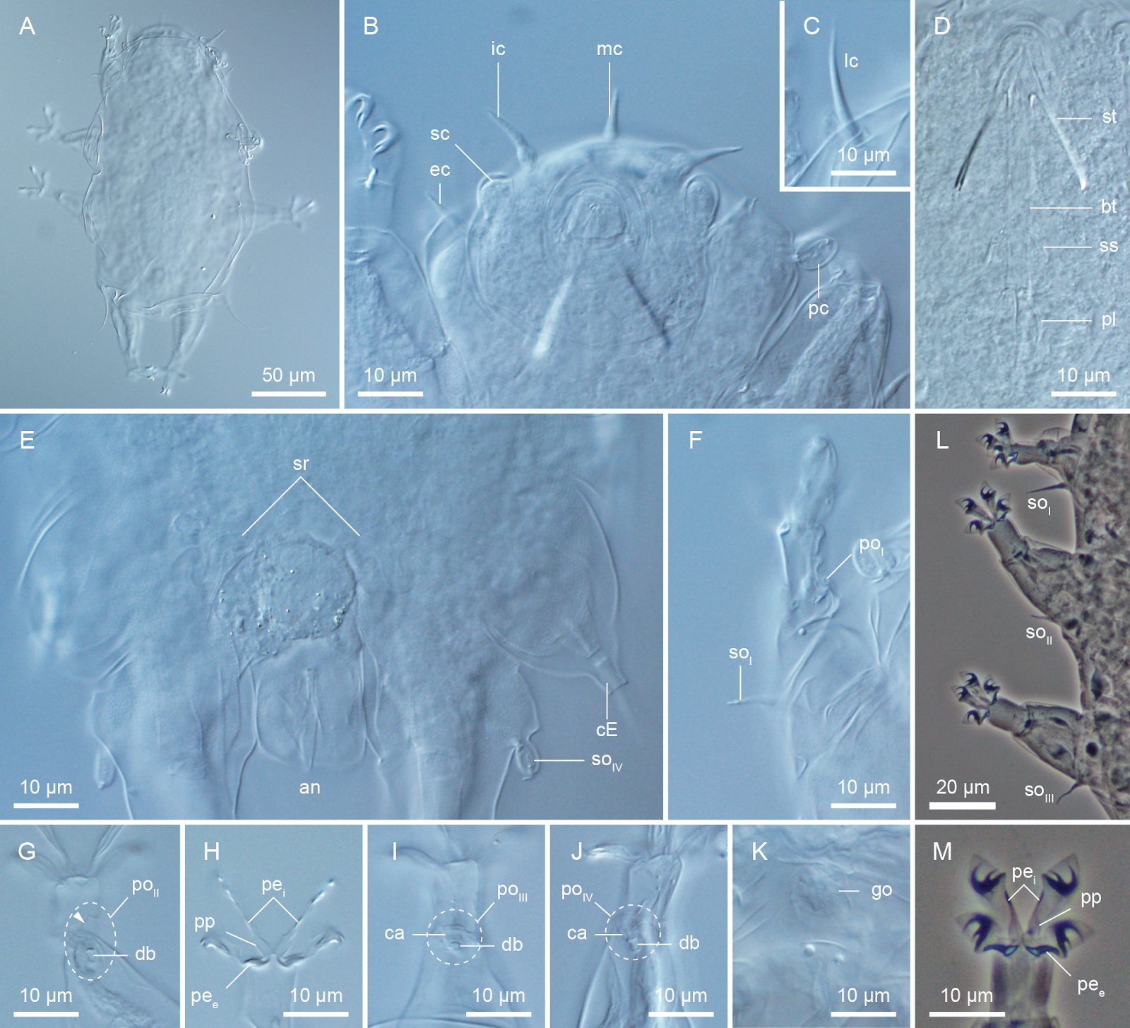
DIC and PhC micrographs of Cyaegharctus kitamurai, adult female. (A) Habitus (dorsal view), (B) cephalic region (ventral view), (C) lateral cirrus. (D) buccal apparatus, (E) caudal region (ventral view) (epicuticle pillars visible), (F) leg I sensory organ and pocket organ, (G) leg II pocket organ (arrowhead indicates protruding portion), (H) leg II digits and claws, (I) leg III pocket organ, (J) leg IV pocket organ, (K) female gonopore, (L) legs I–III sensory organs, (M) leg III digits and claws. (A)–(J) holotype KUZ Z2624, (K) paratype KUZ Z2625, (L), (M) paratype KUZ Z2626. Abbreviations: an, anus; bt, buccal tube; ca, cavity; cE, cirrus E; db, dense body; ec, external cirrus; go, gonopore; ic, internal cirrus; lc, lateral cirrus; mc, median cirrus; pc, primary clava; peᵢ,ₑ, peduncles of internal and external digits; pl, placoid; poₗ–ₗᵥ, legs I–IV pocket organs; pp, proximal pad; sc, secondary clava; soₗ–ₗᵥ, legs I–IV sensory organs; sr, seminal receptacles; ss, stylet support; st, stylet. Fujimoto & Jimi (2020).

The scanning electron micrographs of a four claw juvenile confirmed the results of light microscopy and also provided further detail. However, this scanning electron micrograph specimen also seems to have its outer epicuticle detached. If the outer epicuticle is attached to the underlying layer, a pattern is recognised on the surface of the body due to the pillar layer, but no such indentations were found, suggestive of the detached state of the outer cuticle. The proximal part of each leg has an inflated appearance not recognised in light microscopy. The view of the cephalic region revealed the three-dimensional morphology and the arrangement of the cephalic appendages and also confirmed the presence of terminal pores on the cephalic cirri and the primary clavae. The conical shape of the secondary clavae seems not as evident as in light microscopy probably due to the overlying outer epicuticle. The large anus is not on the ventral surface and rather direct posteriorly. The leg sensory organs were recognised and those of legs I and IV revealed to have terminal pores. The pocket organs were recognised on all legs, however, with slightly different degrees of protruded appearances. The protruded state might be an artefact caused during specimen preparation since specimens on microscope slides do not always have these appearances or the pocket organs are capable of moving. In the latter case, since no muscles seem to be attached to the structures, a passive movement is likely. Regarding claw/digit morphology, the peduncles (internal structures) were not recognised but the three hooks of the internal claws and the single-pointed external claws were recognised.

Scanning electron micrographs of Cyaegharctus kitamurai, four-claw juvenile paratype KUZ Z2627. (A) Habitus (lateral view), (B) cephalic region (frontal view), (C) cephalic region (ventral view), (D) anus, (E) leg I sensory organ, (F) leg II sensory organ, (G) leg III sensory organ, (H) leg IV sensory organ, (I) leg IV pocket organ, (J) leg III digits and claws. Abbreviations: ah, accessory hook; an, anus; cE, cirrus E; ec, external cirrus; fe, femur; ic, internal cirrus; lc, lateral cirrus; mc, median cirrus; pc, primary clava; ph, primary hook; poₗ, ₗᵥ, legs III and IV pocket organs; pp, proximal pad; sc, secondary clava; sh, secondary hook; soₗ–ₗᵥ, legs I–IV sensory organs; ti, tibia. Fujimoto & Jimi (2020).

Fujimoto and Jimi consider the dense body inside the pocket organs of Cyaegharctus kitamurai to be related to the van der Land’s body, often situated at the base of the primary clavae and leg IV sensory organs and suggest that the pocket organs are chemoreceptors. However, only one previous study, in 1981, investigated the function and ultrastructure of Arthrotardigrade sensory organs by transmission electron microscopy and, with our poor knowledge on Arthrotardigrade sensory organs, this remains a matter of speculation. Another possibility for the new structure is a secretory organ. However, no gland was recognised in its vicinity and there is no evidence supporting this hypothesis. To understand the true functions and evolutionary significance of the pocket organs, comparative ultrastructure studies of Arthrotardigrades (including Cyaegharctus kitamurai) are necessary.

See also...

https://sciencythoughts.blogspot.com/2018/11/macrobiotus-hannae-new-species-of.htmlhttps://sciencythoughts.blogspot.com/2018/09/bryodelphax-arenosus-new-species-of.html
https://sciencythoughts.blogspot.com/2018/03/macrobiotus-shonaicus-new-species-of.htmlhttps://sciencythoughts.blogspot.com/2017/09/bryodelphax-kristenseni-new-species-of.html
https://sciencythoughts.blogspot.com/2017/08/estimating-possibility-of-all-life.htmlhttps://sciencythoughts.blogspot.com/2017/06/macrobiotus-polypiformis-new-species-of.html
Follow Sciency Thoughts on Facebook.

Sunday, 3 November 2019

Eruption on Iōjima Island, Japan.

The Kikai Volcano on Iōjima Island in Japan's Kagoshima Prefecture erupted at about 5.35 pm on Saturday 2 November 2019, producing an ash column over a kilometre in height. Nobody is thought to have been injured by the event, as the island is only sparsely populated, but the Japan Meteorological Agency has raised the alert level around the volcano, warning people not to approach within a kilometre of the summit in case of flying rocks or pyroclastic flows.

 Eruption on Iōjima Volcano, Japan, on 2 November 2019. Sify News.

Iōjima is a 5 km long volcanic island that forms the exposed tip of the much larger, but mostly submerged, Kikai Volcano, with a maximum elevation of about 700 m above sea level. The island is thought to be a remnant of a much larger volcanic complex destroyed in a huge eruption about 6300 years ago, that caused devastation across much of southern Kyushu.

The approximate location of Iōjima Island. Google Maps.

Iōjima lies at the northeast end of the Ryukyu Island Arc, which sits on top of the boundary between the Eurasian and Philippine Plates. The Philippine Plate is being subducted beneath the Eurasian Plate, in the Ryukyo Trench, to the Southeast of the Islands. As it is drawn into the interior of the Earth, the tectonic plate is partially melted by the heat of the Earth's interior, and liquid magma rises up through the overlying Eurasian Plate to form the volcanoes of the Ryukyu Islands and Kyūshū.

 The movement of the Pacific and Philippine Plates beneath Japan. Laurent Jolivet/Institut des Sciences de la Terre d'Orléans/Sciences de la Terre et de l'Environnement.

See also...

https://sciencythoughts.blogspot.com/2019/01/eruption-on-mount-shindake-kuchinoerabu.htmlhttps://sciencythoughts.blogspot.com/2015/06/kuchinoerabu-island-evacuated-following.html
https://sciencythoughts.blogspot.com/2014/08/eruptionand-pyroclastic-flow-on-mount.htmlhttps://sciencythoughts.blogspot.com/2013/08/eruptions-on-sakurajima.html
https://sciencythoughts.blogspot.com/2013/07/eruption-on-suwanosejima.htmlhttps://sciencythoughts.blogspot.com/2012/02/mapping-subductioin-zone-beneath-taiwan.html


Follow Sciency Thoughts on Facebook.

Monday, 21 January 2019

Eruption on Mount Shindake, Kuchinoerabu Island, Japan.

The Mount Shindake volcano on Kuchinoerabu Island erupted on Thursday 17 January 2018, producing an ash column that rose to about 6 km above the island. The eruption occurred at about 9.20 am local time, and through hot rocks some way from the crater, causing about 80 of the islands 109 residents to seek temporary refuge in a shelter, though the risk was later judged to have passed and people returned to their homes. This is the latest in a series of eruptions on the volcano that began in October last year.

An ash cloud over Mount Shindake on 17 January 2019. Japan Meteorological Agency.

Japan has a complex tectonic environment with four plates underlying parts of the Islands; the Pacific in the east and the Othorsk in the North, there are the Philippine Plate to the south and the Eurasian Plate to the West. All of these plates are moving in different directions, and some subducting beneath the islands, leaning to a complex tectonic situation where earthquakes and volcanoes are common.

The movement of the Pacific and Philippine Plates beneath eastern Honshu. Laurent Jolivet/Institut des Sciences de la Terre d'Orléans/Sciences de la Terre et de l'Environnement.

Kuchinoerabujima lies at the northeast end of the Ryukyu Island Arc, which sits on top of the boundary between the Eurasian and Philippine Plates. The Philippine Plate is being subducted beneath the Eurasian Plate, in the Ryukyo Trench, to the Southeast of the Islands. As it is drawn into the interior of the Earth, the tectonic plate is partially melted by the heat of the Earth's interior, and liquid magma rises up through the overlying Eurasian Plate to form the volcanoes of the Ryukyu Islands and Kyūshū.

See also...

https://sciencythoughts.blogspot.com/2018/09/typhoon-jebi-leaves-at-least-ten-dead.htmlhttps://sciencythoughts.blogspot.com/2018/07/volcanic-activity-on-asamayama-japan.html
https://sciencythoughts.blogspot.com/2018/07/magnitude-60-earthquake-rattles-tokyo.htmlhttps://sciencythoughts.blogspot.com/2018/07/fifteen-known-deaths-as-floods-and.html
https://sciencythoughts.blogspot.com/2018/06/shinmoedake-volcano-eruption-on-friday.htmlhttps://sciencythoughts.blogspot.com/2018/06/magnitude-59-earthquake-in-osaka.html
Follow Sciency Thoughts on Facebook.

Friday, 11 January 2019

Hana hanagasa and Hana hanataba: Two new species of Stoloniferous Octocorals from the northwest Pacific.

Octocorals are colonial Corals, Anthozoa, lacking the extensive mineralised skeletons of Stony Corals. Each polyp of the colony has only eight tentacles, giving the group its name, though these often have numerous side branches, giving them a feathery appearance. Gorgonians are a group of Octocaorals that tend to form fan shaped colonies in shallow environments with strong currents. The Stolonifera is a group within the Octocorallia comprising Corals with polyps which are not set within a common coenenchymal tissue, but rather arise from a branching, ribbon-like stolon which spreads over the substrate, typically another Coral or Sponge. These Corals are less well studied than other Soft Corals, and relationships within the group are poorly understood.

In a paper published in the journal ZooKeys on 15 October 2018, Yee Wah Lau of the Molecular Invertebrate Systematics and Ecology Laboratory at the University of the Ryukyus, Frank Stokvis and Leendert van Ofwegen of the Naturalis Biodiversity Center, and James Davis Reimer, also of the the Molecular Invertebrate Systematics and Ecology Laboratory, and of the Tropical Biosphere Research Center at the University of the Ryukyus, describe two new species of Stoloniferous Octocorals from the northwest Pacific Ocean.

The first species is placed in a new genus, named Hana, which means ‘flower’ in Japanese, and given the specific name hanagasa, in reference to a flowered head-dress worn during traditional dances in Okinawa, in both cases this refers to the polyps of the species, which are somewhat floral in appearance. This species typically forms colonies of 50-100 polyps arising from ribbon-like stolons about 0.5 mm in diameter. Each polyp lives within a calyce 2.5-3.0 mm in height, into which they can completely withdraw. The polyps are spaced irregularly on the stolen, with the gap between them ranging from about 0.3 mm to about 2.5 mm. The species was found living on hard Coral rock on the northwest coast of Okinawa Island and southeast coast of Iheya Island, both in the Ryukyu Island chain.

Hana hanagasa from Okinawa. Lau et al. (2018). 

The second species is also placed in the genus Hana, and given the specific name hanataba, meaning a bouquet in Japanese, again in reference to the floral appearance of the polyps. This species typically forms colonies of 30-100 polyps, again rising dwelling within calyxes 2.5-3.0 mm high and arising from a branching stolon 0.5 mm in width. It was found dwelling off the southeast coast of Palau and off the Taiwanese atoll of Dongsha.

Hana hanataba from Ngemelis Island, Palau. Lau et al. (2018). 

See also...

https://sciencythoughts.blogspot.com/2018/04/adelogorgia-osculabunda-adelogorgia.htmlhttps://sciencythoughts.blogspot.com/2017/06/sinularia-mesophotica-new-species-of.html
https://sciencythoughts.blogspot.com/2017/04/flagelligorgia-gracilis-new-species-of.htmlhttps://sciencythoughts.blogspot.com/2017/02/muricea-subtilis-new-species-of.html
https://sciencythoughts.blogspot.com/2014/12/a-new-species-of-soft-coral-from.html
Follow Sciency Thoughts on Facebook.

Sunday, 17 December 2017

Terpios hoshinota: Tracking the progress of the Black Death Sponge on Okinoerabu-jima Island.

Sponges, Porifera, form an important part of many benthic marine communities, both for their contribution to the structure of reefs and their forming of symbiotic relationships with a variety of other organisms, including Prokaryotes, Shrimp, Worms, Hydroids, Zoantharians, and Fish. However not all Sponges are beneficial to the communities that host them. The Heteroscleromorph Demosponge Terpios hoshinota forms symbiotic relationships with a variety of Cyanobacteria, spreading rapidly by photosynthetic growth. This enables it to grow at a rate of several milometers per day, forming a thin black or grey crust that rapidly overgrows and kills Hard Corals such as Lobophylia, Montipora, Acropora, Merulina, and Goniastrea

The first known outbreak of Terpios hoshinota was recorded on Guam in 1973, since when it has spread to the Northern Mariana Islands, Western Caroline Islands, the Philippines, American Samoa, southern Taiwan, the Great Barrier Reef, Sulawesi, Java, the Maldives, Mauritius and the Ryukyu Islands. The first outbreak of the Sponge in the Ryukyu Islands hit the island of Tokunoshima in 1985-86, where it rapidly spread across the reefs of the Yonama Coast, eventually covering 87.9% of the reefs, and gaining the name 'Black Death Sponge'. 

 A colonoy of Terpios hoshinota overgrowing a Coral on Guam. Florida Museum.

The Sponge was detected on the reefs of Okinoerabu-jima Island, about 50 km to the southwest of Tokunoshima in 2010, raising concerns about the fate of the reefs there, however a survey carried out at this time found that Terpios hoshinota had completely disappeared from Tokunoshima, where reefs were now dominated by Hard Corals of the genus Acropora, suggesting that the appearance of the Sponge does not necessarily represent the end of a Coral Reef community.

In a paper published in the journal Zoological Studies on 19 April 2017, Masashi Yomogida, Masaru Mizuyama, and Toshiki Kubomura of the Molecular Invertebrate Systematics and Ecology Laboratory at the University of the Ryukyus, and James Davis Reimer, also of the Molecular Invertebrate Systematics and Ecology Laboratory, and of the Tropical Biosphere Research Center at the University of the Ryukyus, describe the results of a long-term study of the Terpios hoshinota outbreak on Okinoerabu-jima Island, based upon a series of surveys carried out between March 2010 and September 2014.

Yomogida et al. carried out a series of transect studies on the Yakomo Coast of Okinoerabu-jima Island, with each survey examing the surface covering of an area of reef measuring 10 m by 1 m. Each survey divided the covering of the reef into nine categories: (1) Terpios hoshinota, (2) Macroalgae (Seaweed) except the Sponge Weed Ceratodictyon spongiosum, (3) the Sponge Weed Ceratodictyon spongiosum, (4) Cyanobacterial mats, (5) living reef-building Corals, (6) dead Coral, (7) other benthic organisms, including Soft Corals, Giant Clams, Sea Cucumbers, and Sea Urchins, (8) sand and gravel, and (9) anything that could not be identified.

(A) Location of Okinaerabu-jima Island, Kagoshima, Japan in the northwestern Pacific and, (B) map of Yakomo coast on Okinoerabu-jima Island. Red dotted box shows the Terpios hoshinota survey area, white dotted lines show the approximate area of Terpios hoshinota along the coast, and red solid lines approximate locations of permanent transects. Google Earth/Yomogida et al. (2017).

Terpios hoshinota covered over 24% of the reef at the outset of the study (March 2010), and remained this high until October of that year, but fell to 17.6% coverage in December 2010. In June 2011 the species underwent a catastrophic die-back, falling to a covering of only 0.02% of the reef. Sponge levels remained low for the next year, having reached only 0.3% coverage by May 2012, but did eventually begin to recover, reaching 11.4% coverage by September 2014.

Coverage of the reef by Macroalgae remained below 10% in all surveys except one, in May 2012, when it reached 13.6%. Cyanobacteria were completely absent from the reef in all surveys except one, in October 2011, when it covered 39.9% of the reef. Sand and gravel remained the dominant coverings of the reef throughout the survey, with coverage varying between 50.2% and 89.4%; none of the other categories ever climbed above a 5% coverage on the reef.

Clearly some event significantly reduced the coverage of Terpios hoshinota in 2011, and came close to removing the Sponge from the reef altogether. Yomogida et al. suggest that the most likely culprit was Typhoon Songda, which passed close to the island on 28 May 2011, and which is recorded as having generated windspeeds of up to 139 kilometres per hour, and wave heights of up to 10.22 m. This event could have removed the Sponge encrustation either by directly tearing it from the reef or covering it in sand or other soft sediments.

This suggests that typhoons could play a major role in inhibiting the ability of Terpios hoshinota to dominate ecosystems, and are likely to have been the cause of the disappearance of the Sponge from Tokunoshima Island. However Yomogida et al. also note that tropical storms may also play a role in the dispersal of Terpios hoshinota, as the larvae of Cyanobacteria-hosting Sponges tend to have rather limited dispersal capacities, suggesting that something else has aided the apparent rapid dispersal of this species. They also note that Terpios hoshinota is now found in both tropical and subtropical seas, and that tropical storms are a feature of only subtropical seas, with areas such as Indonesia and the Maldives, where the Sponge has become established, not effected by these storms.

Yomogida et al. also note that an outbreak of Terpios hoshinota on Pagan Island in the Mariana group was strongly linked to a volcanic eruption on that island, with a large patch of the Sponge appearing with the onset of volcanic activity in 2010, and disappearing when volcanic activity stopped in 2012. They suggest that this might be connected to the deposition of volcanic ash into the waters around Pagan Island, which would have increased the levels of nutrients, particularly iron, to the Sponge and its symbiotic Cyanobacteria. This raised the possibility that Human activities may be facilitating the spread of Terpios hoshinota, if these activities result in extra nutrients being released into the water.

See also...

http://sciencythoughts.blogspot.co.uk/2017/10/plenaster-craigi-new-species-of.htmlhttp://sciencythoughts.blogspot.co.uk/2015/03/preservation-of-cellular-structures-in.html
http://sciencythoughts.blogspot.co.uk/2014/12/two-new-species-of-homoscleromorph.htmlhttp://sciencythoughts.blogspot.co.uk/2014/12/a-new-species-of-sponge-from-late.html
http://sciencythoughts.blogspot.co.uk/2014/12/thirteen-new-species-of-deepwater.htmlhttp://sciencythoughts.blogspot.co.uk/2014/11/calcifying-endosymbiotic-bacteria-in.html
Follow Sciency Thoughts on Facebook.

Sunday, 19 November 2017

Navigobius kaguya: A new species of Dartfish from Japan and the Philippines.

Dartfish, Ptereleotrinae, are small Goby-like Perciform Fish closely related to Wormfish, and found exclusively in marine environments. Three members of the genus Navigobius have previously been described from Japan, Vietnam and Brunei Darussalam (Borneo), but specimens likely to belong to this genus have been observed in the aquarium trade with origins from as far west as the Maldives.

In a paper published in the journal Zootaxa on 13 November 2017, Anthony Gill of the Macleay Museum and School of Life and Environmental Sciences at the University of Sydney and Ichthyology at the Australian Museum, Yi-Kai Tea of Newtown in New South Wales and Hiroshi Senou of the Kanagawa Prefectural Museum of Natural History, describe a new species of Navigobius from waters off the Ryukyu Islands, Japan, and Luzon Island, the Philippines.

The new species is named Navigobius kaguya, in reference to the Moon Princess Kaguya from Japanese folklore, in reference to markings on the dorsal fins of the fish, which resemble Moon phase charts. The species is described from two female specimens, one caught at a depth of 42 m between Ie-jima Island and Okinawa-jima Islands, in the Ryukyu Archipelago, and one caught at a depth of 55-65 m, off the coast of Zambales Province on Luzon Island, the Philippines. These specimens are 52 and 49 mm long, respectively, and are an orangish or pinkish yellow-grey colour, lighter on the underside, with yellow, white and purple markings. 

 Navigobius kaguya, female, off coast of Ida, Zambales Province, Luzon, Philippines.
SK Tea in Gill et al. (2017).

See also...

http://sciencythoughts.blogspot.co.uk/2016/11/opistognathus-ensiferus-new-species-of.htmlhttp://sciencythoughts.blogspot.co.uk/2016/10/grammatonotus-brianne-new-species-of.html
http://sciencythoughts.blogspot.co.uk/2016/08/periophthalmus-pusing-new-species-of.htmlhttp://sciencythoughts.blogspot.co.uk/2016/05/callionymus-alisae-new-species-of.html
http://sciencythoughts.blogspot.co.uk/2015/10/symphysanodon-andersoni-second-specimen.htmlhttp://sciencythoughts.blogspot.co.uk/2015/04/philometrid-nematodes-from-perciform.html
Follow Sciency Thoughts on Facebook.