Showing posts with label Monotremes. Show all posts

Wednesday, 14 October 2020

Tachyglossus aculeatus: Understanding the shoulder muscle architecture in the Echidna.

Echidnas (Tachyglossidae) represent only one of two living families of Monotremata, an early-diverging lineage of egg-laying Mammals. Generally classified as fossorial specialists, Echidnas possess robust pectoral girdle and limb bones, a broad manus, and long, spade-like claws. However, within the forelimb they also display many anatomical features reminiscent of earlier Mammalian relatives. These include additional bones within the pectoral girdle (an interclavicle fusedwith the clavicle, a coracoid fused to the scapula, and an epicoracoid), a laterally-facing hemi-sellar glenoid, lack of a scapular spine, and similar general humeral morphology. Although the subject of few biomechanical studies, their unusual anatomy and phylogenetic placement makes musculoskeletal function in Echidnas of particular interest. Not only can they offer an independent case study on how anatomy is shaped by evolutionary adaptation and constraint, but they can also provide insight on the evolution of the Mammalian musculoskeletal system and ancestral Mammalian function. Echidnas have a characteristic sprawling gait, with rolling of the trunk and a slow, pace-like walk. Walking kinematics show long-axis rotation to be the principal motion of the humerus, unlike most Therian Mammals where the predominant motion is flexion-extension within the parasagittal plane, but likely similar to some earlier relatives of Mammals (e.g., the non-Mammalian Pelycosaurs and Cynodonts). Moment arm estimations based on muscle attachment sites likewise suggest that the shoulder is optimized for internal rotation and adduction. There appears to be little intrinsic muscular stabilization of the pectoral girdle and shoulder joints, due to the robust bony and articular morphology in Monotremes. However, it is unknown whether the muscles themselves are structurally specialised for the Echidna’s lifestyle and locomotion.

A primary determinant of muscle function is muscle architecture, or the arrangement of fibers within muscle. Fiber length is linked to muscle shortening ability and velocity, while muscle physiological cross-sectional area determines how much force a muscle can generate. Gross muscle anatomy has been described qualitatively for the Echidna, but muscle architecture remains unexplored in Monotremes. Studies on forelimb muscle architecture of other Mammals demonstrate how certain muscles have become specialized for different lifestyles and locomotion. For example, the physiological cross-sectional areas of the teres major muscle and the subscapularis muscle in the fossorial Mole, Scalopus aquaticus, are disproportionately large compared to body mass, to produce the forceful humeral abduction and internal rotation necessary for their unusual digging style. The physiological cross-sectional area of the subscapularis muscle is likewise large in the fossorial Badger, Taxidea taxus, with short fibers suggesting this muscle is specialised for stabilisation of the humerus while digging. The physiological cross-sectional area of the subscapularis muslce is also large in the arboreal Pine Marten, Martes martes, to counteract laterally-directed reaction forces in the limb during climbing.

In a paper published in the Journal of Mammalian Evolution on 14 March 2020, Sophie Regnault and Philip Fahn-Lai of the Museum of Comparative Zoology and Department of Organismic and Evolutionary Biology at Harvard University, Rachel Norris of the School of Animal and Veterinary Science at the University of Adelaide, and Stephanie Pierce, also of the Museum of Comparative Zoology and Department of Organismic and Evolutionary Biology at Harvard University, document the shoulder and proximal forelimb muscles and architectural parameters of the Short-beaked Echidna, Tachyglossus aculeatus, providing a detailed examination of muscle form and function in Monotremes.

Muscle data from the Echidna are further compared to other fossorial and non-fossorial Mammals, as well as sprawling non-Mammals, to determine musculoskeletal specialisations associated with the Echidna’s digging lifestyle and sprawling gait. Based on prior work Regnault et al. predicted that the Echidna would show high physiological cross-sectional areas for humeral adductors such as the pectoralis muscle (important for supporting sprawling Animals during stance) and internal rotators such as the teres major muscle (important for the highly specialised fossorial Mole). Regnault et al. also hypothesised convergent specialisations in homologous muscles with fossorial Therians (e.g. the subscapularis muscle). However, as many muscles differ in position, attachments, and inferred function between Echidnas and Therian Mammals, Regnault et al. further anticipated other muscles to reveal differences reflective of the Echidna’s mixture of plesiomorphic and derived anatomical features.

Four adult Short-beaked Echidnas, Tachyglossus aculeatus, were used in this study. One specimen was contrast-stained to obtain a 3D overview of whole muscle anatomy, and three specimens were dissected for architectural properties. The specimens were collected in South Australia, and provided by the University of Adelaide. Cause of death in all cases was suspected impact with a vehicle. The specimens were intact with no grossly observable injuries to the pectoral girdle/forelimb. All specimens had been collected an unknown time after death and stored frozen at −18°C.

The mean architectural parameters and physiological cross-sectional area were calculated for each Animal’s right and left sides. These values were then normalised against each Animal’s body mass (in grams). A mean, body-mass normalised value was then calculated for each muscle. Body mass-normalised parameters allowed broad comparisons to be made between species of different size. Finally, these architectural parameters were also scaled to an Echidna of body mass 3.31 kg, to contextualise architecture in real-world terms and match the mass of the contrast-stained specimen displayed in the gross musculoskeletal anatomy.

 
Rendering of the skeleton of the Short-beaked Echidna, Tachyglossus aculeatus, produced from μCT scans of specimen SEP42 before contrast-staining, shown in (a) left lateral view, (b) ventralview, and (c) dorsal view. Relevant anatomical landmarks are highlighted. Regnault et al. (2020).

Body mass-normalized values of physiological cross-sectional area and fascicle length were compared to normalised muscle architecture parameters calculated from other published Mammal species: the fossorial Mole, Scalopus aquaticus, the fossorial Badger, Taxidea taxus, the cursorial Hare, Lepus europaeus, the arboreal Pine Marten, Martes martes, and the terrestrial Opossum, Didelphis virginiana. Additionally, parameters were compared with two sprawling non-Mammals: the Tegu Lizard, Salvator merianae, and the Alligator, Alligator mississipiensis. Physiological cross-sectional area PCSA was recalculated from the muscle belly masses, pennation angles, and muscle fascicle lengths reported in previous studies, to ensure the same method and scaling was applied to all species. The mean value of each parameter was used from each Animal, apart from the Alligator, where data from an individual of similar mass to the 3D modeled Echidna were used. Muscle architecture parameters were scaled by body mass reported in each study.

The R package ‘dispRity’ was used to compare variance in the mean body mass-normalised architectural parameters (fascicle length and physiological cross-sectional area) of the different species. The disparity metric is equivalent to the mean Euclidean distance between the observed muscle points and a ‘centroid’ (or mean point) on an x-y plot of physiological cross-sectional area vs. fascicle length. To capture the spread as well as the disparity of each species’ muscle architecture, the interquartile range of observed muscle point distances to the centroid were also reported. The disparity metric is calculated as disparity between the single value (e.g. mean body-mass normalised of physiological cross-sectional area/fascicle length) for each muscle of a species i.e. disparity between muscles.

Differentiation of individual muscles via contrast-staining was generally good, despite relatively less diffusion of contrast agent to some very deep parts of the pectoral girdle muscles (e.g., the origin of the  subcoracoideus muscle). For ease of description, muscles are classified here into functional groups inferred from estimates of glenohumeral muscle moment arms from a previous study, but note that many muscles cross additional joints and/or have multiple inferred actions. As such, muscle functional group here is classified only according to the largest moment arm produced by that muscle at the glenohumeral joint.

The humeral internal rotators originate and insert near one another. The latissimus dorsi muslce (also called the latissimus spinalis muscle) has a muscular origin from the caudal scapula and vertebral column, with an aponeurotic extension caudally along the vertebral column, and inserts on the edge of the distal humeral entepicondyle. The teres major muscle arises alongside the scapular origin of the latissimus dorsi muscle, and inserts proximally on the crest of the lesser tubercle of the humerus. The subscapularis muscle originates below these, in the subscapular fossa on the external caudal aspect of the scapula, and inserts on the lesser tubercle of the humerus. The teres minor muscle originates above the glenoid fossa, and also inserts on the lesser tubercle. Together, the humeral internal rotator group had the greatest combined muscle mass, principally through the bulky latissimus dorsi and subscapularis muscles.

 
Attachment sites of muscles on the Echidna’s left shoulder girdle bones (scapulocoracoid, clavicle-interclavicle, and epicoracoid), viewed from the Animal’s (a) left lateral aspect, (b) ventral aspect, (c) medial aspect, and (d) dorsal aspect. Regnault et al. (2020).

The humeral external rotators, supraspinatus and infraspinatus muscles, originate cranially on the scapula. The infraspinatus muscle originates from the external aspect, in the infraspinous fossa (separated from the subscapular fossa by the crest of triceps longus muscle), while the supraspinatus muscle originates from the internal scapular surface. Both insert on the greater tubercle of the humerus. The The clavodeltoideus muscle also acts principally to externally rotate and extend the humerus; this muscle originates along the length of the clavicle’s ventral surface, and inserts below the greater tubercle on the cranial aspect of the humerus. 

 
Attachment sites of muscles on the Echidna’s left humerus, viewed from the bone’s (a) proximal dorsal aspect, (b) distal dorsal aspect, (c) proximal ventral aspect, and (d) distal ventral aspect. Regnault et al. (2020).

Of the humeral abductors, the acromiodeltoideus muscle originates from the acromion and inserts on a small area on the dorsal aspect of the proximal humerus. This muscle appears to have a split or subdivision in the segmented scan and in dissected specimens. The spinodeltoideus muscle originates on the dorsal edge of the scapula and, enveloped within the clavodeltoideus muscle, inserts near the acromiodeltoideus muscle on the humerus. The coracobrachialis muscle is divided into a long and short head, which originate on the ventral aspect of the coracoid and insert along the ventral aspect of the humerus. The scapular heads of the triceps brachii longus muscles originate from a crest between the infraspinous and subscapular fossae on the external scapula. These two heads are joined by the accessory and lateral heads originating from the caudal aspect of the proximal humerus and the medial head originating from the distal dorsal surface of the humerus. All five heads of the triceps brachii muscle insert on the olecranon process of the ulna.
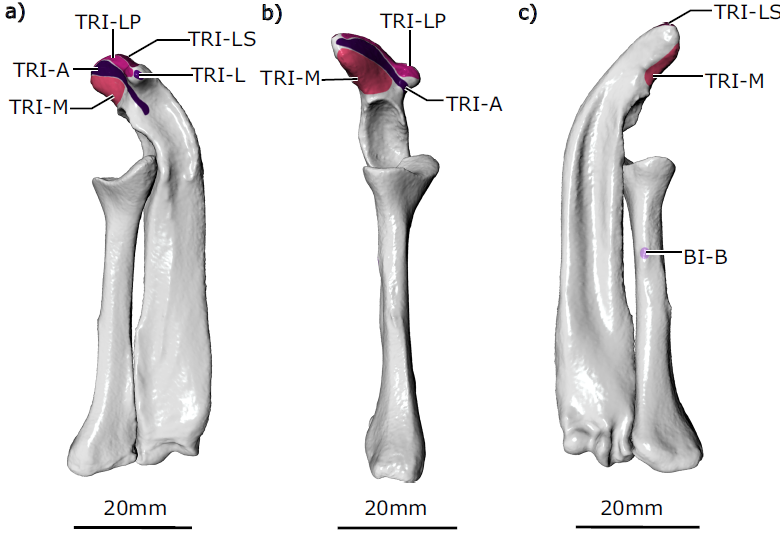
 
Attachment sites of muscles on the Echidna’s left radius and ulna, viewed from Animal’s (a) cranial aspect, (b) preaxial aspect, (c) caudal aspect. Regnault et al. (2020).

The supracoracoideus muscle and both heads of the biceps brachii muscle act to adduct the humerus; all originate from the ventral epicoracoid and coracoid. The biceps brachii brevis muscle inserts on a small crest on the caudal/ventral aspect of the radius. The biceps brachii longus muscle possesses a longer tendinous insertion and was difficult to trace precisely on the contrast-stained scans, but from dissections appeared to insert on the ulna. The supracoracoideus muscle inserts on the ventral aspect of the humerus’ greater tubercle.

The pectoralis muscle is usually considered a humeral adductor, although in the echidna its moment arms suggest it is capable of producing other equally forceful actions as a humeral internal rotator and extensor. The pectoralis muscle is not subdivided; it originates along the presternum (or manubrium) and three sternal elements, and inserts next to the supracoracoideus muscle on the crest of the greater tubercle of the humerus.

The subcoracoideus muscle appears to be the only muscle whose principal action is humeral flexion. This muscle originates on the internal surface of the epicoracoid and inserts on the caudal humerus, along the lesser tubercle and crest of the lesser tubercle. None of the muscles examined here appear to act principally as humeral extensors (based on relative size of their moment arms), though several are capable of producing humeral extension alongside other movements, e.g. the pectoralis muscle and clavodeltoideus muscle.

Many of the muscles crossing the shoulder generally had little to no external tendon, attaching directly to bone or via short internal tendons or superficial aponeuroses. However, a few muscles did have an appreciable tendon, including the  spinodeltoideus muscle (20 mm), the biceps brachii longus and brevis muscles (16 and 12 mm), and the coracobrachialis longus muscle (7 mm).

 
Deep (left) and more superficial (right) contrast-stained and segmented shoulder muscle anatomy of the Short-beaked Echidna, Tachyglossus aculeatus, shown in (a) left lateral view, (b) ventral view, and (c) dorsal view. Regnault et al. (2020).

Many muscles appeared to have fascicles oriented parallel to the muscle’s long axis, with fascicles running along much of the muscle’s length. On closer examination, some of these muscles possessed fascicles inserting directly onto bone, but other fascicles elsewhere within the same muscle inserted at a slight angle onto short internal tendons or superficial aponeuroses (e.g. the pectoralis and latissimus dorsi muscles). Mean pennation angles from these muscles, calculated from angle measurements made at three different muscle regions, ranged from 2 to 13°. Other muscles were clearly parallel-fibered throughout (the acromiodeltoideus,  biceps brachii longus, coracobrachialis brevis, supracoracoideus, m. teres minor, and triceps medialis muscles), with pennation angles of 0°. Finally, a third of the examined muscles were more clearly pennate, with mean pennation angles between 15 and 22° (the coracobrachialis longus, infraspinatus, spinodeltoideus, subscapularis, supraspinatus, teres major, and triceps brachii accessorius muscles).

Many of the muscles cluster around similar fascicle lengths, between approximately 20–45 mm, though the latissimus dorsi muscle can be clearly seen to possess the longest mean fascicle length at 78 mm. The long, near parallel structure of fascicles in many of the Echidna’s shoulder muscles, described above, is reflected in calculated ratios of fascicle length to muscle belly length; for all muscles examined, fascicle lengths measured between 52 and 88% of total muscle belly length. Compared to fascicle length, the spread of physiological cross-sectional areas appears more diverse. The muscles with the largest physiological cross-sectional areas were generally those with the largest masses (the latissimus dorsi, the subscapularis, the combined heads of the triceps brachii, and the combined heads of mcoracobrachialis muscle), though many of these muscles had some degree of fascicle pennation that would also have contributed toward increasing physiological cross-sectional area.

In general, the Echidna shoulder muscles exhibited much less architectural variation compared with the other Mammals. The Echidna has the lowest observed disparity (0.72), followed by the Tegu (0.97), Alligator (1.07), Badger (1.39), Pine Marten (1.47), Opossum (1.63), Hare (1.97), and Mole (2.38).

Relative to each muscle’s length and mass, the Echidna possesses muscles with relatively long fascicles and small-to-intermediate physiological cross-sectional areas. These values are similar to the range exhibited by Tegu muscles (fascicles between 54 and 93%), and many of the Mole muscles (fascicles between 63 and 100%, excluding the supraspinatus and infraspinatus muscles). In contrast to the Echidna, the Alligator, Opossum, Hare, and Pine Marten muscles have more variable relative muscle fascicle lengths and physiological cross-sectional areas.

The main aim of Regnault et al.'s study was to characterise the architecture of the shoulder and proximal forelimb muscles in the Monotreme Echidna, to better understand forelimb function and evolution in this relatively under-studied but phylogenetically important group of Animals. Regnault et al. anticipated that the Echidna would show muscle architectural specialisations related to its lifestyle and locomotion. Namely, they hypothesised increased force-generating capacity (via large physiological cross-sectional area) for humeral internal rotators and adductors, which have been identified as important in the fossorial sprawling Echidna in a previous study, and would be convergent with other fossorial Mammals (e.g. Moles) and sprawling non-Mammals (e.g. Crocodilians).

Both Echidnas (Monotremata: Tachyglossidae) and Moles (Placentalia: Talpidae) are thought to be humeral rotation diggers. Although separated by long evolutionary distances they share some anatomical similarities, including a robust humerus with convergent ‘hourglass’ morphology, bulky pectoral girdle, and broad spade-like manus with restricted joint mobility. Many muscles appear to act similarly in the Mole and Echidna, particularly to produce internal humeral rotation. Counter to their expectations, Regnault et al. found that Echidna body mass-normalised muscle architecture parameters were not appreciably convergent to the mole or other fossorial Mammals examined. Unlike the Mole, teres major muscle in the Echidna appeared unremarkable in terms of physiological cross-sectional area. The subscapularis muscle was relatively large and had a larger physiological cross-sectional area than the other Echidna shoulder muscles, but even this muscle did not show the same extreme specialisation relative to body mass as in the Mole or Badger. The pectoralis was another muscle Ragnault et al. anticipated might show architectural specialisation, due to its importance as a humeral adductor in sprawling animals. However, it also did not have a large physiological cross-sectional area or long muscle fascicles relative to other muscles in the Echidna shoulder nor when compared with the pectoral muscle(s) of other sprawling and upright Animals.

In fact, the Echidna’s shoulder and proximal forelimb muscle architecture appears fairly unspecialised. Physiological cross-sectional area and fascicle length values showed little spread, and measures of disparity placed the Echidna lowest amongst all the Animals compared by Regnauly et al., closest to the Tegu Lizard than to any of the other Mammals. Of all the Echidna muscles examined, the latissimus dorsi muscle was probably the most divergent, with the longest mean fascicle length and one of the largest physiological cross-sectional areas. This is likely linked to the muscle’s overall anatomy: the latissimus dorsi muscle inserts very distally on the humerus in Monotremes compared with other Animals. Even so, its mean fascicle length does not reach the same extremes as the latissimus dorsi muscle in the Hare, Opossum, or Mole, and neither is its physiological cross-sectional area especially notable when considered against the range of muscle physiological cross-sectional areas exhibited by other Animals. In contrast to the Echidna, all other Mammals examined by Regnault et al. showed much more variation in shoulder muscle physiological cross-sectional areas and fascicle lengths, with the Mole exhibiting the greatest disparity. The muscle parameters used in the calculation of disparity are scaled by body mass, so in part, increased disparity (e.g. Mole, Hare) can be a reflection of an increase in the proportion of the measured forelimb muscle masses (5.4% and 3.4% of body mass in the Mole and Hare, respectively, compared to 1.8% in the Echidna). In these cases, increased disparity results from some forelimb muscles maintaining similar relative mass, while there are dramatic increases in masses of other specific and apparently functionally-relevant muscles (e.g. the teres major and  pectoralis muscles in the Mole, and the triceps brachii and pectoralis muscles in the Hare). The latter appear to be driving the relative increase in overall forelimb muscle mass (the two muscles in each example accounting for over 55% of measured forelimb muscle mass for those species).

When architectural parameters are expressed relative to each muscle’s size, another pattern then emerges. In terms of muscle architecture relative to muscle size, the Echidna (and Tegu) again shows little variation. Moles are more variable; however, this appears to be mostly driven through modulation of muscle size. Once muscle size is accounted for, many Mole muscles have similar relative fascicle lengths and physiological cross-sectional areas as in the Echidna. Regnault et al. show that pennation angle and fascicle length become the determinants of physiological cross-sectional area/muscle mass, and of these, fascicle length appears to be the primary source of diversity. Muscle pennation can help to pack in more short fibers/fascicles, but does not appear a prerequisite for achieving high physiological cross-sectional area/muscle mass, e.g. the coracobrachialis muscle in the Alligator has one of the highest physiological cross-sectional area/muscle mass and no pennation. The Echidna shows only a 3.5-fold difference between its shortest and longest fascicle length; Tegus are similar at 3.6, and moles 5.3 (when excluding the supraspinatus and infraspinatus muscles). In contrast, the other Therian Mammals and the Alligator show much greater variation in fascicle length (up to 17.5 times in the Hare), and consequently much more variation in both fascicle length/muscle length and physiological cross-sectional area/muscle mass.

Why are the Echidna shoulder muscles so architecturally similar? Perhaps muscle specialisations are unnecessary. Muscles with short fascicles in highly pennate arrangements, with large resultant physiological cross-sectional areas, are capable of generating large forces but do not shorten much; these muscles are inferred to have specialised roles as joint stabilisers. Examples include the supraspinatus and infraspinatus muscles in Therian Mammals. These stabilising muscles are important in Therians, to compensate for their highly mobile pectoral girdle and glenohumeral joints. However, the Echidna (like earlier Mammaliaforms) has less mobility at the scapula and glenohumeral joint; the robust and interlocking bones and joint morphologies of the Echidna’s pectoral girdle and forelimb may explain why its muscles have not acquired ‘stabiliser’ muscle architecture. Relative to muscle size, the Echidna’s muscles instead have long fascicles and small-to-moderate physiological cross-sectional areas (similar to the Tegu Lizard). Such muscles are capable of relatively greater and more rapid shortening, and are suited for force production over a wide range of the muscle’s range of motion. The Echidna’s low architectural diversity can thus be interpreted as a lack of muscle specialisation into distinct roles (e.g., joint stabilising, or high force production over narrow working ranges for specific movements), and/or a generalised need for muscles with wide working ranges (e.g. potentially as an adaptation for fossoriality).

It is notable that muscle architectural parameters in the Echidna appear to be broadly conservative. Extant Mammals, and particularly Eutherians (Placentals), exploit a huge variety of ecological niches and locomotory strategies, underpinned by modifications of their forelimb into fins, wings, and other specialised structures. The wide variety of physiological cross-sectional areas and fascicle lengths exhibited by the Eutherian shoulder muscles sampled thus far suggests that the remarkable versatility of the Placental forelimb extends to the level of muscle architecture. Metatherians (Marsupials) appear more constrained in forelimb versatility, perhaps because their forelimb structure is linked to their reproductive strategy of climbing to the pouch after birth. Although Regnault et al. examined data from only one Marsupial in their study (the Opossum), its muscle architecture parameters are less diverse than the Eutherian Mammals, and so appear to reflect the generalised constraint on Marsupial forelimb anatomy. Although Monotremes possess a mosaic of plesiomorphic and more derived forelimb anatomical features, no developmental constraints of the forelimb have been identified comparable to Marsupials, and so Monotremes might be expected to exhibit more muscle architectural diversity than the Echidna does.

If the Echidna’s architecture does indeed reflect lack of muscle specialisation into distinct roles (rather than reflecting a fossorial lifestyle), one possibility may be that diversification of Mammalian muscle architecture occurred in the lineage leading to stem Therians or Eutherians, after their divergence from Monotremes, alongside increased diversification of the forelimb more generally. Previous studies have offered evidence that the architecture of many homologous shoulder muscles is statistically similar between Tegu Lizards and Opossums. They interpreted the surprising similarity in functional characteristics as either conservation from the Amniote state or convergence towards a small-bodied ecological generalist phenotype, and link differences to a postural shift from‘sprawling’ to ‘parasagittal’ limb posture and movement in Therians. Though Reghault et al. do not directly compare homologous muscles in their study, they note that the Echidna, phylogenetically bracketed by those two species, also appears to support the notion of conserved muscle architecture in terms of overall diversity. Rehnault et al. found the spread of Echidna muscle architecture values closest to the Tegu, which might be expected given that Monotremes diverged from other Mammals prior to the hypothesised acquisition of parasagittal posture.

In conclusion, Regnault et al. find that the muscle architecture of the Echidna pectoral girdle and proximal forelimb is functionally conservative. Muscle physiological cross-sectional areas and fascicle lengths do not appear modified for specific roles (e.g., stabilisation) to the same extent as in other fossorial Mammals, or indeed any of the other Animals sampled apart from the Tegu. Rather, all the Echidna’s muscles show similar relative architectural parameters, suited for low-to-moderate force production over a wide working muscle range. Regnault et al.'s species comparisons using existing literature data also suggest that the increase in diversity in Eutherian forelimb structure and function may be mirrored by increasing disparity of muscle architecture in these species, though more data are needed to test this. Diversity in muscle architecture in the studied species appears to be primarily driven through modulation of effective fascicle length (and in some cases, relative muscle size). Muscle architectural studies such as ours are therefore of key importance, not only for better understanding forelimb function in extant Animals like the Echidna and other Mammals, but also for their contribution to reconstructing function in extinct taxa.

See also...


Follow Sciency Thoughts on Facebook.

Follow Sciency Thoughts on Twitter.


Sunday, 11 October 2020

The definitive Mammalian middle ear appears to have evolved independently in Monotremes and Therian Mammals.

In non-Mammalian vertebrates, the jaw joint is formed between the quadrate (or palatoquadrate) of the upper jaw and the articular part of Meckel’s cartilage, a rod of cartilage that runs through the lower jaw. This is known as the primary jaw joint. In Mammals, this function is carried out by a new joint between the dentary and squamosal bones, known as the temporomandibular joint or TMJ in Humans, and is referred to as the secondary jaw joint. In mammals, the bones of the original primary jaw joint have been incorporated into the ear and play a role in hearing. In addition to forming a joint with the articular as part of the primary jaw joint, the Amniote quadrate also articulates with the cranial base. During the evolutionary transition that gave rise to Mammals, the connection between the quadrate and the cranial base simplified. The robust quadrate of Reptiles moved from being attached to up to five separate skeletal elements, able to bear the mechanical force of feeding, to become the diminutive Mammalian incus, suspended by a ligament from a single cranial base bone, the petrosal, in an air-filled cavity allowing sound transmission. At the same time, the Meckel’s cartilage lost its permanent nature, separating the incus and neighbouring malleus from the rest of the jaw in adults. Early Mammal-like Reptiles had a permanent Meckel’s cartilage and joints between the quadrate and articular, and posteriorly between the quadrate and cranial base, similar to extant Reptiles. In Mammaliaforms, such as Morganucodon, both a primary quadrate-articular and a secondary dentary squamosal joint were present, in addition to a joint between the quadrate (incus) and the paraoccipital process of the petrosal. This petrosal and incus joint precedes detachment of the middle ear from Meckel’s cartilage in Mammal evolution. A connection between the future middle ear bones and the cranial base is therefore a feature of fossil Mammaliaforms. In extant Mammals, the proposed homologue of the paraoccipital process is the crista parotica, which forms as a cartilaginous spur off the petrosal and is derived from neural crest cells, distinct to the rest of the petrosal and otic capsule, which are mesodermally derived. Modern Mammals have separated the middle ear from the jaw in adults, and the ossicles (malleus, incus and stapes) are now suspended by ligaments from the cranial base to allow free vibration during sound transmission from the ear drum to the inner ear. Palaeontological evidence indicates that the evolution of the definitive Mammalian middle ear occurred at least twice, once in the lineage that gave rise to Monotremes and once in the Therian (Marsupial and Eutherian) Mammals, while new developmental data suggests that the two groups of Therian Mammals may have each independently acquired the definitive Mammalian middle ear (it has become common to use the term Eutherian Mammals rather than Placental Mammals, as Marsupials have a yolk-sac placenta).

Marsupials and Monotremes, exhibit extreme altriciality, greater than is seen in any Eutherian. This has profound consequences for early feeding as the bones that form the Mammalian jaw joint, the dentary and squamosal, have not fully ossified by the time of birth/hatching. The dentary-squamosal joint forms prior to birth in Eutherian Mammals, and begins to function in the embryo. In the Mouse, gestation is approximately 20 days, with breakdown of Meckel’s cartilage, to separate the lower jaw from the ear bones, following during early postnatal stages. In contrast, the Opossum, Monodelphis, has a short gestation of just 13 days, and is born before development of the dentary-squamosal articulation, which forms between 14 and 20 days after birth. Monotremes hatch out of the egg after 10 days post-oviposition. The formation of the dentary-squamosal joint in Monotremes has recently been followed and shown to form from 10 days after hatching in the Platypus. Breakdown of Meckel’s cartilage in both Marsupials and Monotremes occurs relatively late postnatally, with a robust Meckel’s still evident in nest young Platypuses. There is, therefore, a significant gap between birth and the advent of a functional Mammalian jaw joint in both Marsupials and Monotremes.

The feeding strategies of new-born Mammals vary in extant members of each group of Mammals. Compared to Eutherian Mammals, Marsupials rely on placental support for a relatively short period of time and consequently receive the nutrition required for their development via a lengthy and sophisticated lactation. During their early postnatal life Marsupials attach to the mother’s teat and use the comparatively early developed tongue musculature to suck. In the Grey Short-tailed Opossum, Monodelphis domestica, pups are born after 13 days of embryonic development, which is followed by around 14 days permanently attached to the mother’s teat, after which they detach intermittently from the mother but continue to suckle. Weaning occurs around postnatal day 60. In contrast, young Monotremes do not obtain milk in quite the same way as Therian Mammals due to the absence of teats in the mother. Instead young Monotremes suck up milk vigorously from the flattened but protuberant nipple-like areola on the mother’s abdomen.In the case of Echidnas, these areolae are within the pouch.

Given the lack of a jaw joint at birth, it has been proposed that Marsupials and Monotremes use the connection between the middle ear bones and cranial base to permit feeding prior to the formation of the articulation between the dentary and squamosal and cavitation of the middle ear. 

In a paper published in the journal eLife on 30 June 2020, Neal Anthwal of the Centre for Craniofacial and Regenerative Biology at King’s College London, Jane Fenelon of the School of BioSciences at the University of Melbourne, Stephen Johnston of the School of Agriculture and Food Sciences at the University of Queensland, Marilyn Renfree, also of the School of BioSciences at the University of Melbourne, and Abigail Tucker, also of the Centre for Craniofacial and Regenerative Biology at King’s College London, present the results of a study which analysed the articulations that link the lower jaw to the cranial base (cranio-mandibular joints) in Monotremes (Platypus, Ornithorhyncus anatinus, and Short-beaked Echidna, Tachyglossus aculeatus) as they develop from hatching, and compare them to a Marsupial (Grey Short-tailed Opossum, Monodelphis domestica), and a Eutherian (Mouse, Mus musculus), with additional comparison to the Gecko, Guinea Pig and Bat.

 
Schematic of cranial-mandibular jaw articulation showing the roles of the quadrate/incus and articular/malleus in the hearing and jaw joint modules in (A) Reptile Gecko, (B) Mammal Opossum, (C) Mammal-like Reptile Morganucodon. Anthwal et al. (2020).

Anthwal et al. show that in early post-hatching life the Monotreme incus and cranial base fuse, and later form an articulation, creating a double cranio-mandibular articulation, similar to the jaw anatomy of fossil Mammal-like Reptiles. This close association of the incus and cranial base is also observed at embryonic stages in Eutherians and is reflected in Mouse cell lineage studies. In contrast, Opossums at birth utilise a cushion of extra-cellular matrix-rich mesenchyme in between the incus and petrosal to provide an articulation point. Marsupials and Monotremes, therefore, have different strategies for coping with an early birth. Anthwal et al.'s research suggests that the incus retains a transient lower jaw support role across extant Mammal groups but at differentstages of pre and postnatal development.

It has been suggested that the joint between the malleus and incus might act as the jaw joint early on in Marsupial postnatal development, thereby recapitulating the Reptilian function of these bones in Mammals. Alternatively, it has been suggested that the actual articulation point in Marsupials is between the incus and the cranial base. Less information is available regarding Monotreme development, however, the incus has been described as being in cartilaginous connection with the cranial base during early post hatching development. The development of the malleus and incus, and incus and cranial base, was therefore investigated across the three groups of Mammals, with the Gecko as an outgroup.

In the Ocelot Gecko, Paroedura picta, the quadrate and articular (the homologous elements to the incus and malleus respectively in non-Mammal Amniotes) form a clear synovial joint in the embryo at mid-gestation. In Mice, Mus musculus, the malleus and incus are initially formed from a single cartilaginous condensation that separates, by the formation of a joint, at embryonic day 15.5. At birth, therefore, the incus and malleus are evident as distinct cartilages. In Monodelphis domestica, the malleus and incus are still connected at birth at the dorsal end by a ridge of cartilage. Anthwal et al. observed a similar connection between the malleus and incus in the Echidna, Tachyglossus aculeatus, just after birth. Like the Opossum, the middle ear ossicles were fused dorsally, indicating that they function as a unit. These findings demonstrate that, like Opossums, Monotremes do not use the primary jaw joint as the craniomandibular articulation before the development of the dentary-squamosal joint.

 
Timing of the development of the quadrate-articular/malleus incus, and cranio-incudo joints. Histological sections stained with alcian blue and picrosirius red. (A) The primarily jaw articulation is formed by 35 days of post-oviposition (35dpo) during in ovo development in Geckos. (B) The malleus-incus joint, the homologue of the quadrate-articular joint, is formed during in utero development in mice, and is fully formed at birth (Postnatal day (P) 0). (C)-(D) The malleus incus joint is still partially fused in 4 day postnatal (P4) opossum pups (C) and 1 day post-hatching Echidna young (P1) (D). (E) During development the Gecko quadrate forms a joint with the opisthotic (structurally equivalent to the Mammalian petrosal). (F) At birth there is no articulation between the crus breve of the incus and the surrounding crista parotica of the petrosal in Mice (P0). (G) The crus breve of the incus sits in close proximity to the petrosal in Opossums at P4 (G). (H)-(I) The incus is fused with the petrosal in both P1 Echidna (H) and the P2 Platypus (I). Green arrows highlight quadrate-articular/malleus-incus interaction. Red arrows highlight Incus/Q-petrosal/opisthotic interaction. Abbreviations a. articular; c.b.i crus breve of the incus; (i) incus; m. malleus; opo, opisthotic; ptr. petrosal; q. quadrate. Scale in (A) is 200 μm, same scale in (E). Scale bar in (I) is 100 μm, same scale in (B)-(D), (F)-(H). Anthwal et al. (2020).

Anthwal et al. therefore investigated the relationship between the incus and the petrosal in the cranial base in Mice, Opossums, Platypus and Echidna, comparing the interaction to the developing joint between the quadrate and opisthotic in embryonic Geckos. In many Reptiles, as shown in the Gecko, the quadrate (incus homologue) forms a synovial joint with the opisthotic (also known as the otoccipital) in the cranial base during embryonic development. The opisthotic/otoccipital is architecturally equivalent to the petrosal of Mammals. In Mice, the crus breve (short process) of the incus nestled in a fossa created by the crista parotica of the petrosal, but was separated by a region of mesenchymal cells, highlighting the lack of a clear articulation point between the two elements. The incus at birth, therefore only articulated with the adjacent middle ear bones, the malleus and stapes. Similar to the Mouse, the crus breve in neonatal opossums, fitted into a fossa created by the crista parotica, but abutted the petrosal on the inferior aspect of the crista parotica. The incus and petrosal were therefore positioned much closer than in the Mouse.

The relationship between the incus and crista parotica in the two Monotreme species was significantly different from the Therian Mammals. In both Platypus, Ornithorhynchus anatinus, and Echidna, Tachyglossus aculeatus, the incus appeared to be fused with the crista parotica at birth. The lower jaw, via Meckel’s cartilage, would therefore be physically connected to the upper jaw, via the incus at this timepoint. The relatively small size of the incus in both monotremes is striking, as is the extended and tapered crus breve of the incus in the Opossum.

To investigate the monotreme relationship between the incus and crista parotica further Anthwal et al. followed development of these two cartilages from birth to functional use of the dentary-squamosal joint, but before complete cavitation of the middle ear space. Due to the scarcity of available specimens very little is known about Monotreme ear and jaw development. In adult Platypuses, the incus appears in contact with the crista parotica, forming a fibrous articulation. Similarly, in the adult Echidna, the incus has been described as tightly attached to the petrosal.

At 2 days and 6.5 days the Platypus incus was fused to the crista parotica by immature chondrocytes. Between 10 days and 30 days the connection was difficult to make out, with the two cartilages almost completely integrated together. Strikingly, by 80 days, when the dentary-squamosal joint would have started to become functional, the incus and crista parotica were no longer fused, with the two distinct cartilages abutting each other. At this stage, in contrast to the other stages investigated, the ear ossicles and petrosal had begun to ossify. However, the regions forming the malleus-incus joint, and the incus-petrosal articulation remained cartilaginous. A cartilaginous articular surface between the incus and petrosal was maintained at 120 days, a period when the young would have started to leave the burrow. A similar move from early fusion, to articulation was observed in the Echidna. No evidence of a synovial capsule, however, was identified at any stage.

 
Development of the incus-petrosal joint in Monotremes. (A)-(B) The Platypus incus is fused to the petrosal by immature chondrocytes at 2 days (A) and 6.5 days (B) post-hatching. (C) At 10 days post-hatching, the fusion persists, with mature chondrocytes forming the connection. (D) A similar morphology is seen at 30 post-hatching. (E) At 80 days post-hatching the incus and petrosal are no longer fused, but instead the two cartilages abut each other. (F) At 120 days post-hatching the incus and petrosal have begun to ossify, but the region of articulation in between the two elements remains cartilaginous. (G)-(H) In echidna the incus is fused to the petrosal by immature chondrocytes at 3 days (G) and 10 days (H) post-hatching. (I)-(J) By 18 days post-hatching the two elements are separated but remain abutted (I), This connection remains though to 55–65 days post-hatching (J). Abbreviations: i: incus; ptr. petrosal. Scale bar is 100 μm. Anthwal et al. (2020).

The fusion of the incus and crista parotica coincides with the period when the young would have been feeding from milk, while the move to an articulation was associated with periods when the dentary-squamosal was fully formed and functional. After separation of the incus and petrosal, there was a period where two cranial-mandible articulations were evident in the Platypus, between Meckel’s cartilage and the petrosal, via the malleus and incus, and between the dentary and squamosal.

Middle ear cavitation occurred very late in the monotreme specimens analysed, with only the 120 day Platypus showing partial cavitation around the hypotympanum, but this did not extend upwards to where the ossicles are housed. Hearing, thus, must be a very late developing sense in the Platypus.

Limited expression analysis has been performed in monotremes, with no previous expression data performed in the ear or jaw during development. In order to further understand the change in the relationship between the incus and petrosal, immunohistochemistry staining was carried out in Echidna samples 0 and 3 days post hatching.

In the fused incus-petrosal region of 0-day-old Echidna, the expression of both a master regulator of cartilage development, Sox9, and a principal component of cartilage extra cellular matrix, Collagen Type 2, were continuous between the incus and the crista parotica of the petrosal, as well as between the incus and the malleus. Since the connection between these elements is lost later in post-hatching development, IF for beta-catenin was carried-out. Nuclear localised beta-catenin is a readout of canonical Wnt signalling, and is known to negatively regulate chondrocytes differentiation and promote joint formation. Few betacatenin positive cells were observed within the cartilage of the middle ear and petrosal at 0 days, though beta-catenin was strongly expressed in the neuro-epithelium of the inner ear. At post-hatching day 3, the incus and crista parotica were still fused, although the cells joining the two elements resembled fibrocartilage or immature chondrocytes. Expression of Sox9 was still strong and continuous throughout all elements, however Collagen Type 2 expression was weaker in the fusion region, possibly indicating a change in cartilage type from hyaline cartilage to fibrocartilage. Interestingly nuclear beta-catenin, suggestive of active Wnt signalling, was observed in two stripes, in the chondrocytes between the incus and petrosal, and within the malleus-incus joint, indicating suppression of cartilage fate in these regions. Upregulation of Wnt signalling between the incus and petrosal therefore, may play a role in formation of a joint between these two, initially fused, structures.

 
Fusion of the Incus with the petrosal in Echidna pouch young. (A) Alcian blue/picrosirius red staining on the fusion between the incus and petrosal observed in the newly hatched Echidna. (B) Immunofluorescence staining against the regulator of chondrogenesis Sox9 (red) (B), (B’) and the marker of mature cartilage Collagen type 2 (green) (B), (B”) demonstrates that the cartilaginous incus and petrosal bones are fully fused at post-hatching day 0/1 (P0/1). (C) Immunohfluorescence against b Catenin (green) shows no activity within the cartilages at this timepoint. Expression is observed in the neuroepithelium of the inner ear. (D) Alcian blue/picrosirius red staining on the fusion between the incus and petrosal observed in 3 day post-hatching Echidna (P3) shows that the elements are now fused by fibrocartilage. (E) Immunofluorescence staining against the regulator of chondrogenesis Sox9 and the marker of mature cartilage collagen type 2 (E), (E”). Sox9 is still continuously expressed between the elements (E), (E’), but collagen type 2 is down regulated in the incus-petrosal and incus-malleus articulation region (E), (E”). (F) Immunofluorescence against b Catenin shows nuclear localisation within the incus-petrosal and incus-malleus articulation regions, indicating active canonical Wnt signalling, an important step in suppression of chondrogenesis during joint formation. Abbreviations: i. incus; m. malleus; ptr. petrosal. Anthwal et al. (2020).

While the fusion between the incus and petrosal in Echidna and Platypus could be explained by the evolutionary distance between Monotremes and Therian Mammals, it has also been suggested that the incus is transiently attached to the cranial base in 7-week-old Human fetuses. This suggests that the potential for fusion may be a default state in Mammals. In order to examine this, Anthwal et al. next undertook fate mapping experiments in the Mouse, and investigated the relationship between the incus and petrosal in other Eutherian Mammals during embryonic development.

Sox9 expressing cells were fate mapped by tamoxifen induction at embryonic day 14.5 in Sox9CreERT2; tdTomato Mice, which were then collected at postnatal day 0. At this stage Sox9 (green) was expressed in the petrosal and incus and suspensory ligaments, overlapping with the red fluorescent protein marking the Sox9 lineage cells. In addition, the red Sox9 lineage cells were found in the Sox9 negative mesenchymal cells, in the gap between the petrosal and incus. A pre-cartilaginous bridge is therefore evident in the mouse between the incus and the crista parotica. Next, expression of Sox9 was investigated at embryonic day 14.5. The incus, and the crista parotica are both neural crest derived, while the rest of the petrosal is mesodermal. Anthwal et al. therefore looked at the expression of Sox9 (red) in Mesp1Cre;mTmG mice, where mesoderm-derived tissue can be detected by anti-gfp fl IF. Since tissue processing and wax embedding removes endogenous fluorescence, the membrane red fluorescent protein that is expressed in the non-mesodermal tissue of Mesp1Cre;mTmG mice was not detectable in these slides. Consequently, all red signal was Sox9 immunofluorescence staining. Sox9 protein was expressed continuously between the incus and the petrosal. The incus Sox9 expression domain was continuous with the expression domain of the neural crest -derived crista parotica, which in turn was fused to the mesodermal portion of the petrosal. Since the incus does not fuse with the petrosal in the mouse, despite the expression of Sox9 between the elements, we next looked at the mRNA expression of joint markers Gdf5 and Bapx1 between the incus and petrosal of mice by in situ hybridisation. Gdf5 was expressed in the mesenchyme between the incus and petrosal, as well as in the malleus-incus joint. Bapx1, which specifies both the malleus-incus joint and the quadrate-articular joint, was not expressed in between the incus and the petrosal. In the Mouse, therefore there is a potential for the incus and crista parotica to fuse but they are prevented from doing so by the upregulation of the joint marker Gdf5.

 
Mouse fate mapping studies demonstrate developmental fusion between incus and petrosal. (A) Genetic tracing of chondrogenic Sox9 expression cells by inducible reporter mice at postnatal day 0 (P0). Sox9 lineage cells (red) (RFP) are observed in the mesenchyme and developing ligaments between the crus breve of the incus and the petrosal. Sox9 protein (green) is not expressed in the mesenchyme surrounding the incus at P0 (arrowhead). (B) Genetic tracing of mesoderm lineage cells (green) (GFP) and immunohistochemistry against Sox9 protein (red) at embryonic day 14.5 (E14.5). Sox9 expression at E14.5 confirms that the incus and petrosal are formed from a continuous chondrogenic mesenchyme, and that the incus joins with the petrosal at the crista parotica, which is not of mesodermal origin. (C)-(E) Expression by in situ hybridisation of joint markers in sagittal section of E14.5 mouse middle ears. Gdf5 mRNA is expressed with the malleus-incus joint, and between the incus and the petrosal (D), potentially acting to inhibit the Sox9 expressing mesenchyme between the ear and the cranial base from differentiating into cartilage. The middle ear joint marker Bapx1 is not expressed between the incus and the petrosal (E). * indicates space between of incus and petrosal in (C)-(E). Abbreviatios: i. incus; m. malleus; ptr. petrosal. Scale bar in (A), (B)  100 μm. Anthwal et al. (2020).

Very close associations between the incus and crista parotica during development were also observed in other Eutherian Mammals via PTA stained microCT, suggesting that interactions between these two elements are observed as a feature prenatally in Eutherian Mammals, similar to post-hatching monotremes. The function of this prenatal connection between the upper and lower jaw is unclear but may act as a brace to buffer movement during this period.

Next Anthwal et al. investigated the articulation between the incus and petrosal observed in the developing Opossum. It was originally suggested that the Marsupial incus forms a joint with the crista parotica, although this was disputed in Monodelphis. A previous study found no evidence of a joint but did show the mesenchyme between the crista parotica and incus as being condensed. Anthwal et al. therefore investigated the extra cellular matrix components of the mesenchyme surrounding the Opossum incus in more detail. It was noted that mesenchyme surrounding the crus breve and superior portion of the body of the incus had a more intense staining with alcian blue compared to those regions around the inferior border of the incus and the other ossicles. This pattern was observed throughout ossicle development. In order to further characterise the differences in the extra cellular matrix in the different regions of the middle ear mesenchyme, immunohistochemistry for versican was carried out. Versican is a large proteoglycan with side chains of glycosaminoglycans, such as hyaluronic acid. Proteoglycan complexes act to attract water, and are held in place by collagen fibres to stiffen the matrix in hyaline cartilage, and act to lubricate articular cartilage. Versican is required during the initial condensation of mesenchyme but is absent from mature cartilage, where aggrecan is expressed. Versican expression is maintained in the joint region during limb cartilage development, acting to inhibit maturation of the mesenchyme to form cartilage.

 
Specialist mesenchyme supports incus-petrosal connection in juvenile opossums. (A)-(F) Mesenchyme surrounding the crus breve of the incus is rich in the proteoglycan Versican (Vcan) at postnatal day (P)5 (A), (D) and P10 (B), (E). During cavitation of the middle ear at P28 versican rich mesenchyme is concentrated between the crus breve of the incus and the petrosal (C), (F). (G) At P5 the proteoglycan-rich regions surrounding the crus breve have a significantly greater cell density than the regions with less proteoglycan. *p=0.0152 unpaired two-tailed t-test. Error bars = 1 standard deviation. Abbreviations: c.b.i crus breve of the incus; i. incus; ptr. petrosal. Scale bar in (A) is 100 μm, same scale in (B)-(F). Anthwal et al. (2020).

Versican was strongly expressed in the mesenchyme surrounding the short arm of the incus at 5 days, 10 day and 27 days, correlating with the region of strong alcian blue expression. The high level of versican around the crus breve therefore suggests a role for the extra cellular matrix in providing a buffering function in this region. Cell density of the mesenchyme was measured in regions with strong alcian blue/versican staining and compared against the cell density of regions with low alcian blue/versican staining. Unpaired two-tailed t-test demonstrated that the regions with high alcian blue had a significantly higher (p=0.0152) cell density than those regions with lower alcian staining. 

Versican is processed by ADAMTS family members for clearing and remodelling. While the full-length form of versican is thought to have a structural role, the cleaved form has an active role in signalling, influencing morphogenesis and tissue remodelling. Interestingly when we analysed the cleaved form of versican, using antibodies against DPEAAE, the expression was largely reciprocal to that of uncleaved versican, with lower levels specifically around the crus breve. This suggests that versican around the incus is protected from cleavage allowing it to maintain its structural role. The lack of cleaved versican around the crus breve, suggests the lack of a signalling role in this region, in agreement with the low level of expression of CD44, a cell surface receptor and binding partner of versican-hyaluronan complexes. CD44 was not associated with the mesenchyme around the crus breve, but was instead restricted to the perichondrium of the cartilaginous elements and periosteum of the skeletal elements of the ear. 

The incus of adult Mammals plays a key role in hearing. Anthwal et al.'s data here suggest that the incus also plays a transient role supporting the lower jaw against the cranial base during both Marsupial and Monotreme postnatal development. The incus and petrosal were found to be fused at hatching in both Monotremes. During this early fusion period, the puggle would be feeding exclusively on milk and Meckel’s cartilage could therefore act as a flexible elastic strut to facilitate jaw movement.

 
Summary of involvement of middle-ear ossicles in jaw articulation during development. (A) The location of the jaw articulation in developing living Mammals and in the extinct Mammal-like Reptile Morganucodon. Arrows indicate jaw articulation points. Eutherian Mammals are born with a functional dentary-squamosal joint (TMJ), while young Marsupials and Monotremes use the middle ear bones due to a lack of this joint, which develops later. During postnatal development Monotremes show evidence of a double jaw articulation, similar to fossil Mammal-like Reptiles such as Morganucodon. (B) The connections between the middle ear ossicles and the cranial base in developing mammals. The connections between the incus and cranial base differ in young Marsupials and Monotremes. The fusion followed by a joint seen in Monotremes is also observed in pre-natal Eutherians. Neonatal Marsupials support the incus with a specialised middle-ear mesenchyme. Anthwal et al. (2020).

Interestingly, a potential role of the ear ossicles in jaw support was also observed in Eutherians during prenatal development. Fate mapping and gene expression studies in Mice indicated that the crus breve of the incus and the crista parotica were formed from a continuous region of Sox9 expressing chondrogenic cells, separated by expression of the joint marker Gdf5. Furthermore, the incus and cranial base temporarily fuse during the development of the Human middle ear region, and abut during bat development. Together these data indicate that the relationship of the incus to the cranial base is not a derived feature of Monotremes, and that the common Mammal-like Reptile ancestors of both Monotremes and Therian Mammals may have formed an articulation between the quadrate/incus and petrosal through fusion of the elements followed by joint formation though Wnt and Gdf5 signalling.

The current study indicates that the first pharyngeal arch-derived incus forms a continuous field of chondrocytes with the second arch-derived crista parotica, which in turn is fused with the mesoderm-derived body of the petrosal. The borders between these developmentally distinct populations are, therefore, not always reflected by the mature anatomy.

For young Monotremes and Marsupials, the middle ear must function as part of the mandible postnatally until the dentary-squamosal bones have formed. This is similar, but not identical to the situation in Cynodont ancestors of Mammals. In these animals, the quadrate/incus articulated with a number of cranial elements, including the quadratojugal, to stabilise the jaw articulation. These connections and many elements like the quadratojugal have been lost in extant Mammals in order to free the incus and increase its mobility during sound transmission. The mechanical requirements for feeding placed upon the middle ears in Monotremes and Marsupials during early life have resulted in the fusion of the incus and petrosal in monotremes, and the elongated contact supported by a proteoglycan matrix in Marsupials. These adaptations allow for stabilisation of the middle ear before the development of the dentary-squamosal joint and separation of the middle ear from the mandible, but do not compromise the effectiveness of the middle ear in later life.

The crus breve of the incus is elongated in the developing Opossum compared with other species analysed. In order to feed by suckling in the absence of a dentary-squamosal joint Anthwal et al. propose that this anatomy allows for an increased surface contact with the cranial base during postnatal development, which, in combination with the proteoglycan-rich surrounding mesenchyme, acts to stabilise the mandible against the rest of the head. It is noted that many adult Marsupials have a relatively elongated crus breve of the incus compared to Eutherian species, for example the Bare-tailed Woolly Opossum, Caluromys philander, and the Grey Short-tailed Opossum, Monodelphis domestica. Even when Eutherian Mammals have a longer crus breve, such as in Talpid Moles, the process is thinner and more finger-like compared to that of Marsupials. This may be a consequence of the developmental requirement for an elongated short process to facilitate feeding before the development of the mature Mammalian jaw articulation.

In the majority of adult Marsupials, including Monodelphis, the incus is suspended from the cranial base by suspensory ligaments, and the crus breve extends into a fossa. One interesting exception is the Marsupial Mole, in which the crus breve has a connective tissue attachment to a lamella on the petrosal. This results in the middle ear ossicles being affixed to the cranial base, an adaptation to a fossorial niche found in other Mammals such as in True Moles. In light of the current study, the absence of an incudal fossa in the Marsupial Mole may be interpreted as a retention of the juvenile petrosal morphology (paedomorphy).

In adult non-Mammalian Amniotes the homologue of the incus, the quadrate, and cranial base are strongly attached by fibrous syndesmoses or cartilaginous synchondroses, and Anthwal et al. show that a synovial joint appears to form in Geckos during development. In the neo-natal Opossum neither type of connection is observed. In neonatal Marsupials, the connection between the incus and petrosal has been described as being an ‘immature syndesmosis’, which acts as a ‘supportive strut’ during sucking. Anthwal et al. demonstrate a specialised condensed mesenchyme surrounds the incus of Opossum postnatal juveniles. They show that this condensed mesenchyme is rich in the proteoglycan versican. In contrast expression studies in Human foetuses demonstrate that versican is restricted to the perichondrium of Meckel’s cartilage, with high hyaluronic acid levels within the joints but not surrounding the incus. This concentration of versican around the crus breve therefore may be a feature of Monodelphis, and perhaps Marsupials in general.

The versican-rich mesenchyme may act to either stabilise the incus by increasing the tension of the surrounding mesenchyme during feeding, ‘lubricate’ the articulation between the incus and cranial base by increasing the hydration of the extra cellular matrix, or both. In keeping with this role, versican is dynamically expressed at the pubic symphysis during pregnancy in Mice, during which time the Mouse pubic symphysis forms a fibrous joint or syndesmosis. Significantly, there is little cleaved versican (DPEAAE) around the crus breve of the incus, suggesting a mechanical, rather than a signalling role. Overall it is likely that this mesenchyme is supporting the incus, rather than enabling mobilisation, with the high level of uncleaved versican acting to increase fibroviscocity while also elevating hydration of the extra cellular matrix. In this way, the mesenchyme around the incus acts as a cushion during the mechanical stress of suckling.

Meckel’s cartilage persists to at least 50 days post-hatching in the platypus. At this timepoint, juvenile Monotremes have two connections between the lower and upper jaw. The first connection is through the middle ear, which in juveniles remains attached to the mandible and articulates with the cranial base via the incus. The second is the later developing novel Mammalian jaw joint. Only much later in the life of the young does it appear that the connection between the middle ear and mandible is lost, and the malleus and incus act as a definitive Mammalian middle ear. The connection of the incus to the cranial base appears to be maintained in the adult Echidna and Platypus. This would be expected to impact on the movement of the incus, and therefore the efficiency of hearing, reflected in the poor hearing reported for Monotremes.

This novel finding of a double cranial articulation in the juvenile has significant implications for the evolution of the middle ear and jaw joint in Mammals. Fossil evidence indicates that Mammalian ancestors had a persistent connection between the middle ear ossicles and the jaw, as evidenced by the presence of an ossified Meckel’s element, or a dentary groove and post dentary trough, supporting a persistent Meckel’s cartilage. For these Animals, the connection of the middle ear with the jaw took one of two forms, in each case the Mammalian secondary jaw joint was present. The first was a more basal mandibular middle ear where the incus and malleus were firmly attached to the cranial base and dentary respectively. More derived fossils had a partial, or transitional Mammalian middle ear, where the middle ear was medially inflected away from the dentary, presumably allowing for improved vibration, but the malleus was still connected to the jaw, via Meckel’s cartilage. In these fossils with a partial Mammalian middle ear, little is understood of the rear of the ossicular chain, where the incus meets the petrosal, due to the poor and rare preservation of middle ear ossicles in the fossil record, a consequence of their small size. For example, only recently has a Multituberculate with a complete incus been described. Anthwal et al.'s data suggest that even in these transitional Mammals with a partial Mammalian middle ear, the incus would have still articulated with the cranial base via the crista parotica, at least at some point during the Animal’s life history.

The definitive Mammalian middle ear appears to have evolved independently in Monotremes and Therian Mammals. Due to the absence of evidence we do not know if the incus articulation in Animals with a partial Mammalian middle ear varied in a lineage specific manner, with the Therian lineage resembling juvenile Marsupials, and Monotremaformes resembling juvenile Platypuses and Echidna, or if both lineages had similar articulations. The data from transgenic reporter Mice, along with data from Humans suggests that the Monotreme-type fusion and articulation of the incus with the cranial base may have been common in Mammal like-Reptiles. As such, the developing Monotreme, with a double jaw articulation and a fused or articulated incus and petrosal, provides an exciting model for the study of the developmental basis of Mammalian evolution.

See also...


Online courses in Palaeontology. 

Follow Sciency Thoughts on Facebook.

Follow Sciency Thoughts on Twitter


Friday, 9 October 2020

The temporomandibular joint disc Is a common ancestral feature in all Mammals, including Monotremes.

The temporomandibular joint is the one of the most used joints in the body, articulating the upper and lower jaw in Mammals. A fibrous articular disc sits between the skeletal elements of the joint and acts as a cushion. Temporomandibular joint development occurs by the coming together of two membranous bones: the condylar process of the dentary bone in the mandible and the squamosal bone in the skull. The interaction of the condylar with the squamosal induces the formation of a glenoid (or mandibular) fossa on the latter. The articular disc sits between the two within a synovial capsule. The temporomandibular joint disc attaches to the superior head of the lateral pterygoid muscle anteriorly, and to ligaments posteriorly including the disco-mallear ligament that runs thought the capsule of the middle ear, joining the malleus to the temporomandibular joint disc. The temporomandibular joint articulates the jaw in all Mammals and is referred to as the squamosal dentary joint in those Mammals without a fused temporal bone. In non-Mammals the upper and lower jaw articulate via the endochondral quadrate and articular, known as the primary jaw joint. Temporomandibular joint developmental anatomy reflects its evolutionary history as this novel jaw joint forms after the development of the primary joint, which, in Mammals, is integrated into the middle ear. In recent years, a number of studies have advanced the understanding of middle ear evolution in the context of anatomical development, but little work has sought to understand the temporomandibular joint in an evolutionary and comparative developmental biology context. This is despite the crucial role that the formation of the temporomandibular joint has in mammalian evolution.

An important part of the temporomandibular joint is the disc that cushions its action. The origin of the disc is uncertain. The insertion of the lateral pterygoid muscle into the disc on the medial aspect, and the presence of the disco-malleolar ligament, has led to speculation that the disc represents a fibrocartilage sesamoid within a tendon. According to this hypothesis, this tendon, originally associated with the musculature of the articular (homologous to the malleus) of the primary jaw joint, would have become trapped as the dentary and squamosal moved together to create the Mammalian jaw joint. However, studies in mice indicate that the disc develops from a region of flattered mesenchyme cells adjacent to, or possibly part of, the perichondrium of the developing condylar cartilage. Formation of the disc condensation is dependent on Ihh gene signaling from the cartilage. and Fgf signaling via Spry 1 and 2 genes from the adjacent muscles. Therefore, the disc may have its origins in either a tendon, the novel secondary cartilage of the condylar process, or a combination of the two.

Interestingly the disc is absent in extant Monotremes. Monotremes and Therian Mammals (Marsupials and Eutherians) are evolutionary distant, with the common ancestor of the two subclasses being a Mammal-like Reptile from around 160 million years ago. Monotremes have a number of 'Reptile; like anatomical features such as a cloaca, external embryonic development in an egg, a straight cochlea in the inner ear and a sprawling posture. The absence of a disc in both Echidna and Platypus suggests that the disc evolved after the split between Monotremes and Therian Mammals, and is therefore a Therian novelty. Alternatively, absence of the temporomandibular joint disc in extant Monotremes might be due to a secondary loss linked to the loss of teeth, and associated changes in the muscles of mastication. Extant adult Monotremes are edentulous, possibly due to the expansion of the trigeminal during the evolution of electroreceptivity limiting the available space for tooth roots within the maxilla.

The juvenile Platypus has rudimentary teeth that regress, while the Echidna shows only thickening of the dental epithelium during development. In contrast, fossil Monotremes have a Mammalian tribosphenic dentition, a structure unique to the Mammal lineage that allows occlusion of upper and lower molar teeth for grinding of food in addition to crushing and shearing during mastication. his indicates that extant Monotremes evolved from Animals with the ability to chew in the Mammalian manner, involving lateral and rotational movements. The presence or absence of a disc in such fossils is difficult to ascertain due to lack of preservation of soft tissue. In support of mastication playing a role in disc formation, edentulous Therian Mammals, or those lacking enamel, often lack a disc. These species include some (but not all) Baleen Whales, Giant Ant Eaters and Sloths.

In a paper published in the journal Frontiers in Cell and Developmental Biology on 19 May 2020, Neal Anthwal and Abigail Tucker of the Centre for Craniofacial and Regenerative Biology at King’s College London, present the results of a study which examined the development of the temporomandibular joint in Monotremes and made comparison with Mouse developmental models where muscle development is perturbed, in order to discriminate between these two scenarios.

Platypus, Ornithorhynchus anatinus, and Short-beaked Echidna, Tachyglossus aculeatus, slides were imaged from the collections at the Cambridge University Museum of Zoology. All museum samples have been studied in previously published works. CT scans of adult Platypus were a gift of Anjali Goswami of the Natural History Museum, London.

If the temporomandibular joint disc is a therian novelty, then no evidence of a disc would be expected in extant Monotremes during development of the temporomandibular joint. The development of the jaw joint was therefore examined in museum held histological sections of developing post-hatching Platypus and compared with the Mouse.

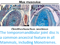
As other authors have previously described, in embryonic day 16.5 Mice the disc anlage is observed as thickened layer of mesenchyme connected to the superior aspect of the condylar cartilage. At postnatal day 0, the disc has separated from the condylar process and sits within the synovial cavity of the jaw joint. In a Platypus sample estimated to be 6.5 days post-hatching, the temporomandibular joint had been initiated, but the joint cavity had not yet formed. Close examination of the superior surface of the condylar cartilage revealed a double layer of thickened mesenchyme in the future fibrocartilage layer of the condylar. The outer layer is similar to that known to develop into the articular disc in Therian Mammals. This thickened mesenchyme persisted in older samples, estimated to be 10 days post-hatching, where the synovial cavity of the temporomandibular joint was beginning to form above. In the most mature Platypus sample examined (around 80 days post-hatching) the fibrocartilage layer of the condylar process was thick and had a double-layered structure. The outer layer was connected via a tendon to the lateral pterygoid muscle. At this late stage of postnatal development, the Platypus puggle would have been expected to start leaving the burrow and to be eating a mixed diet, although full weaning does not occur until around 205 days post-hatching. In the mature Platypus, the condylar process sits within a glenoid fossa, which was not fully formed at earlier stages. A disc-like structure lying over the condylar and connected to the adjacent muscles was therefore evident in the Platypus postnatally but did not lift off the condylar at any stage.

 
Comparison of Mouse, Mus musculus, and Platypus, Ornithorhynchus anatinus, developing jaw joint reveals the presence of a jaw joint disc anlage in early post-hatching Platypus despite absence of the disc in adults. (A), (B) Histological sections of Mouse jaw joint disc development at embryonic day 16.5 (A) and postnatal day 0 (B). (C)–(D’) Histological sections of estimated post-hatching day 6.5 jaw joint at two different levels (C), (D) Note that the separation between the disc anlage and condylar in (D) is probably a processing artifact. (E), (E’) Histological sections of estimated post-hatching day 10 jaw joint. (F) Histological section of mature jaw joint in a juvenile Platypus estimated post-hatching day 80. (G) mCT scan of jaw joint region of adult Platypus. Abbreviations: G.F., glenoid fossa; Cdy., condylar process; Cdy. Fibro., condylar fibrocartilage; Synv., synovial cavity of the jaw joint. Anthwal & Tucker (2020).

Next Anthwar and Tucker examined the development of the temporomandibular joint in a museum derived young Short-beaked Echidna puggle specimen with a crown-rump length of 83mm, which they estimate to be around 18 days post-hatching. The temporomandibular joint is not fully developed. The condylar process possessed a thick, doubled fibrocartilage outer layer, much as was observed in the Platypus. The outer fibrocartilage layer was connected by connective tissue to the lateral pterygoid muscle. Clear disc-like structures were therefore present during development in both extant Monotremes.

 
Examination of the developing jaw joint reveals the presence of a jaw joint disc anlage in post hatching day 18 Short-beaked Echidna, Tachyglossus aculeatus. (A), (B) Histological staining at the forming jaw articulation in echidna young estimated to be 18 days post-hatching at two different level. Fibrocartilage disc anlage superior to the condylar and connected by tendon to the lateral pterygoid muscle is observed. (B’) High-powered view of boxed region in (B) showing the connection between the muscle and the developing disc anlage. Abbreviations: Cdy., condylar process; m. lat. ptry., lateral pterygoid muscle. Anthwar & Tucker (2020).

Taken together, the developmental evidence suggests that extant Monotremes initiate a layer of fibrocartilage connected to the lateral pterygoid muscle, similar to the initiation of the TMJ disc in therian mammals. However, unlike in Therian Mammals, the monotreme fibrocartilage failed to separate from the condylar to form an articular disc in the temporomandibular joint. Interactions with musculature, both mechanical and molecular, have been suggested to be responsible for the proper formation of the temporomandibular joint disc. Lack of mechanical force or changes in signaling from the muscle in Monotremes might therefore result in the disc remaining attached to the condylar. In order to examine how changes in muscle might influence disc development, Anthwar and Tucker next examined disc development in the Mesp1Cre;Tbx1flox conditional mutant Mouse (Tbx1CKO). This mouse has a mesoderm specific deletion of the T-box transcription factor Tbx1, resulting in severely perturbed development of the pharyngeal arch mesodermderived muscles of the head, resulting in their significant reduction or absence. 

Anthwar and Tucker used alcian blue/alizarin red stained histological sections to investigate the development of the temporomandibular joint disc in TbxCKO Mice at embryonic day 15.5. This is the stage when future disc mesenchyme is first observed. In wildtype embryos, the future disc mesenchyme was observed as a condensation attached to the superior surface of the condylar fibrocartilage. A distinct disc-like mesenchyme was also observed superior to the condylar of the Tbx1CKO. This mesenchyme and the fibrocartilage layer of the condylar cartilage both appeared thicker in the Tbx1CKO compared to its wildtype littermate. At embryonic day 18.5, the wildtype temporomandibular joint disc had separated from the condylar process and sat within a synovial joint cavity. In the Tbx1CKO an upper synovial cavity had formed, similar to the wildtype, but there was little evidence of the earlier disc with no separation from the condylar. Instead, a thickened band of fibrocartilage was observed on the superior surface of the condylar process. The lateral pterygoid muscle was either massively reduced or absent in the Tbx1CKO, while other muscles, such as the temporalis, were present but much reduced in volume.

 
Muscle-disc interactions are required for the maturation and separation of the jaw joint articular disc. (A), (B) The disc anlage is observed at embryonic day 15.5 in both wildtype Mice (A) and Mesp1Cre;Tbx1fl/fl Mice with a hypomorphic muscle phenotype (B). (C), (D) By E18.5 the disc has separated from the condylar process in wildtype Mice (C), but not in Mesp1Cre;Tbx1fl/flMice. Abbreviations: Cdy., condylar process; Cdy. Fibro., condylar fibrocartilage. Anthwar & Tucker (2020).

The absence of an articular disc in Monotremes has been thought to be either a secondary loss related to the absence of a mature dentition, or the disc being a later acquisition in the Therian clade. The data presented here show that a mesenchyme similar to the a range of mandibular movements during chewing, including rolling, yaw and front to back movements. It is not clear if these species had evolved an articular disc, since fibrocartilage is rarely fossilised. However, the synovial secondary jaw joint was likely present in stem Mammals such as Morganuconodon, and the lateral pterygoid has been proposed to have inserted into the condylar of the dentary forming the secondary articulation in basal Mammaliforms. When this is considered alongside the presence of the first stages of disc formation during Monotreme development, it is likely that the common stem Jurassic Mammal-like Reptilian ancestor of both Monotremes and Therian Mammals had a disc. The data presented here confirms an essential biomechanical component in disc development. Therefore, Anthwar and Tucker were able to consider when during Mammalian evolution these forces were able to act to enable disc formation. For example, it is probable that many late Triassic and early Jurassic Mammaliaforms such Hadrocodium possessed an articular disc, since they possessed a well-formed squamosal dentary joint and occluding teeth capable of grinding food between the cusps of tribosphenic teeth during mastication.

One hypothesis for the origin of the articular disc is that it formed from the tendon of a muscle of jaw closure of the primary jaw joint interrupted by the formation of the novel Mammalian jaw joint. The tendons and skeleton of the front of the head are derived from the cranial neural crest, whereas the musculature is mesoderm derived. Interactions between the mesoderm and neural crest co-ordinate the muscular skeletal development of the head. A striking piece of evidence for the tendon origin of the disc is the expression in the developing articular disc of Scleraxis, a specific regulator of tendon and ligament development. If the disc is derived from a tendon, then it may be thought of as a fibrocartilage sesamoid. Such sesamoids are found in joints and in tendons that are subject to compression, like the tendons that pass around bony pulleys such as the flexor digitorum profundus tendon in quadrupeds, the patella tendon and ligament and the cartilago transiliens in Crocodilians. Fibrocartilages also form at the enthesis of long bones. Interestingly, it has been demonstrated that much like the temporomandibular joint disc, enthesis fibrocartilage cells are derived from Hh responsive cells and that these cells are responsive to mechanical loading. To support the tendon origin of the temporomandibular joint disc, Anthwar and Tucker's data show that the formation of the disc is dependent on interactions between the skeletal and muscle components of the temporomandibular joint. Such tissue interaction is also a key process in the formation of tendons and ligaments.

The mechanism by which the disc fails to separate from the condylar in Monotremes is not yet clear. Hh signaling is known to be involved in both the initiation of the disc, and the later separation from the condylar. It is still possible that the role in Hh in separation of the disc is a Therian innovation, and as such the reason that Monotremes fail to do so is a lack of the later Hh dependent developmental program for disc separation. However, the absence of the disc in Therian edentates, such as some Whales and Giant Anteaters, strongly suggests that the loss is secondary. The absence of teeth and associated changes in jaw function in Monotremes lends itself to the hypothesis that related changes in the lateral pterygoid muscle are responsible for the failure of disc maturation. Secondary loss, through changes in interactions between the developing disc and muscles, is supported by the failure of the disc to elevate off the condylar in Tbx1CKO Mice that fail to form the lateral pterygoid muscle. These interactions may be either, or a combination of, biomechanical stimulation acting in addition to the compressive force of the TMJ joint, or molecular signaling from the muscles, such as Fgf and Tgf-beta signaling pathways that are known to act in the muscle-tendon-bone/cartilage axis. The source of this signal is likely the lateral pterygoid muscle, which acts to abduct, protrude and laterally move the jaw. These movements are of decreased importance in extant tooth-less monotremes due to feeding modalities that do not rely on chewing with an occluded dentition. As such the formation and maturation of the disc is unlikely to be directly dependent on the presence of teeth, and its absence in edentates is instead a function of the associated changes in musculature. This is supported by the fact that the temporomandibular joint disc forms during embryonic development in Mice, quite some time before the eruption of the teeth at the end of the second postnatal week. Baleen Whales vary in the presence or absence of temporomandibular joint discs, and indeed temporomandibular joint synovial cavities. Significantly, the toothless Gray Whale has no disc and the lateral and medial pterygoid muscles are fused and function as the medial pterygoid, a situation also reported in the adult Platypus. In addition, although they have a full carnivore dentition, the Marsupial Tasmanian Devil has a poorly developed lateral pterygoid muscle and completely lacks the temporomandibular joint disc. Evidence that disc maturation is, at least in part, dependent on biomechanical, rather than molecular signaling, cues is found in the disrupted development of the disc in mice after ex utero surgical manipulation, where the jaw is sutured closed at embryonic day 15.5 but the muscle is unaffected.

Monotremes appear to have two distinct layers in the disc remnant attached to the upper surface of the condylar cartilage, whereas the Tbx1CKO Mouse has only one continuous fibrocartilage by embryonic day 18.5. This may reflect the near total absence of the lateral pterygoid muscle in the Mouse mutant, compared to its presence in a reduced form in Monotremes. Unfortunately, due to the rarity of fresh material, it is not possible to further examine the mechanistic aspects of temporomandibular joint development in edentulous monotreme species at the present time.

In conclusion, Anthwar and Tucker demonstrate that during development, Monotremes show evidence of initiation of a fibrocartilage articular disc, despite all adult Monotremes not having an articular temporomandibular joint disc. Maturation and separation of the disc is dependent on interaction with the developing musculature, either through biomechanical stimulation or molecular signals, as demonstrated by the failure of disc maturation and separation in Mouse mutants with hypomorphic cranial muscles. Therefore, toothed ancestors of Monotremes likely had a temporomandibular joint disc. Anthwar and Tucker's research suggests that changes in the cranial musculature that occurred as a consequence of a move toward edentulous dietary niches resulted in absence of the temporomandibular joint disc in Monotremes, a parallel loss occurring in edentulous Therian Mammals. Finally, the presence of the disc anlage in Monotremes indicates that the Mammal-like Reptile ancestor of all modern Mammals likely possessed a disc to cushion the novel jaw articulation.

 
Maturation of the jaw joint articular disc in Mammals is dependent on muscle interactions. In toothless Mammals and in Tbx1CKO Mice, reduction or loss of jaw musculature results in changes in muscle-disc interaction and so the disc does not separate from the mandibular condyle to sit within the synovial joint capsule. Anthwar & Tucker (2020).

See also...


Follow Sciency Thoughts on Facebook.

Follow Sciency Thoughts on Twitter