Wednesday 31 December 2014

Magnitude 3.9 Earthquake off the coast of Long Beach, California.

The United States Geological Survey recorded a Magnitude 3.9 Earthquake at a depth of about 1.1 km roughly 20 km off the coast of Long Beach, California, slightly before 3.30 pm local time (slightly before 11.30 pm GMT) on Tuesday 30 December 2014. There are no reports of any damage or injuries relating to this quake, but people have reported feeling it across much of the Los Angeles area.

The approximate location of the 30 December 2014 Long Beach Earthquake. Google Maps.

California is extremely prone to Earthquakes due to the presence of the San Andreas Fault, a tectonic plate margin that effectively bisects the state. The west of California, including Santa Barbara and Los Angeles, is located on the Pacific Plate, and is moving to the northwest. The east of California, including Fresno and Bakersfield is on the North American Plate, and is moving to the southeast. The plates do not move smoothly past one-another, but constantly stick together then break apart as the pressure builds up. This has led to a network of smaller faults that criss-cross the state, so that Earthquakes can effectively occur anywhere.

undefined
The extent of and movement on the San Andreas Fault. Geology.

Witness accounts of Earthquakes can help geologists to understand these events and the underlying structures that cause them. If you felt this quake (or if you were in the area but did not, which is also useful information) then you can report it to the United States Geological Survey here.


See also...

The United States Geological Survey recorded a Magnitude 4.4 Earthquake at a depth of 9.9 km, roughly 9 km to the northwest of Westwood, at about 6.25 am local time (about 1.25 pm GMT) on...

The United States Geological Survey recoded a Magnitude 3.0 Earthquake at a depth of 6.7 km in Orange County, California, roughly 40 km east of Los Angeles, at about 1.55 pm local time...


Los Angeles was shaken by a Magnitude 3.2 Earthquake at a depth of 12.8 km, slightly to the north of Los Angeles International Airport, slightly after 7.50 pm on Friday 26 April 2013 local time (slightly after 2.50 am on Saturday 27 April, GMT), according to the United...


Follow Sciency Thoughts on Facebook.

The effect of non-lethal Carbosulfan exposure on Rainbow Trout.


Carbosulfan, 2,3-dihydro-2,2-dimethyl-7-benzofuranyl (di-n-butylaminosulfenyl) methyl carbamate, is a carbamate pesticide which was banned in for use the European Union in 2007, but is still manufactured for export, and is widely used in countries such as Mexico, Brazil, India and Sri Lanka. It is used on a wide variety of crops, particularly Citrus fruits, Corn, Soya beans and Rice. Carbamate pesticides, like organophosphates, act by inhibiting the enzyme acetylcholinesterase by reacting with its active sites, i.e. they are neurotoxins which halt synaptic transmission in the nervous system. Such toxins readily enter the aquatic ecosystem, where they are harmful to Fish and aquatic invertebrates, but break down rapidly in the environment, which can make them hard to detect by analysis of water samples.

In a paper published in the Turkish Journal of Fisheries and AquaticSciences on 24 September 2014, Erol Capkin of the Department of Marine Sciences andTechnology Engineering at Karadeniz Technical University, Halis Boran of the Department of Fisheries and Aquatic Sciences at Recep Tayyip Erdoğan University and Ilhan Altinok, also of the Department of Marine Sciences and Technology Engineering at Karadeniz Technical University, discuss the results of an experiment in which Rainbow Trout, Oncorhynchus mykiss, were exposed to sub-lethal levels of Carbosulfan exposure over a period of 60 days, with enzyme activity in the blood and liver monitored during this period and a 24 day recovery period.

 A commercially availble brand of Carbosulfan. Juanco SPS Ltd.

The exposure of the Fish to the toxin was gauged at levels known to be sublethal, and none of the Fish died during the procedure. Fish exposed to Carbosulfan showed a number of abnormalities compared to Fish in a control group, including loss of balance, darkening in colour and reduced feeding. There was some improvement during the second week of exposure, suggesting that the Fish were able to adapt to Carbosulfan exposure to some extent. The exposed showed a much reduced growth rate compared to the control group, gaining only 3% bodyweight over the course of the experiment, compared to 39% in the unexposed Fish.

The exposed Fish showed reduced protein levels in the liver, a symptom of metabolic degradation (the liver is the principle organ used for the removal of toxins from the body by vertebrates). They also showed reduced levels of acetylcholinesterase activity in the blood and liver, with the liver affected more than the blood. The level of enzyme activity in the blood had returned to normal 18 days after exposure ceased, while that in the liver recovered after 21 days.


A Rainbow Trout,  Oncorhynchus mykiss. Wikimedia Commons.

From this Capkinet al. conclude that freshwater Fish are useful biomarkers for the presence of the toxin in aquatic ecosystems, even if it is not present at levels high enough to be lethal. They suggest that it should be possible to monitor for the presence of Carbosulfan by taking blood samples from wild Fish, a relatively simple procedure which can be carried out in the field.

See also…
The Swedish naturalist Carolus Linnaeus originally described a single Trout species, Salmo trutta, to deascribe small Samonid Fish across Europe, western Asia and North Africa in 1758. Over the next two...

In the late 1990s and early 2000s it became apparent that the Oriental White-backed Vulture, Gyps bengalensis, Long-billed Vulture, Gyps indicus, and Slender-billed Vulture, Gyps tenuirostris, were undergoing rapid population declines across Bangladesh, India, Nepal and Pakistan, loosing...
http://sciencythoughts.blogspot.co.uk/2014/08/arsenic-levels-in-soil-around-cattle.html Arsenic levels in soil around cattle-dipping sites in the Vhembe District of Limpopo Province, South Africa.                               Arsenic-based cattle-dips were used in South Africa from their first becoming available in1893 until their...
 
Follow Sciency Thoughts on Facebook.

Reconstructing cranial endocasts of Palaeozoic Ray-finned Fish.


Palaeontologists have been interested in the endocasts of vertebrate skulls (moulds of the interior of the skull made by sediment) since at least the nineteenth century, due to the possibility that these can reveal details of the animal’s brain. Unfortunately the brains of some vertebrates are very different to the skull cavities, with those of Sharks and Lungfish occupying as little as 1% of the available space, though in other groups, such as Birds and Mammals, the endocast records the shape of the brain far more accurately. Early Ray-finned Fish, Actinopterygians, are thought to fall into the latter category, and their endocasts have been studied extensively since the early 20th century, although this has been problematic, as the best method available for most of that time has been to make serial sections of the skull, a destructive technique that can only be used on the most abundant of fossils, and which leaves only the palaeontologist’s notes and interpretation for examination by future generations of scientists.

In a paper published in the Journal of Paleontology in July 2014, Sam Giles and Matt Friedmann of the Department of Earth Sciences at the University of Oxford present the results of a study of two early Ray-finned Fishes made using computed tomographic X-ray scanning to build up a picture of the cranial endocasts without destroying the original skulls.

The first Fish selected was Mimipiscis toombsi, from the Late Devonian Gogo Formation of Western Australia. This species had its endocast studied by traditional methods by Brian Gardiner of King's College, London in the 1980s. The specimen chosen for this study came from the collection of the Natural History Museum in London, and comprises a 25 mm long by 15 mm with specimen, which was removed from its original matrix via acid preparation, and now has its dorsal surface preserved in a thick resin block. Although some original sediment remains and is preserved in the resin along with the skull, the brain cavity is largely devoid of infilling, creating a void space which shows up well in CT scans.

Photograph of the Mimipiscis toombsi used in the study as it is preserved. Giles & Friedmann (2014).

Giles and Friedmann found that the endocast of Mimipiscis toombsi was long and narrow, with elongate olfactory tracts. The forebrain comprises only 20% of the total length (25-30% is more typical) and is separated from the midbrain by a constriction. The forebrain is narrow, barely wider than the olfactory tracts, which diverge almost immediately after the olfactory bulbs, unlike the situation seen in most early Ray-finned Fish, where the two olfactory nerves share a common canal for most of their length. The midbrain is about twice the width of the forebrain, with poorly developed optic lobes. The hindbrain takes up about 60% of the length of the skull (50% is more typical).

Reconstruction of the endocast of Mimipiscis toombsi, in ventral view. (1) Three-dimensional rendering of endocast; (2) interpretive drawing of endocast. Anatomical abbreviations — bhc, buccohypophysial canal; b.oc.a, canal for branch of occipital artery; ccc, communication between cranial cavity andnotochordal canal; mcv,canal for middle cerebral vein; oof, otico-occipital fissure; opt.f, optic fenestra; spio, canal for spinooccipital nerve; s.v, saccus vasculosus; v.fon, vestibular fontanelle; I, canal for olfactory nerve; IV, canal for trochlear nerve; V, canal for trigeminal nerve; Vprof, canal for profundus nerve; VI, canal for abducens nerve; VII, canal for facial nerve;VIIlat, canal for lateralis branch of facial nerve; IX, canal forglossopharyngeal nerve; X, canal for vagus nerve. Giles & Friedmann (2014).

The second Fish chosen was Kentuckia deani, from the Early Carboniferous Stockdale Formation of Kentucky. Two specimens were examined, both from the collection of the Museum of Comparative Zoology at Harvard University. The first is 30 mm in length and preserved within a nodule, which has been broken in half to reveal the internal structure of the skull. The second is 15 mm in length and also within a nodule, which has been prepared so as to expose the upper surface of the skull.

(2) Kentuckia deani, 15 mm specimen; (3) Kentuckia deani, 30 mm specimen, part; (4) Kentuckia deani, 30 mm specimen, counterpart. Giles & Friedmann (2014).

Giles and Friedmann found that both endocasts of Kentuckia deani were less slender than that of Mimipiscis toombsi, with proportions closer to those of other, more familiar, ray finned fish. The forebrain makes up about 25% of the total length, with a single tract carrying both olfactory nerves for most of their length. The midbrain is twice the width of the forebrain, with well-developed optic lobes; the optic nerves also share a single opening. The hindbrain makes up about 50% of the total length.

Reconstruction of the endocast of Kentuckia deani, in left lateral view. (1) Three-dimensional rendering of endocast; (2) Interpretive drawing of endocast. The pocket of the lateral cranial canal has been ‘cut’ to avoid obscuring parts of the labyrinth. Anatomical abbreviations —acv, canal for anterior cerebralvein; aur, cerebellar auricle; c.c,crus commune; die, diencephalon; ica, canal for internal carotid artery; l.c.c, lateral cranial canal; sac, sacculus; s.su, sinus superior; s.v, saccus vasculosus; I, canal for olfactory nerve; III, canal for oculomotor nerve; IV, canal for trochlear nerve; V, canal for trigeminal nerve; VI, canal for abducens nerve; VII, canal for facial nerve; VIIlat, canal for lateralis branch of facial nerve. Dotted lines infigure indicate unresolved regions. Giles & Friedmann (2014).

See also…

Lungfish are an ancient group of Vertebrates more closely related to the Tetrapods (terrestrial Vertebrates) than to other groups of Fish. They get their name from their ability to breath air, which is useful in anoxic or seasonal waters; all modern species are freshwater and most can no longer use their gills to extract...

The early Gnathostomes (jawed vertebrates) have traditionally been split by taxonomists into four groups, the Placoderms, the Chondrichthyans, the...

Follow Sciency Thoughts on Facebook.

Nine new species of flightless Bess Beetles from Central America.


Bess Beetles, Passalidae, are large (usually over 20 mm), wood-feeding Beetles found predominantly in the tropics. They have a subsocial lifestyle (unusual in Beetles), breeding in colonies in rotting logs, where the adults care for the young, by processing food for them; the wood consumed the adults is broken down by gut microbes, producing edible droppings consumed by both adults and young. They are also capable of quite complex communication, which adults of some species having been demonstrated to produce as many as fourteen distinctive signals by rubbing their legs against their wing-cases.


In a paper published in the journal Zootaxa on 3 December 2014, EnioCano of the Museo de Zoología “Alfonso L. Herrera” and Departamento deBiología Evolutiva at the Universidad Nacional Autónoma de México, the Universidaddel Valle de Guatemala and the Museo de Historia Natural and Escuela de Biología at the Universidad de San Carlos de Guatemala described nine new species of Bess Beetles from Central America, all of which are placed in the flightless genus Ogyges.

The first new species described is named Ogyges cavei, in honour of the entomologist Richard Cave for his work in Central America. The species is described from two female specimens, one collected from cloud forest between 1800 and 2100 m in the Olancho Department of Honduras and the other collected from cloud forest at about 2000 m in the Comayagua Department, also of Honduras (the two locations being widely separated. The first specimen is 40.48 mm in length and 14.41 mm wide, the second specimen is 38.91 mm long and 13.64 mm wide. Both are a glossy black colour, with distinct grooves on the wing-cases.

Ogygescavei, female specimen in lateral view. Cano (2014).

The second new species is named Ogyges handali, in honour of Commander Schafik Handal for his efforts to bring peace in Central America. The species is described from 49 specimens collected from fragments of cloud forest in the Cerro Montecristo in Guatemala, between 1640 and 1900 m; it is thought likely that this species will also be present in Honduras and El Salvador. The specimens are 30.54–35.58 mm in length and 9.7–11.54 mm in width, and black or reddish brown in colour.

Ogyges handali in lateral view. Cano (2014).

The third new species is named Ogyges laurae, in honour of the myrmecologist (scientist who studies Ants) Laura Sáenz. The species is described from eleven specimens collected in the Cerro La Picucha cloud forest in the Olancho Department of Honduras, at altitudes of 1800-2100 m. These range from 18.71 to 22.91 mm in length and from 7.24 to 8.10 mm in width and are light brown or black with a bluish iridescence (sheen) in colour.

Ogyges laurae in lateral view. Cano (2014).
  
The fourth new species described is named Ogyges llama, in honour of the Leaf Litter Arthropods of Mesoamerica (LLAMA) project at the NationalScience Foundation led by Jack Longino and Bob Anderson, which collected several of the specimens used in this study. The species is described from 44 specimens collected in the Cerca de San Pedro Sula in the Cortés Department of Honduras, at altitudes of about 1580 m. These specimens are 19.30 to 24.49 mm in length and 7.55 to 8.69 mm in width and have a black upper surface with a bluish iridescence and a dark brown lower surface.

Ogyges llama in lateral view. Cano (2014).

The fifth new species is named Ogyges menchuae, in honour of Rigoberta Menchú Tum, who was born in Laj Chimel and who won the NobelPrize for Peace in 1992. The species is described from 35 specimens collected from the Cerro El Amay cloud forrest of Laj Chimel in the Quiché Department of Guatemala at altitudes of 1800to 2100 m. The specimens are 29.36 to 33.15 mm in length and 10.12 to 11.72 mm in width and black in colour.

Ogyges menchuae in lateral view. Cano (2014).

The sixth new species described is named Ogyges mutenroshii, named in honour of the character Muten Roshi (Master Roshi) from the manga and anime series ‘Dragon Ball’ by Akira Toriyama. The species is described from two specimens of indeterminate sex collected from cloud forest in the the Cerro Azul Meambar National Park in the Comayagua Department of Honduras at altitudes of 1656 m and 1550 m. The specimens are 34.56 and 33.19 mm in length and 12.05 and 11.52 mm in width, and black in colour.

Ogyges mutenroshii in lateral view. Cano (2014).

The seventh new species is named Ogyges ratcliffei, in honour of the entomologist Brett Ratcliffe, the leader of Team Scarab, for his work on Dynastine Beetles. The species is described from a single specimen collected from cloud forest in the Reserva La Picucha in the Olancho Department of Honduras, at an altitude of between 1800 and 2100 m. The specimen is female, 33.02 mm long and 11.95 mm wide and black with a blue iridescence.

Ogyges ratcliffei, female specimen in lateral view. Cano (2014).

The eighth new species described is named Ogyges sandinoi, in honour of Augusto César Sandino, a Nicaraguan national hero who was born in the Segovia Mountains. The species is described from five specimens collected from cloud forest at Cerro Jesus on the border between Honduras and Nicaragua at an altitude of 1600 m. The specimens are 30.02–31.63 mm long and 10.23–11.19 mm wide, blackin colour, with some dark brown areas on the underside.

Ogyges sandinoi, male specimen in lateral view. Cano (2014).

The final new species described is named Ogyges toriyamai, in honour of the Japanese artist Akira Toriyama, creator of the manga and anime series ‘Dragon Ball’. The species is described from four specimens collected from cloud forest in the Cerro Azul Meambar National Park in the Comayagua Department of Honduras. The specimens are 28.29–31.06 mm long and 9.69–11.21 mm wide and black with a blue iridescence.

Ogyges toriyamai, male specimen in lateral view. Cano (2014).

See also…

Beetles first appear in the fossil record in the Early Permian. And by the Triassic were already beginning to dominate Insect faunas, with many modern groups in existence by the end of the Cretaceous. However...

There are currently eight species of Scarab Beetles in the genus Scapanoclypeus, which is known from Namibia and western South Africa. They are placed within a group of Tanyproctine Scarabs which are found only in Africa.


http://sciencythoughts.blogspot.co.uk/2014/08/a-new-species-of-scarab-beetle-from.html A new species of Scarab Beetle from the Elandsberg Mountains of the Western Cape, South Africa.                                                    Scarab Beetles of the genus Trichostetha occur across southern Africa, reaching their greatest diversity in...
 
Follow Sciency Thoughts on Facebook.

A new species of Olingo from the cloud forests of Colombia and Ecuador.


Olingos, Bassaricyon, are small members of the Racoon family, Procyonidae, found in Central and South America. They are not well understood, as they live in the canopy of dense forests where they are not easily observed, and are easily mistaken for the related Kinkajou, Potos flavus.

In a paper published in the journal ZooKeys on 15 August 2013, a team of scientists led by Kristofer Helgen of the Division of Mammals at the National Museum of Natural History in Washington DC describe a new species of Olingo from the cloud forests of Colombia and Ecuador.

The new species is named Bassaricyon neblina, which means ‘fog’ or ‘mist’ in Spanish, a reference to the cloud forests where it lives; Helgen et al. also suggest the common name Olinguito, meaning ‘Little Olingo’. The species was discovered during a genetic study intended to determine the relationships between the four previously described species of Olingo and other members of the Racoon Family, using DNA from museum specimens.

Surprisingly, despite all the specimens referred to the new species having previously been assigned to other species, the new species emerged as a distinct lineage, which was the sister group to all the other species (i.e. all the other species were more closely related to one another than to the new species). More surprisingly still, all the specimens found to belong to the new species were found to have been collected in cloud forests at altitudes of 1500-2750 m, while all the other specimens were from below 2000 m, suggesting a clear difference in habitat preference. They were also smaller and more slender than members of other species, with darker coats.

The Olinguito, Bassaricyon neblina, in life, in the wild. Taken at Tandayapa BirdLodge, Ecuador. MarkGurney in Helgen et al. (2013).

The Olinguito is found in cloud forests between 1500 m and 2750 m in montane cloud forests on the slopes of the Western and Central Andes in Colombia and Western Andes in Ecuador.

Distribution map for Bassaricyon neblina. Helgen et al. (2013).

See also…

Glyptondonts were large, heavily armored mammals related to Armadillos that evolved first appeared in South America in the Miocene, spread to North America in the Pliocene and became extinct at about the same time...

Follow Sciency Thoughts on Facebook.

Tuesday 30 December 2014

Magnitude 5.0 Earthquake off the coast of Albania.

The United States Geological Survey recorded a Magnitude 5.0 Earthquake at a depth of about 14.4 km, roughly 10 km off the coast of northern Albania, slightly before 9.35 pm local time (slightly before 8.35 pm GMT) on Monday 29 December 2014. There are no reports of any damage or injuries associated with this event, but people have reported feeling the quake as far away as Tirana, 54 km to the southeast of the epicenter.

The approximate location of the 29 December 2014 Albanian Coast Earthquake. Google Maps.

The coastal region of Albania, and the other states of the western Balkan Peninsula, forms the eastern margin of the Adriatic Plate, a piece of the African Plate that has broken away and is now wedged into the southern part of the Eurasian Plate. This is being squeezed by the impact of Africa into Europe from the south, which is pushing western Italy, which sits on the Eurasian Plate, to the east, and Greece and Turkey, which sit on the Aegean and Anatolian Plates, to the west. This squeezing leads to uplift around the margins of the Adriatic Plate, in the Apennine Mountains of central Italy and the mountain ranges of the west Balkan Peninsula.

Outline map showing the approximate positions of the Eurasian (EU), Adriatic (AD) and African (AF) Plates. Di Bucci & Mazzuli (2003).

See also...


The United States Geological Survey recorded a Magnitude 3.9 Earthquake at a depth of 20.5 km in...


The United States Geological Survey recorded a Magnitude 4.9 Earthquake at a depth of 10 km in the Sarandë District of southern...


The United States Geological Survey recorded a Magnitude 4.6 Earthquake at a depth of 28.5 km, roughly 4 km to the south of the...


Follow Sciency Thoughts on Facebook.

Two new species of Pinworms from Sulawesi Soft-furred Rats.


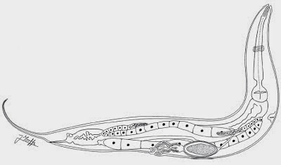
Pinworms, Oxyuridae, are parasitic Nematodes infecting the digestive tracts of Mammals. They have short life cycles, typically undergoing several generations in a year, with eggs being released in the host’s faecal matter to infect new hosts. Some species of Pinworm appear to be quite cosmopolitan, infecting a range of hosts, but most species have a close relationship with a specific host species and rarely cross-infect other species.

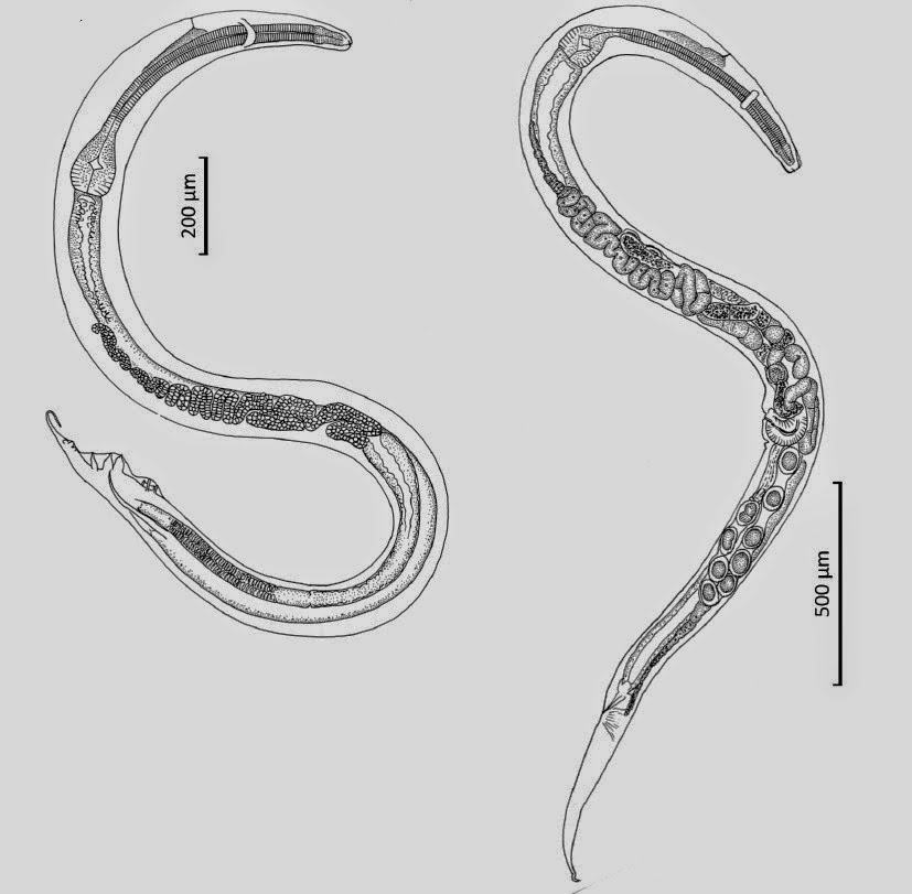
In a paper published in the Raffles Bulletin of Zoology on 18 September 2014, Kartika Dewi of the Zoology Division at Museum Zoologicum Bogoriense, Hideo Hasegawa of the Department of Biology at OitaUniversity and Mitsuhiko Asakawa of the Department of Parasitology at Rakuno Gakuen University describe two new species of Pinworms from Sulawesi Soft-furred Rats, Eropeplus canus, trapped at Lambanan in the South Sulawesi Administrative District. The Sulawesi Soft-furred Rat is an indigenous species to Sulawesi (i.e. found nowhere else), known only from a few mountainous localities (altitudes between 1800  and 2300 m) and considered to be Vulnerable under the terms of the International Union for the Conservation of Nature’s Red List of Threatened Species. Both of the new species are placed in the genus Syphacia, seven species of which have previously been recorded from Indonesia; the cosmopolitan Syphaciamuris, which infects a variety of Rats (Rattus spp.), plus four species infecting indigenous Rodents on Sulawesi and one species each infecting Rodents on Halmahera and Papua.

The first new species is placed in a new subgenus, Rumbaisyphacia, which is a combination of ‘rumbai’, an Indonesian word meaning ‘fringe’ and the name of the genus (i.e. ‘fringed Syphacia’) and given the specific name kumis, meaning ‘moustache’ in Indonesian; both names refer to a fringe of papillae around the mouthparts. The species is described from twenty two specimens, eleven of each species. The females are considerably larger than the males, at 3.21–4.12 mm as opposed to 1.51–1.72 mm.

Syphacia (Rumbaisyphacia) kumis, from Eropeplus canus in south Sulawesi, Indonesia. (1) male, lateral view; (2) cephalic end of male, apical view. Dewiet al. (2014).

The second new species is also placed in a new subgenus, Segienamsyphacia, from ‘Segienam’, which means ‘hexagonal’ in Indonesian, a reference to the mouthparts of the female, and given the specific name yuniae in honour of Yuni Apriyanti, who prepared the specimens for observation by SEM. This species is also described from eleven male and eleven female specimens. The females of this species are also bigger than the males (though not as much so as in Syphacia (Rumbaisyphacia) kumis) at 2.34–2.56 mm in the females and 1.00–1.25 mm in the males.

Syphacia (Segienamsyphacia) yuniae, from Eropeplus canus in south Sulawesi, Indonesia. (11) male, lateral view; (12) cephalic end of male, apical view. (18)Female, lateral view; (19) cephalic end of female, apical view. Dewi et al. (2014).

Dual infections of Pinworms are rare in Rodents, with most species having a single Pinworm parasite, and where more than one species is present this is generally thought to be an exceptional situation in which unhealthy animals have been opportunistically infected by non-specific parasites. However all of the Sulawesi Soft-furred Rats inspected were suffering from infections of both Pinworm species, and did not appear to be particularly unhealthy. Dewiet al. suggest that as additional resource partition by the parasites, with the Pinworms probably infecting different microhabitats of utilizing different nutrients.

SEM of mouth opening Syphacia (Rumbaisyphacia) kumis showing setiferous apical margin of pharynx. Dewi et al. (2014).

See also…

Parasitic Nematodes of the superfamily Heterakoidea are typified by having three lips, an esophagus with a valved bulb, thick shelled eggs and a pre-anal sucker on the males. They are typically parasites of the digestive tracts of small vertebrates, which do not require an intermediate host (i.e. the species only needs to infect one species of hosts, rather than infecting different...
The science of palaeoparasitology involves the study of parasite remains from palaeontological and archaeological sites. This rarely involves the recovery...
 Nematodes are probably the most numerous and specious group of animals on Earth, though relatively few species have actually been described. The title of...


Follow Sciency Thoughts on Facebook.

Four new species of Hangingflies from the Middle Jurassic of Inner Mongolia.


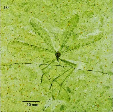
Hangingflies, Bittacidae, are large members of the Scorpionfly order, Mecoptera, which resemble the more widespread Craneflies (members of the True Fly order, Diptera) both in morphology and lifestyle, although the two groups are easily separated, as Hangingflies have two pairs of wings and Craneflies only one. Hangingflies uncommon today, with about 270 species known worldwide, but are thought to have been far more abundant and diverse in the Jurassic, before Flowering Plants and associated Insects came to dominate terrestrial ecosystems.

In a paper published in the journal ZooKeys on 19 December 2014, Sulin Liu, Chungkun Shih and Dong Ren of the College of Life Sciences at Capital Normal University describe four new species of Hangingflies from the Middle Jurassic Jiulongshan Formation of Inner Mongolia. The Jiulongshan Formation comprises a series of grey tuffaceous sandstones and sandy mudstones which has produced a large number of extremely detailed fossils of a wide variety of insects, including Moths, Hangingflies, Wasps, True Flies, Lacewings and many extinct groups.

The first new species described is placed in the genus Mongolbittacus, which to date has been known only from a single species from the Jiulongshan Formation, and given the specific name speciosus meaning ‘showy’. This species is described from a single male specimen, preserved in lateral view as part and counterpart from within a split block. This specimen is 8.8 mm in length and preserved with all four wings held open and separated from one-another.
Mongolbittacus speciosus, photo of part. Scale bar is 3 mm. Liu et al. (2014).

The second new species is also placed in the genus Mongolbittacus, and is given the specific name oligophlebius, meaning ‘few veins’ in reference to the low number of crossveins on the the wings. This species is described from two specimens, both preserved as part and counterpart from split blocks. The first is only partially exposed, with only one forewing and the forepart of the body visible, the second is preserved with the limbs folded over the body.

Mongolbittacus oligophlebius, first specimen, photo of part. Scale bar is 3 mm. Liu et al. (2014).

The third specimen is placed in the genus Exilibittacus, which is also known to date from a single species from the Jiulongshan Formation, and given the specific name foliaceus, meaning ‘leafshaped’, in reference to the shape of the wings. This species is described from a single female specimen, 12.9 mm in length, preserved in dorsal view on a split block.

Exilibittacus foliaceus, female specimen in dorsal view. Scale bar is 3 mm. Liu et al. (2014).

The final new species is also placed in the genus Exilibittacus, and is given the specific name plagioneurus, meaning ‘oblique veined’, in reference to the oblique crossveins on its wings. The species is described from a single female specimen, preserved in dorsal view as part and counterpart from a split block.

Exilibittacus plagioneurus, dorsal view of part. Scale bar is 3 mm. Liu et al. (2014).

See also…

Hangingflies are long-legged Scorpionflies (Mecoptera), a group of insects related to the True Flies. Scorpionflies get their name from the reproductive organs of the males of some species, which resemble the tails of Scorpions. Despite this fierce appearance most species are harmless herbivores, though it is thought that fleas are highly...
Earwigflies (Meropeidae) are a small group within the Mecoptera (Scorpionflies), known only from four fossil and two modern species. Three of the fossil species are from the Triassic of Kyrgyzstan, and the fourth from the Jurassic of Siberia, while the two modern species, Merope tuber and Austromerope poultoni are from North America and Australia respectively. Little is known of the biology of these Insects, the larvae having never...
The Scorpionflies (Mecoptera) are a group of insects related to the True Flies. They get their name from the reproductive organs of the males of some species, which resemble the tails of Scorpions. Despite this fierce appearance most species are harmless...


Follow Sciency Thoughts on Facebook.