Showing posts with label Denmark. Show all posts

Wednesday, 6 May 2026

Two English 'Lamb of God' coins discovered in Denmark.

Two 'Lamb of God' coins issued by the English King Æthelred the Unready around the year 1009 AD have been discovered in Denmark, according to a press release issued by the National Museum of Denmark on 29 April 2026. The coins were both uncovered by metal detectorists, one in the north of Jutland and one in the south.

An English 'Lamb of God' coin discovered by a metal detectorist in northern Jutland. Søren Greve/National Museum of Denmark.

The Lamb of God coins were a special edition coin produced by Æthelred the Unready as part of an attempt to obtain divine protection for his kingdom, along with a series of religious ceremonies, fasts, and penances. Unlike regular Saxon coins, which typically had the king's head on one side and a cross on the other, they had a 'Lamb of God' sign on one side, which comprised a Lamb pierced by a cross, a symbol for Jesus, and a Dove on the other, a symbol of the Holy Spirit.

An English 'Lamb of God' coin discovered by a metal detectorist in southern Jutland. Søren Greve/National Museum of Denmark.

The crisis which provoked these measures was the invasion of England by Viking raiders, mostly from Denmark. However, this was did not prove to be an effective method of defence, with southern England being ravaged by the armies of Thorkell the Tall between 1009 and 1013, and Sweyn Forkbeard launching a full-scale invasion in 1013 which forced Æthelred into exile. Sweyn Forkbeard died in 1014, allowing Æthelred to briefly regain his thrown, though he lost it again in 1016 to Sweyn's son, Cnut.

While the coins failed to save Æthelred's reign, they were apparently very popular with the invading Vikings. Of the 30 known examples, only 4-5 have been found in England, with the remainder discovered in Scandinavian and Baltic countries, the majority with piercings which suggest the Vikings wore them as pendants.

Having conquered England in 1016, Cnut succeeded to the throne of Denmark in 2018, following the death of his brother Harald II. In 1026 Olaf Haraldsson, King of Norway, mounted an invasion of Denmark while Cnut was in England, starting a series of wars which led to Olaf's death in 1030, and Cnut installing his wife, Ælfgifu of Northampton, as regent of Norway. 

Having unified England, Denmark, and Norway into a single state (referred to as the North Sea Empire by modern historians), Cnut set about consolidating and unifying his empire. This included the introduction of silver coinage on the English model to Denmark and Scandinavia. Prior to this, coins had not been directly used as legal tender in this area, silver measured by weight used as the standard medium of exchange. However, the innovation appears to have been quickly adopted, with coins accepted as an easier way to do business.

See also...

Sunday, 1 March 2026

Denmark officially recognised as having ended mother-to-child transmission of HIV and Syphilis.

Denmark has been officially recognised as having eliminated mother-to-child transmission of HIV and Syphilis according to a press release issued by the World Health Organization on 27 February 2026. In doing so it becomes the first member of the European Union and the third European country to achieve this status (behind Moldova and Belarus, both of which were confirmed as having ended mother-to-child transmission of both diseases in 2016). 

Flag of Denmark. Tomasz Sienicki/Wikimedia Commons.

The World Health Organization is aiming for all countries to achieve the triple elimination of mother-to-child transmission of three critical diseases, HIV, Syphilis, and Hepatitis B, although to date only a single country has passed this milestone, the Maldives in October 2025. Denmark's progress makes it one of 23 countries to have ended mother-to-child transmission of at least one of these diseases, alongside Anguilla, Antigua and Barbuda, Armenia, Belarus, Belize, Bermuda, Brazil, Botswana, Cayman Islands, Cuba, Dominica, Jamaica, Malaysia, Maldives, Montserrat, Namibia, Oman, Republic of Moldova, Saint Vincent and the Grenadines, Sri Lanka, St. Kitts and Nevis, and Thailand.

The confirmation of Denmark's status comes following assessments by the World Health Organization's Regional Validation Committee for Europe, and Global Validation Advisory Committee in 2025. In order to be confirmed, it was necessary to demonstrate that 95% of all pregnant women in Denmark were tested for the diseases, and that new infant infections were below 50 in 100 0000 births. Denmark currently has 5950 people living with HIV, with less than 0.1% of pregnant women affected. It is possible to completely suppress the disease with antiretroviral treatments, preventing development of symptoms and all forms of transmission, including mother-to-child. In 2024, Denmark reported 626 cases of Syphilis, 524 in men and 102 in women; infections with this disease can be eliminated with antibiotics. Denmark is still working towards ending mother-to-child transmission of Hepatitis B.

Human Immunodeficiency Virus, or HIV, is a form of Lentivirus which causes infections in Humans, spread through sexual intercourse or exchange of blood. Notably, the Virus infects the cells of the Human immune system, where it is hard for that immune system to attack, and eventually leads to a breakdown of the immune system known as AIDS (Acquired Immune Deficiency Syndrome), during which the body becomes vulnerable to a wide range of infections, including many by micro-organisms which are not usually pathogenic. 

Untreated, HIV invariably leads to AIDS, and AIDS is invariably fatal, but, as with Syphilis, HIV infections can go through long phases of dormancy, leaving infected people unaware that they have the disease. Neither an effective vaccine nor a cure for HIV has yet been developed, however, it is possible to suppress the infection with a combination of anti-viral drugs, allowing patients to lead relatively normal lives, as long as their supply of antivirals is not interrupted. Children born to mothers with HIV are not automatically infected, as the Virus is usually unable to cross the placenta, but there is a high chance of infection during birth if the mother is not receiving treatment.

Scanning electron microscope image of an HIV virion. Hockley et al. (1988)

Syphilis is a sexually transmitted disease caused by the Spirochaete Bacterium Treponema pallidum. The symptoms of Syphilis can be variable, and it can go through long periods of dormancy, making the disease difficult to diagnose without modern testing methods. Syphilis infection in pregnant women leads to a raised risk of miscarriage, and infants born with the disease are prone to physical deformities, damage to the liver and spleen, and neurological problems. Syphilis can be treated with a range of antibiotics, including penicillin, although the development of a vaccine has proved difficult.

Scanning electron microscope image of Treponema pallidum, the causative agent of Syphilis. Centers for Disease Control and Prevention/Wikimedia Commons.

See also...


Tuesday, 23 January 2024

Knife blade with (joint) oldest known Runic inscription found in Denmark.

Archaeologists from Museum Odense in Denmark have discovered what is thought to be the joint oldest Runic inscription, in an Iron Age burial ground at Vimose, to the east of Odense on Funen Island. The knife was found beneath an urn burial, and has been dated to about 150 AD. It inscribed Runic letters about 0.5 cm high, which spell out the word 'hirila', meaning a small sword. The knife is thought to be about the same age as a bone comb with a Runic inscription which was found at the same site in 1865, and which until now has been the oldest known object with a Runic inscription.

A small knife with a Runic inscription discovered by archaeologists on Funen Island, Denmark. Rogvi Johansen/Museum Odense.

Literacy is thought to have been extremely rare in Denmark at the time when the knife was made, and an object with a written inscription would probably have been a significant status symbol. The precise origin of Runic script is unclear with speculation that it might have derived from Phoenician or Etruscan scripts among others. The oldest confidently dated objects with such inscriptions come from Funen Island and date to about 150 AD, although a few objects with less clear dating could potentially be older, and the Roman writer Tacitus described a Runic script in about 98 AD.

A bone combe from the Vimose burial ground, with a Runic inscription reading 'Harja', a male name. National Museum of Denmark/Wikimedia Commons.

See also...

Tuesday, 9 January 2024

Timorebestia koprii: A giant stem-group Chaetognath from the Early Cambrian Sirius Passet Lagerstätte of Peary Land, North Greenland.

The Cambrian Explosion was marked by a sudden radiation of numerous Animal taxa, and the simultaneous expansion into numerous ecological roles. Exactly why this started remains unclear, but the evolution of predatory behaviour and the subsequent arms race between predators and prey is considered to be a significant part of the subsequent rapid evolutionary divergence. The 'explosion' is now recognised to have had three distinct steps; the appearance of Worm-like organisms in the terminal Ediacaran, the appearance of hard parts in the earliest Cambrian, and an rapid evolutionary diversification which produced the majority of modern phyla in Cambrian Stage 3.

Chaetognaths, or Arrow Worms, may have been the first group of Bilaterian Animals to specialise in hunting within the water column, with the Paraconadonts of the Earliest Cambrian now recognised as the grasping spines of early Chaetognaths. Amiskwia sagittiformis, a Burgess Shale fossil first described by Charles Doolittle Walcott in 1911, shows a strong similarity to modern Chaetognaths, possessing a roughly tubular body with paired lateral and tail fins. Despite this, it was for a long time thought unlikely to be a Chaetognath, as no specimen had ever been found with grasping spines, considered to be a key feature of the group. However, recent studies of these fossils have shown that they have an internal jaw apparatus, similar to that seen in Gnathiferans, a group which phylogenetic studies have suggested are closely related to the Chaetognaths, with the two forming a single clade, the Chaetognathifera. This combination of Chaetognath-like paired lateral and tail fins with an Gnathiferan-like internal jaw apparatus, raises interesting questions about the origin of these groups; where the two traits present in the ancestors of all Chaetognathiferans? Or was Amiskwia sagittiformis a member of one lineage that had had convergently gained a trait associated with the other? Understanding the answer to this question could have implications for our understanding of not just the origins of the four modern phyla, the Chaetognatha, Gnathostomulida, Micrognathozoa, and Rotifera.

In a paper published in the journal Science Advances on 3 January 2023, a team of scientists led by Tae-Yoon Park of the Division of Earth Sciences at the Korea Polar Research Institute, and the University of Science and Technology, describe a new, giant, stem-group Chaetognath from the Early Cambrian Sirius Passet Lagerstätte of Peary Land, North Greenland, and discuss its implications for the origin of the group.

The new species is named Timorebestia koprii, where 'Timorebestia' means 'fear-inducing beast' (it also appears to be a reference to Robert Burn's line 'timorous beastie', from the poem 'To a Mouse', though this is not stated), and 'koprii' derives from KOPRI, the acronym for the Korea Polar Research Institute. The species is descibed from 13 specimens collected from several horizons at the main Sirius Passet Lagerstätte locality; specimens were found over 12 m of exposure, although most were from within a particular fossiliferous interval of between 5 m and 7 m.

Holotype (MGUH 34286) of Timorebestia koprii. (A) to (C) Entire specimen. (D) and (E) Jaw apparatus in the anterior region of trunk. (A) Wavelength-dispersive x-ray spectrometry map of carbon on the specimen surface. (B) Polynomial texture mapping visualization using specular enhancement, illuminated from top left. (C) Interpretative drawing. (D) Carbon map of jaw apparatus indicating some indistinct enrichment of carbon within it. (E) Polynomial texture mapping image illuminated from top left of jaw apparatus. (F) Interpretative drawing of jaw apparatus based on tracing of multiple illumination angles. Abbreviations: Bp, basal plate; Lb, lateral bars; Jw, jaw; Ja, jaw apparatus; G, gut; Tm, transverse muscles; Fr, fin rays; Ps, posterior structure; Lm, longitudinal muscles. Park et al. (2024).

Timorebestia koprii is a wide bodied 'Amiskwiiform' with lateral fins running along the majority of the length of its trunk and a well-developed rounded caudal fin; these fins are rayed, with no division between the trunk and caudal fins. It's fore-end has a distinct tapering 'head' with paired antennae. An internal jaw apparatus is preserved as a pair of blunt anterior elements, and a single anterior plate. longitudinal bands of discrete muscles can be seen in the trunk, as well as additional outer circular or transverse muscles, although these are much more sparse. A digestive tract can be seen running from the head to just in front of the caudal fin.

Additional specimens of Timorebestia koprii (A) MGUH 34287, the largest preserved individual imaged with high dynamic range based on multiple images taken with different incident illumination angles while submerged in water. (B) Interpretative drawing. (C) MGUH 34288, another very large individual preserving less detail in low angle illumination. (D) High dynamic range image. (E) Interpretative drawing. (F) MGUH 34289, laterally preserved specimen. (G) Interpretative drawing. Abbreviations: An, antennae; Cr, caudal region; Hd, head; Mu, muscles; Mgc, mineralized gut contents; G, gut; Tr, trunk. Park et al. (2024).

Timorebestia koprii shows considerable variation in size, with the smallest being about 22 mm in length, while the largest is an incomplete specimen with a preserved bodylength of 206 mm, plus 92 mm antennae, giving a total preserved length of 298 mm, and an estimated original length in excess of over 300 mm. This is remarkable, as most extant Chaetognaths are only a few millimetres in length, while the largest living species, Pseudosagitta gazellae, reaches about 10 cm in length, similar to the largest described fossil species to date, Capinatator praetermissus, from the Burgess Shale of British Columbia.

Digital 3D model of Timorebestia koprii. Reconstruction showing internal and external anatomy (red, musculature; blue, ventral ganglion; black, jaw apparatus; green, gut). (A) Lateral view. (B) Dorsal view. (C) Ventral view. (D) Ventral view excluding musculature. Park et al. (2024).

Timorebestia koprii shares a general bodyplan with Amiskwia sagittiformis, which is not seen in any extant group of Gnathiferans. Since Amiskwia sagittiformis was the first discovered organism with this bodyplan, Park et al. refer to these Animals as 'Amiskwiiforms'. They also note the presence of another, as yet unnamed stem-Chaetognath in the Sirius Passet Lagerstätte, which does have external grasping spines, and is interpreted as being closer to the crown group than either Timorebestia koprii or Amiskwia sagittiformis. Thus it is assumed that an internal jaw apparatus is the ancestral state in Chaetognaths and Gnathiferans. The accessory pair of transverse elements within these internal jaws resembles the uncus elements of Rotifers (which are closely related to Chaetognaths and Gnathiferans). The external transverse or circular muscles seen in Amiskwiiforms are absent in modern Chaetognaths, however, Rotifers and Mictognanozoans have strong circular muscles and Gnathostomulids have numerous, but much thinner circular muscles, similar to those seen in Timorebestia koprii, suggesting that this might be the ancestral trait in Chaetognathiferans. Both Timorebestia koprii and the unnamed Sirius Passet species show phosphatized structures which appear to be a ventral ganglion with lateral neuron somata, something which strongly suggests they are more closely related to Chaetognaths than to other groups. A phylogenetic analysis carried out by Park et al. suggests that Timorebestia koprii and Amiskwia sagittiformis are the earliest (known) branching taxa on the Chaetognath branch of the Chaetognathiferan tree.

Ventral ganglion comparisons and phylogenetic relationships. (A) and (B) Timorebestia koprii MGUH 34290 and an interpretative drawing highlighting the presence of a paired set of bilobed structures (arrowed), mineralized by phosphate interpreted as lateral neuron somata of a ventral ganglion. (C) and (D) A small undescribed Chaetognath from Sirius Passet, MGUH 34299, preserving the ventral ganglion (arrowed) as paired phosphatized structures. (E) to (G) Confocal laser scanning microscope images of the extant Chaetognath Sagitta sp. (E) Histochemical labelling of nuclei (blue) and α-tubulin (green). (F) Same view as in (E), with only immunolocalization nuclei (blue). (G) Magnified view of the ventral nerve centre and the lateral neuron somata (arrowed) enriched in nuclei (blue). (H) Summary of phylogenetic analysis placing Timorebestia koprii on the Chaetognath stem. Schematic reconstructions at the tips indicate relative association of the jaw apparatus, pedal ganglion in Rotifers, and ventral ganglion. Park et al. (2024).

The large size of Timorebestia koprii is surprising; the largest known specimen of Amiskwia sagittiformis is only about 35 mm long, while the previous largest known fossil Chaetognath, Capinatator praetermissus from the Burgess Shale reaches only about 100 mm, comparable to the size of the largest living species, Pseudosagitta gazellae. The large size of Timorebestia koprii makes it one of the largest Early Cambrian pelagic species, which in combination with its complex swimming apparatus and long antennae, would probably have made it a top predator in its environment, an interpretation which is supported by the presence of the Bivalved Arthropod Isoxys volucris within the digestive tract of many specimens.

Reconstruction of Timorebestia koprii in the pelagic ecosystem preserved in Sirius Passet. Other taxa shown in the foreground are Kiisortoqia, Siricaris, Kerygmachela, Pauloterminus, Kleptothule, and Isoxys. Further in the background are two Radiodonts: Tamisiocaris and an Amplectobeluid. Robert Nicholls in Park et al. (2024).

This is surprising, as Chaetognaths are close to the bottom of the food chain in modern oceans, feeding on tiny zooplankton and, in the case of the largest species, very small Fish. However, they are one of (if not the) earliest pelagic predatory groups to appear, with Paraconadonts such as Protohertzina appearing in the oldest Cambrian deposits, so it is perhaps unsurprising that they were for a time the top predators in the Cambrian oceans. The first Panarthropods are thought to have colonised the water column around the transition from Cambrian Stage 2 to Cambrian Stage 3, or roughly 525 to 522 million years ago. The Sirius Passet Lagerstätte is thought to be between 523 and 518 million years old, making it a window into a time when one ecological realm was replacing another, including the replacement of Chaetognaths as top pelagic predators by the emerging Panarthropods.

See also...

Online courses in Palaeontology

Follow Sciency Thoughts on Facebook.

Follow Sciency Thoughts on Twitter.


Tuesday, 13 September 2022

Portfjeldia aestatis: An enigmatic tubular microfossil from the Late Ediacaran Portfjeld Formation of North Greenland.

The Late Ediacaran layers of the Portfjeld Formation of North Greenland produce an array of phosphatised microfossils, including embryo-like forms, comparable to those from the Doushantuo Biota of Weng’an in South China, Acritarchs, Cyanobacteria, and some enigmatic helically coiled threads which have tentatively also been assigned to the Cyanobacteria.

Tubular and cylindrical fossils are comparatively common in the Proterozoic. Macroscopic forms begin in the Mesoproterozoic (1600 to 1000 million years ago) with fossils such as the carbonaceous Tawuia, which first appears in the Chorat Sandstone of Central India and persists into the Ediacaran, with a more diverse assemblage appearing in the Ediacaran, including the carbonaceous Sabellidites, and the mineralised Cloudina. Rod-like and filamentous tubular microfossils also become abundant in the Ediacaran, although these can be hard to classify due to the wide range of preservation methods, including compressions, casts, molds, and replacement fossils in silica and phosphatic materials.

Interpreting such simple fossils is difficult, and suggestions for their origin have included Bacterial colonies, filamentous Algae, and early Animals, possibly related to Annelids or Cnidarians.

A number of nonbranching and helically coiled tubular microfossils from the Portfjeld Formation have previously been described under the name Jiangispirellus groenlandicus, with has annulations, and Spirellus, which lacks annulations and has an enclosing, often calcified sheath, both of which have been interpreted as Cyanobacteria.

In a paper published in the Journal of Paleontology on 2 June 2022, Sebastian Willman and John Peel of the Department of Earth Sciences at Uppsala University, describe a new tubular microfossil from the Portfjeld Formation of North Greenland.

The description is based upon material collected by John Peel and Peter Frykman in July 1978, from an outcrop of the Portfjeld Formation on the north side of Wandel Dal, west of Øvre Midsommersø, the western of the two lakes comprising the Midsommersøer.

Simplified geological map and lithostratigraphic column of the Portfjeld Formation. (1) Geological map showing the sampling site at the western end of Midsommersøer in North Greenland; (2) lithostratigraphic column through the Portfjeld Formation at eastern Midsommersøer where the fossiliferous horizon is located at a lower level than in the fossil locality at western Midsommersøer. Willman & Peel (2022).

The microfossil is named Portfjeldia aestatis, where 'Portfjeldia' is a reference to the Portfjeld Formation, and 'aestatis' means summer, a reference to the location where it was found (Midsommersøer means 'Midsummer-lakes'). Portfjeldia aestatis is a cresent-shaped annulated tubular fossil with two or three slender, slowly expanding, internal tubules. The annulations on the external surface do not appear to correspond to any form of internal segmentation. The tube is not circular in cross section, and has longitudinal groves where it it adpressed against the internal tubules, probably implying that it was flexible in life. The internal tubules run parallel to one-another along the entire length of the preserved outer tubes. The best preserved section of tube, designated as the holotype, is about 400 μm in length, and about 45 μm wide, with the inner tubules being about 20 μm wide with an internal cavity about 10 μm wide.
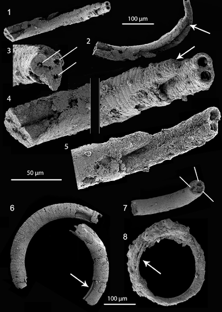
Holotype of Portfjeldia aestatis and other unnamed tubular fossils showing various types of similar internal structures: (1)–(5) Portfjeldia aestatis (PMU 36870/2) from various viewpoints displaying two tubules interpreted as being originally enclosed by a now partly degraded external sheath; (3) enlargement of lower left of (4) with lines indicating three tubes rather than two, indicating a possible triradial symmetry; arrows in (2) and (4) show possible branching and development of daughter tube; (5) possible third tubule originating as a ridge near the other extremity. (6) Broken tube (PMU 39237/1), possibly related to the problematic spiral tube also described; arrow indicates internal groove also visible in (4). (7) Broken tube (PMU 38168/2) showing three possible tubules indicating a triradial structure (white lines). (8) Single whorl of a degraded, annulated helix with internal tubular structure (arrow) (PMU 36876/4). Scale bar 100 μm for (1), (2), (6)–(8) and 50 μm for (3)–(5). Willman & Peel (2022).

Willman and Peel also describe another distinctive tubular microfossil, but decline to name this as only a single specimen is known, and this was found within the helical coil of a specimen of Jiangispirellus groenlandicus, making it impossible to say there is not a relationship between the two. This specimen is almost 2 mm in length, with a diameter of about 20 μm, and a central opening running along its length about 10 μm in diameter.

Problematic helically spiraled tubular organism. (1), (2) Preserved as an internal tube inside an outer Jiangispirellus 'trichome', showing traces of cell wall (PMU 36870/3); (3) remains of a possible branching organism (arrow) within a 'trichome' (PMU 36868/4); (4) preserved 'trichome' of Jiangispirellus groenlandicus with internal secondary phosphatisation (PMU36874/5). Note that no internal septa are preserved, indicating that the phosphatised 'trichome' covered a hollow chamber. Scale bar 100 μm for (1), (3), (4) and 50 μm for (2). Willman & Peel (2022).

Ediacaran deposits have produced a wide range of tubular fossils of varying sizes, and debatable origins. The majority of the larger tubes are likely to have been produced by Metazoans (Animals), and some may have been biomineralised. As such, understanding these tubes is thought to be a key step to understanding the origin of the Animals as a whole. Many of these macroscopic fossils share morphological similarities to the microfossils Willmand and Peel describe from the Portfjeld Formation, being slightly-curved to sinuous, and occasionally branching. However, the macro- and microfossils are not identical in morphology, which, combined with the size difference between the two groups, makes it impossible to say if they are genuinely related or just show convergent morphology.

Microscopic tubular and filamentous fossils have variously been interpreted as Cyanobacteria, Algae, and Fungi. It is seldom even possible to say with any confidence whether these tubes were made by prokaryotic or eukaryotic organisms, due to the overlap in size between the two groups, although a number of 'grades' of structure have been proposed to try to separate different groups of tube-makers.

Tubes of sizes similar to the ones Willman and Peel describe from the Portfjeld Formation are known to be made by extant Cyanobacteria, such as Microcoleus, and Schizothrix, as well as sulphur-oxidizing Bacteria such as Thioploca. Forms such as the extant Trichodesmium even form bundles of filaments, as seen in Portfjeldia aestatis, and Subtifloria, an Ediacaran-Cambrian calcarious fossil comprising bundles of filaments, also interpreted as a Cyanobacteria. However, such bundles of filaments are not usually contained within an outer sheaf, and it is difficult to imagine what the advantage of such a sheaf would be to a photosynthetic Cyanobacterium.

Subtifloria is known from the Late Eidacaran deposits of Shaanxi Province, China, where it forms part of an assemblage that also includes forms interpreted as Oscillatorialean and Rivulariacean Cyanobacteria, some of the most complex members of the group today. The Shaanxi and Portfjeld Ediacaran biotas both contain Obruchevella, a helical microfossil possibly also made by a Cyanobacterium, as well as Jiangispirellus and Spirellus, suggesting a possible link between the two sites. 

Complex modern Cyanobacteria, such as those of the Order Nostocales, develop distinctive resting cells towards the end of their annual growth cycle, which can produce new filaments when conditions improve again, building up Cyanobacterial mats over time. The specimens from Portfjeld appear to be of a much more complicated nature than anything seen in such Cyanobacteria, and may well themselves be part of a larger and mote complex organism, making it unlikely the fossils are of Cyanobacterial origin.

The organism found preserved within the coil of a specimen of Jiangispirellus groenlandicus could be interpreted as a specimen of that organism, but for the presence of an internal cavity. Again, its general morphology is consistent with a Cyanobacterial origin, but its position within the coil of another calcified organism seems unlikely for anything which was reliant on photosynthesis to survive.

Eukaryotic Algae are important members of many modern ecosystems, both aquatic and terrestrial, and come in a wide range of forms. Neoproterozoic Algae can usually be identified either by a branching structure or the presence of more than one type of cell. The earliest form of multicellular Eukaryotic Algae, filamentous Red Algae, probably appeared at around the beginning of the Mesoproterozoic (1600 million years ago), while the first filamentous Green Algae probably appeared at around the beginning of the Neoproterozoic, about 1000 million years ago. The general shape and size of  Portfjeldia aestatis, this appears quite likely to be an Algae, although the internal tubules are unlike anything known from any Algal group, living or fossil.

Ediacaran deposits contain a wide variety of macrofossils which may-or-may-not represent early Animals. However some, such as the bilaterally symetrical Kimberella, are now generally accepted as being true Animals, in addition to which trace fossils have been found at a number of Ediacaran sites, leading to the possibility that fossils from Ediacaran deposits may represent Animals or parts of Animals. 

Mineralised tubular fossils are also common in the Ediacaran, and an Animal origin has been suggested for many of these, such as the Anabaritids, globally distributed tubular organisms that grew by accretion around the aperture and showed a triradial symmetry, which have been suggested as possible stem-group Cnidarians.

Another possibility is that the inner tubules of Portfjeldia aestatis are not part of the same organism as the outer tubes, but some form of cavity dwelling micro-organism which has secondarily occupied the tubes. Such behaviour is found in a variety of different organisms, including Bacteria, Algae, and Fungi. Such organisms will often occupy tubes created by other organisms, although the filamentous shape of Portfjeldia aestatis is unlike any known such organism, which tend to be more mesh-like in organisation, on which basis Willman and Peel consider this to be an unlikely scenario.

Given the possible alternatives, Willman and Peel believe that the most likely explanation is that Portfjeldia aestatis is some form of Algae, albeit one unlike any modern form. 

See also...

Online courses in Palaeontology

Follow Sciency Thoughts on Facebook.

Follow Sciency Thoughts on Twitter.