
The Dinocephalian were a group of large-bodied Therapsids (the wider group which also includes the Cynodonts, the group that gave rise to, and includes the Mammals) known from the Middle Permian of Russia, China, Brazil, South Africa, Zimbabwe, and Tanzania. They had exceptionally thick skulls, with the braincase often entirely enclosed by other elements of the skull. The purpose of this bone is unclear; it may have been used in intra-specific head-butting contests, as in modern ungulates, but ungulates have a thick keratinous (horn) boss that absorbs impact, rather than greatly thickened bone, so the biomechanics of conflict in the two groups would not be the same. This thick layer of dense bone makes it hard for palaeontologists to examine the structure of the skull without recourse to destructive methods, further hampering efforts to understand the biology of these animals.
In a paper published in the journal PeerJ on 10 August 2017, Julien Benoit of the Evolutionary Institute and School of Anatomical Sciences at the University of the Witwatersrand, Paul Manger, also of the School of Anatomical Sciences at the University of the Witwatersrand, Luke Norton, also of the Evolutionary Institute at the University of the Witwatersrand, Vincent Fernandez of the European Synchrotron Radiation Facility, and Bruce Rubidge, again of the Evolutionary Institute at the University of the Witwatersrand, describe the results of a study in which a specimen of the Dinocephalian Moschops capensis was scanned at the European Synchrotron Radiation Facility in Grenoble, France, providing information on the internal anatomy of the skull of this species that had not previously been available.
The specimen, AM4950, was discovered at The Grant 39 site, to the north of Grahamstown in Eastern Cape Province, and is housed in the Albany Museum in Grahamstown. The specimen is estimated to be 265 million years old, and is a subadult, weathered on the left side, but well preserved on the right. It is slightly over 34 cm in length, with the largest adult skulls reaching about 38 cm.

The skull of Moschops capensis AM4950 in lateral view. (A) Photograph of the skull. (B) Reconstruction of the skull (right side, bone transparent) to reveal the neural structures discussed in this paper. (C) Reconstruction of the skull (left side, bone transparent) showing the endocast, bony labyrinth and the angle between the plane of the lateral semicircular canal and the main axis of the skull. Numbers indicate the position of the cross sections in the subsequent figures. EmV, emissary veins; End, endocranial cast; Hyp, hypophyseal fossa; Lab, bony labyrinths; Pin, pineal tube. Luke Norton in Benoit et al. (2017).
Benoit et al. estimate that AM4950 would have had a total body length of about 187 cm and would have weighed about 327 kg. It's braincase has a volume of 61 cm³ (though this will be considerably larger than the actual brain volume as the brain is surrounded by layers of soft tissue and fluid) giving it a braincase-volume to total mass ratio (encephalization quotient) similar to the lowest values found among living animals. The skull above the brain is about 6 cm thick, with the outer 2 cm being dense, osteosclerotic, bone and the lower 4 cm being less dense cancellous bone.
Previous reconstructions of Moschops capensis have suggested that its snout was carried before it, like that of a modern Mammal such as a Horse or Dog, and it has even been suggested that these animals may have been semi-aquatic, but the position of the labyrinth, the organ in the inner ear which is used to tell which way up the head is (and therefore enable us to balance), in AM4950 makes this highly unlikely. Benoit et al. reconstruct Moschops capensis with its head held at an angle of 60 -65° to the horizontal.
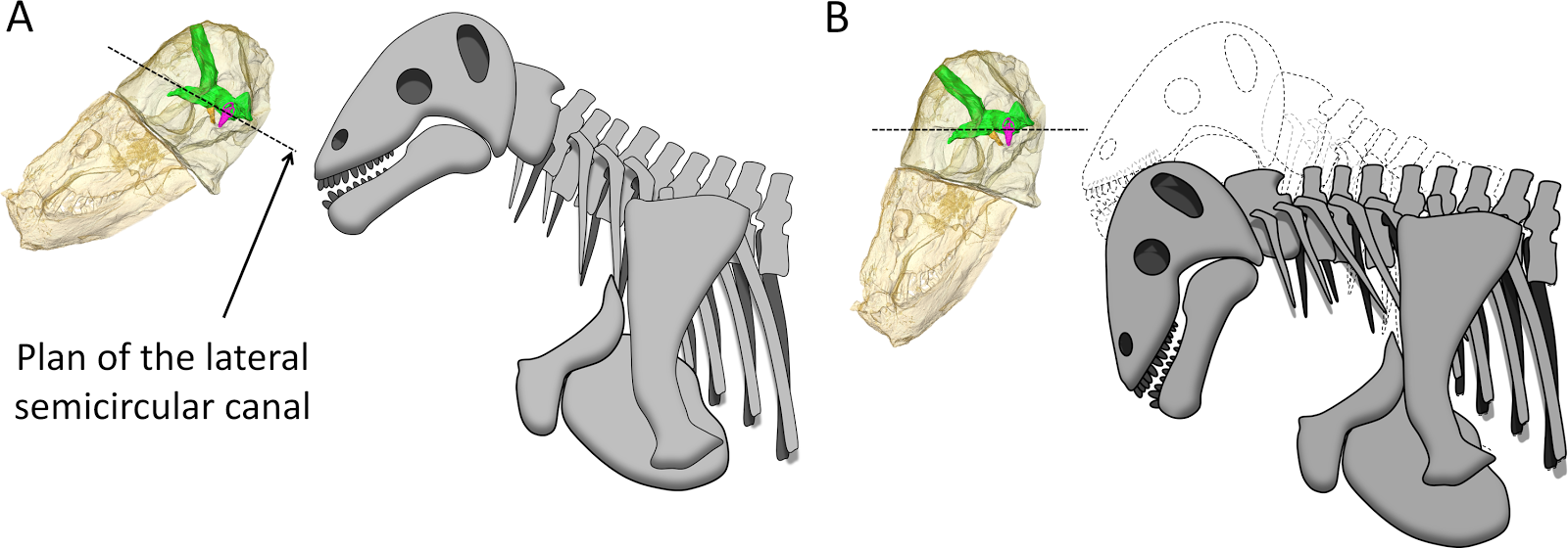
Hypothesized reconstructions of the natural head posture in Moschops capensis. (A) Redrawn after the mounted skeleton at the American Museum of Natural History. (B) Based on the position of the plane of the lateral (horizontal) canal. Julien Benoit in Benoit et al. (2017).
In this position the thickened bone above the braincase would be pointed directly forward, and also directly in line with the straightened spine, an ideal position for fighting by head-butting, strongly supporting the idea that these animals exhibited this behaviour. While to us head-butting does seem a particularly advanced behaviour, it actually implies a degree of social behaviour not generally attributed to early Therapsids In modern species that exhibit such behaviour it is usually associated either with ritualized display fights, observed by females who in turn engage in a degree of mate choice, or with the establishment of territories, which requires a degree of spatial awareness and the ability to understand a simple set of social rules.
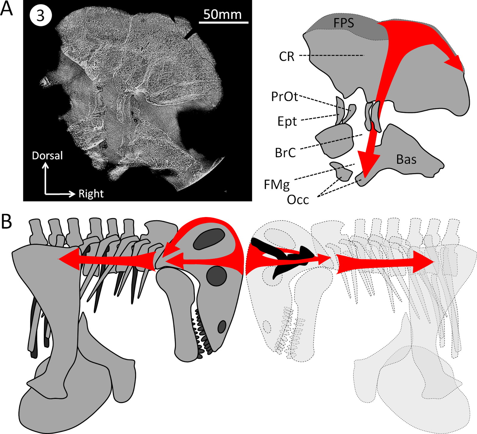
Hypothesized dissipation of the energy during head butting in the skull of Moschops capensis. Arrows indicate the direction of energy transfer. (A) CT section of the skull of Moschops capensis AM4950. (B) The proposed route of the dissipation of energy through the dermatocranium (left) and the braincase (right) in two fighting Moschops. Abbreviations: Bas, basicranium; BrC, braincase; CR, cranial roof; Ept, epipterygoid; FMg, foramen magnum; FPS, frontoparietal shield; Occ, occipital condyles; PrOt; prootic. Julien Benoit in Benoit et al. (2017).
See also...


Follow Sciency Thoughts on Facebook.