Showing posts with label Ascomycetes. Show all posts

Wednesday, 20 March 2024

Zygosporium himachalensis: A fossil Ascomycote Fungus from the Miocene of Himachal Pradesh, India.

The genus Zygosporium contains asexual, largely saprotrophic, Ascomycete Fungi distinguished by dark brown setiform conidiophores (bristle-shaped spore producing bodies) with darkly pigmented, incurved, and swollen vesicles, often stacked, emerging either from the side of the conidiophore or from the mycelium (network of fungal threads).There are 22 described living species within the group, as well as three known fossil species, two from the Siwalik sediments of the eastern Himalayas, and one from the Oligocene of Hungary.

In a paper published in the journal Fungal Biology on 12 March 2023, Sampa Kundu and Mahasin Ali Khan of the Palaeobotany and Palynology Laboratory at the Department of Botany at Sidho-Kanho-Birsha University, describe a new species of Zygosporium from the Late Miocene Middle Siwalik of Himachal Pradesh, in the western Himalayas.

The new species is named Zygosporium himachalensis, where 'himachalensis' means 'from Himachal'. It is based upon a series of specimens found growing on a compressed Monocot leaf recovered from a grey mudstone in the Middle Siwalik in Mandi District of Himachal Pradesh. This location is about 8-10 km from, and probably coeval with Nalad Khad Section, which has been dated to 12-8 million years before the present.

(A) A compressed Monocot leaf segment; (B) Light microscopic image of Zygosporium himachalensis (SKBUH/PPL/HP/M40/S2) showing superficial or partly immersed hyphae with vesicular conidiophores in small or large groups or scattered on fossil Monocot leaf cuticle (SKBUH/PPL/HP/M40); (C), (D), (E), (G). Zygosporium himachalensis showing stacked chained vesicular cells on straight erect unbranched conidiophores; vesicular cells arising from apical, sub-apical, and lateral sides of the conidiophores (vesicular cells marked by blue arrows); (F) Zygosporium himachalensis showing stacked chained vesicular cells on a branched conidiophore (branching point marked by a black arrow; vesicular cells marked by blue arrows). Kundu & Khan (2024).

Specimens of Zygosporium himachalensis have erect, usually unbranched conidiophores, with chains of up to 4 integrated alternatively or suboppositely arranged vesicles arising directly from the mycelium. These are arranged in effuse or sometimes compact irregular-shaped patches on the surface of the leaf.

(A)–(L) Light microscopic images of Zygosporium himachalensis showing septate superficial conidiophore (marked by white arrows) with alternately (marked by blue arrows) and sub-oppositely (marked by green arrow) arranged dark brown to black, swollen, incurved, thick-walled vesicular cells (marked by red arrows); some vesicular cells with a cylindrical, brown, thick-walled, smooth, short stalk cell (marked by black arrows). Kunda & Khan (2024).

See also...

Sunday, 3 March 2024

Talaromyces sedimenticola: A new species of Ascomycote Fungus from the deepest part of the Mariana Trench.

The Challenger Deep forms the deepest part of the Mariana Trench, reaching 10 971 m below sealevel. Curiously, recent studies have shown that the hadal depths of the oceans (areas more than 6 km deep) have a higher microbial carbon turnover than occurs at abyssal depths, between 4 km and 6 km below the surface. This has been supported by metagenomic studies which have shown significantly more genes coding for carbohydrate-active enzymes and peptidase are being expressed at these depths. This has led microbiologists to take an interest in the organisms living in the sediments of the deepest ocean trenches. 

In a paper published in the journal Antonie van Leeuwenhoek on 28 February 2024, Hongbo Zhou of the School of Minerals Processing and Bioengineering and the Key Laboratory of Biometallurgy at Central South University, Liting Xu and Wenxian Liu, also of the School of Minerals Processing and Bioengineering at Central South University, Kaiwen Ta of the Institute of Deep-Sea Science and Engineering of the Chinese Academy of Sciences, Xincun Wang of the State Key Laboratory of Mycology at the Institute of Microbiology of the Chinese Academy of Sciences, Jianwei Guo of the College of Agronomy and Life Sciences at Kunming University, Wenxi Luo, Zhiyuan Peng, and Qiaoni Huang, again of the School of Minerals Processing and Bioengineering at Central South University, and Yuguang Wang, once again of the School of Minerals Processing and Bioengineering and the Key Laboratory of Biometallurgy at Central South University, describe a new species of Ascomycote Fungi from the Challenger Deep.

The new species is described on the basis of two strains isolated from samples collected by the Research Vessel Tan Suo Yi Hao in September 2019, from a depth of 10 063 m below sealevel. A genetic analysis of these strains suggests that they belong to the same species, and, surprisingly, that that species  is a member of the genus Talaromyces, which mostly comprises terrestrial moulds, forming a sister taxon to a clade which includes Talaromyces trachyspermus, which is a serious commercial pest species, frequently infecting packaged fruit juices, and Talaromyces assiutensis, which is found growing within the leaves of Mangroves around the South China Sea. The new species is named Talaromyces sedimenticola, in reference to the environment where it was found.

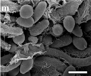
Morphological characteristics of Talaromyces sedimenticola, (k), (l) conidiophores, (m) conidia. Zhou et al. (2024).

While morphologically similar, and genetically close, to other members of the genus TalaromycesTalaromyces sedimenticola shows some remarkable physiological traits, which mark it out as distinctive. It could be grown at temperatures of between 4°C and 50°C, unlike most Talaromyces species, which typically grow between about 28°C and 40°C. It could also survive and grow over a far wider pH range than any other species within the genus, pH 1.5-12, whereas other Talaromyces species could only survive in the pH range 4-8, with the exception of Talaromyces aculeatus, a widespread soil-dwelling form, which can survive over the range pH 1-7. Talaromyces sedimenticola is also remarkably halotollerant, able to flourish on media with 0-14% sodium chloride (weight over volume). More curiously, Talaromyces sedimenticola was unable to metabolise sucrose, tryptone, or monobasic potassium phosphate, all of which can be utilised by other members of the genus, although it was able to utilise other common foodstuffs, such as glucose, maltose, lactose, xylose, soluble starch, glycerol, peptone, ammonium sulphate, potassium phosphate, potassium chloride, and magnesium sulphate. 

See also...

Tuesday, 30 January 2024

Tuber itzcuinzapotl: A new species of edible Truffle from Mexico.

Truffles, Tuber spp., are Ascomycote Fungi which from ectomycorrhizal relationships with a range of forest Plants, including Pines, Oaks, Hickories, and Orchids. They are distinguished for their large, tuber-like ascomata (fruiting bodies), which are formed underground, which often have highly distinctive aromas and flavours, leading to some species being traded as high-value gourmet items.

There are currently 25 species of Truffle known from Mexico, mostly from the temperate forests of the north and the mountains of the Neovolcanic axis. However, none of these are currently traded as foodstuffs, despite Mexico a globally leading countries in terms of the number of edible wild Fungi consumed, with about 500 species, making it second only China, where about 1000 are consumed. However, recent efforts have found that the non-native Black Truffle, Tuber melanosporum, will form ectomycorrhizal relationships with native Mexican Oaks, and several species found in Mexico are considered to have potential for commercial development, including the Pecan Truffle, Tuber lyonii, which is commercially exploited in the US and Canada, and can trade for up to US$400 per kg.

In a paper published in the journal Phytotaxa on 26 January 2024, Javier Isaac de la Fuente of the Colegio de Postgraduados at Campus Montecillo, Wendy Rosales-Rosales of the Instituto Tecnológico Superior de Zongolica of the Tecnológico Nacional de México, César Romero Martínez-González of the Instituto Tecnológico de Ciudad Victoria of the Tecnológico Nacional de México, Magdelana Martínez-Reyes, also of the Colegio de Postgraduados at Campus Montecillo, Andrea Carolina Elizondo-Salas, also of the Instituto Tecnológico Superior de Zongolica of the Tecnológico Nacional de México, and Jesús Pérez-Moreno, agian of the Colegio de Postgraduados at Campus Montecillo, describe a new species of edible Truffle from the Coniferous mixed forests of eastern Mexico.

The new species is named Tuber itzcuinzapotl, where 'itzcuinzapotl' means 'Dog's Zapote' in the Nahua language (a Zapote is a type of fruit). This Truffle produces subglobose fruiting bodies with a light brown, verrucous-granular outer surface, and an gray or pale brown interior, reaching up to 28 mm by 28 mm in size, with a distinctive fruity taste and smell. It is found growing in association with Mexcan Weeping Pines, Pinus patula, in Veracruz State, Mexico.

Tuber itzcuinzapotl (Holotype). Fresh ascomata fruiting body. De la Fuente et al. (2024).

Mexico has a significant culture of wild Fungus consumption, with over 500 types of Fungi consumed by members of all ethnic groups, and in particular rural communities living close to woodland. However, almost all consumed Fungi are epigeal, i.e, found above the ground, such as Mushrooms, with very little exploitation of subterranean species occurring. This is surprising, as Mexico is one of the most biodiverse regions in the world for Oaks, and Oaks are particularly associated with Fungi producing underground fruiting bodies. 

Tuber itzcuinzapotl is known to be consumed by members of the Nahua ethnic group living in the Sierra de Zongolica region of Veracruz State, Mexico, where it is referred to as 'itzcuinzapotl' (the specific name chosen for the species). Local folklore has it that people began to consume these Fungi after observing Dogs digging them up and eating them. Knowledge of the Fungus appeared to be restricted to older women in the community. Such local knowledge of wild foodstuffs is considered to be at risk in the region as traditional cultures are eroded, leading to loss of knowledge and a reduction and homogenisation in the number of foodstuffs consumed by Humans both in Mexico and globaly.

See also...

Sunday, 18 December 2022

Drechslerella daliensis & Drechslerella xiaguanensis: Two new species of predatory Fungi from Yunnan Province, China.

Predatory Fungi are important controls on Nematode populations in many soil ecosystems. The majority of these Fungi belong to the Ascomycote Family Orbiliaceae, which currently includes 116 predatory species, divided into three genera. Arthrobotrys, which contains 67 species, and which traps Nematodes in a network of sticky hyphae, Dactylellina, which contains 34 species and which captures Nematodes with sticky knobs, and Drechslelrella, which contains 15 species, and which produces constricting rings, used to trap Nematodes in a lasso-like action.

The genus Drechslelrella was first described by the Indian mycologist Chirayathumadom Venkatachalier Subramanian in 1963, and has been found in a wide variety of ecosystems, including forest soils, Mangrove sediments, freshwater sediments, brackish water sediments, heavy metal polluted areas and even in tree trunks and animal faeces. They are typically found in the upper part of the soil or overlying humus layer, where the density of Nematodes appears to be highest. The constricting rings produced by these Fungi are made up of three cells, which swell rapidly when a Nematode is detected. 

In a paper published in the Biodiversity Data Journal on 16 December 2022, Fa Zhang of the Institute of Eastern-Himalaya Biodiversity Research at Dali University, and the Center of Excellence in Fungal Research and School of Science at Mae Fah Luang UniversitySaranyaphat Boonmee, also of the Center of Excellence in Fungal Research and School of Science at Mae Fah Luang University, Jutamart Monkai, again of the Center of Excellence in Fungal Research at Mae Fah Luang University, and Xiao-Yan Yang and Wen Xiao, also of the Institute of Eastern-Himalaya Biodiversity Research, and of the Key Laboratory of Yunnan State Education Department on Er’hai Lake Basin Protection and the Sustainable Development Research, at Dali University, describe two new species of Drechslerella from fire-disturbed forest soil from the slopes of Cangshan Mountain, to the west of Dali city in Yunnan Province, China.

The new Fungi were cultivated from soil samples spread on agar, and prompted to produce traps by the introduction of the free-living Nematode Panagrellus redivivus as bait. They were confirmed as new species by the extraction of DNA, and comparison to other species on the GenBank database, using the BLASTn online tool. The phylogenetic relationships of the new species were established using the MrBayes software package.

The first new species is named Drechslerella daliensis, where 'daliensis' means 'from Dali', in reference to the city. This Fungus formed white, cottony colonies on the cultivation medium, reaching a diameter of 50 mm after 18 days. Two types of conidia (spore producing bodies) were produced, both on conidiophores (conidia-bearing hyphae) 125-335 µm in length. The macroconidia were roughly 20-49.5 µm long and 8.5-15 µm wide, smooth, ellipsoid, broadly rounded at the apex and truncated at the base. The microconidia were 6.5-22 µm long and 3.5-7 µm wide, smooth, clavate or bottle-shaped, broadly rounded at the apex and truncated at the base. Drechslerella daliensis was observed to trap Nematodes with constricting rings on stalks 5.5–11 µm long, with an outer diameter of 21–32 µm and an inner diameter of 2–21 µm.

Drechslerella daliensis (holotype, CGMCC3.20131). (a) Culture colony; (b), (c) Macroconidia; (d) Microconidia; (e) Constricting rings; (f), (g) Conidiophores. Scale bars: (a) 1 cm; (b)–(g) 10 µm. Zhang et al, (2022).

The second species is named Drechslerella xiaguanensis, where 'xiaguanensis' means 'from Xiaguan' in reference to the district where the soil samples were collected. This Fungus also formed white, cottony colonies on the growth medium, reaching 50 mm in diameter after 15 days. The conidia of this species were born on conidiophores 145-315 µm in length, with a single type of conidia being produced, these being 33-52 µm in length and 9.5–28 µm in width. Constricting rings were born on stalks 5–11.5 µm long, and had an outer diameter of 19–27.5 µm and an inner diameter of 12.5–20.5 µm.

Drechslerella xiaguanensis (holotype, CGMCC3.20132). (a) Culture colony; (b), (c) Conidia; (d) Germinating conidia; (e) Constricting rings; (f), (g) Conidiophore. Scale bars: (a) 1 cm; (b)–(g) 10 µm. Zhang et al. (2022).

The phylogenetic analysis confirmed that both species could be placed within the genus Drechslerella, with Drechslerella daliensis being recovered as the sister species to Drechslerella hainanensis, known from Hainan, an island province of China in the South China Sea, and Drechslerella xiaguanensis as the sister species to Drechslerella bembicodes, a species from New Zealand.

Maximum Likelihood tree, based on combined ITS, TEF1-α and RPB2 sequence data from 42 Nematode-trapping taxa in Orbiliaceae. Bootstrap support values for Maximum Parsimony (red) and Maximum Likelihood (black) equal or greater than 50% and Bayesian posterior probabilities values (green) greater than 0.90 are indicated above the nodes. New isolates are in blue, ex-type strains are in bold. Zhang et al. (2022).

Zhang et al. note the the network of sticky hyphae used by the genus Arthrobotrys appears to be a more efficient trap than the methods used by Drechslerella or Dactylellina, and that, under normal circumstances, this genus is the most abundant in the upper soil layer, where Nematodes are most abundant. However, in the fire-damaged forest soils, the reverse appeared to be the case, with Arthrobotrys almost absent, while Drechslerella and Dactylellina are abundant. They theorise that in this environment, Arthrobotrys has been killed off by the burning of the upper soil layer, allowing the other two genera to invade rapidly from colonies in deeper soil layers.

See also...


Follow Sciency Thoughts on Facebook.

Follow Sciency Thoughts on Twitter.


Friday, 17 April 2020

Seven new species of Marine Fungi from the Mediterranean.

Marine Fungi are an important and active component of the microbial communities that inhabit the oceans. Fungi in the marine environment live as mutualists, parasites, pathogens and saprobes, and are pivotal to marine food webs because of the recycling of tough organic materials that other organisms cannot break down; besides which, these widely dispersed organisms are a source of novel bioactive compounds. Marine Fungi have been recovered worldwide from a broad range of biotic and abiotic substrata, such as driftwood algae, sponges, corals, sediments, etc. A 'Marine Fungus' is defined as any fungus retrieved repeatedly from marine environment and that reproduces in the marine environment. There are currently about 1680 described Marina Fungal species belonging to 693 genera, 223 families, 87 orders, 21 classes and six phyla. However, considering that the total number of Marine Fungi has been estimated to exceed 10 000 taxa, fungal diversity remains largely undescribed. With more than 900 species, the Ascomycota are the dominant Fungal phylum in the sea.

In a paper published in the journal Diversity on 6 April 2020, Anna Poli, Elena Bovio, Lucrezia Ranieri, Giovanna Cristina Varese, and and Valeria Prigione of the Department of Life Sciences and Systems Biology at the University of Torino describe seven ew species of Marine Ascomycote Fungi from the Mediterranean.

The first new species is placed in a new genus, Parathyridariella, which means 'beside Thyridariella', in reference to a previously described species, to which the new genus is closely related, and given the specific name dematiacea, meaning darkly pigmented, in reference to the colour of the colony on culture media  This species was isolated from a Green Seaweed, Flabellia petiolata, found growing at a depth of 14-15 m off the coast of Ghiaie Beach on the island of Elba, and a Seagrass, Posidonia oceanica, growing at a depth of 5-21 m off the coast of Punta Manara in the Province of Genoa, Italy. Colonies of this Fungus grown on Malt Extract Agar-sea water media reached 28–34 mm in diameter after 28 days at 24 °C, grown on Oatmeal Agar-sea water reached 40-34 mm in diameter after 28 days at 24°C, and on Potato Dextrose Agar-sea water reached 36-49 mm in diameter after 28 days at 24 °C, and 15.5–22.5 mm in diameter after 28 days at 15°C. The species grew actively on Pine wood and cork. The mycelium varies in colour from dark grey/black to dark green, and is dense with radial grooves and concentric rings, and submerged edges; the reverse is dark green. A brown exudate present above the concentric rings. The hyphae are 2.8–4.8 m wide, septate, hyaline to lightly pigmented. Parathyridariella dematiacea produces numerous Chlamydospores (thick-walled hyphal cells which function like spores), but neither sexual morphs or asexual conidiogenesis (spore production) were seen.
Parathyridariella dematiacea, 28-days-old colony at 21°C on Malt Extract Agar-sea water media (A) and reverse (B); solitary (C) and in chain (D) chlamydospores. Scale bars are 10 μ m (C), (D). Poli et al. (2020).

The second new species is also placed in the genus Parathyridariella, and given the specific name tyrrhenica, in reference to the Tyrrhenian Sea, where it was discovered. This species was isolated from a Brown Seaweed, Padina pavonica, (Peacock's Tail), and a Green Seaweed, Flabellia petiolata, both found growing at a depth of 14-15 m off the coast of Ghiaie Beach on the island of Elba. Colonies of this Fungus grown on Malt Extract Agar-sea water media reached 10 mm in diameter after 28 days, at 21° C, grown on Oatmeal Agar-sea water reached 48-50 mm in diameter after 28 days at 24°C, and 26-29 mm in diameter after 28 days at 15°C, and on Potato Dextrose Agar-sea water reached 31–46 mm in diameter after 28 days at 24 °C, and 16–19 mm in diameter after 28 days at 15°C. The species grew actively on Pine wood and cork. The mycelium is funiculose (made up of rope-like strands), yellowish, land ightly ochre at the edges; the reverse is light yellow, lighter at the edges. The hyphae are 5 μm diameter, septate, hyaline to brownish, sometimes wavy or swollen, forming hyphal strands. No reproductive structures were observed.

Parathyridaria tyrrhenica, 28-days-old colony at 21°C on Malt Extract Agar-sea water media (A) and reverse (B); mycelium (C), black and white arrows indicate hyphal strands and wavy hyphae, respectively. Scale bar is 10 μ m. Poli et al. (2020).

The third species described is also placed in the genus Parathyridaria, and given the specific name flabelliae, in reference to the Green Seaweed, Flabellia petiolata, on which it was found growing, at a depth of 14-15 m off the coast of Ghiaie Beach on the island of Elba. Colonies of this Fungus grown on Malt Extract Agar-sea water media reached 37–44 mm in diameter after 28 days, at 21° C, grown on Oatmeal Agar-sea water reached 60 mm in diameter after 28 days at 24°C, and 33–35 mm in diameter after 28 days at 15°C, and on Potato Dextrose Agar-sea water reached 53–64 mm in diameter after 28 days at 24 °C, and 23–24 mm in diameter after 28 days at 15°C. The species grew actively on Pine wood and cork. The mycelium is funiculose (made up of rope-like strands), and whitish with submerged edges; the reverse is brown in the middle, lighter at edges. The hyphae are 2.6-5 μ m wide, septate and hyaline. Parathyridariella flabelliae produces numerous Chlamydospores, which are globose or subglobose, from light to dark brown in colour, and either unicellular (4 x 5 μ m diameter) or multicellular (up to four-celled and 8 x 12 μm diameter), but neither sexual morphs or asexual conidiogenesis (spore production) were seen.

Parathyridaria flabelliae, 28-days-old colony at 21°C on Malt Extract Agar-sea water media (A) and reverse (B); unicellular and multicellular chlamydospores (C). Scale bar is 10 μm. Poli et al. (2020).

The fourth  new species described is placed in the genus Neoroussoella, and given the specific name lignicola, which implies it grows on dead wood.  This species was isolated from a Brown Seaweed, Padina pavonica, (Peacock's Tail), and a Seagrass, Posidonia oceanica, both found growing at a depth of 14-15 m off the coast of Ghiaie Beach on the island of Elba. Colonies of this Fungus grown on Malt Extract Agar-sea water media reached 28–29 mm in diameter after 28 days, at 21° C, grown on Oatmeal Agar-sea water reached 27-40 mm in diameter after 28 days at 24°C, and 14.5-26 mm in diameter after 28 days at 15°C, and on Potato Dextrose Agar-sea water reached 38–45 mm in diameter after 28 days at 24 °C, and 19–29 mm in diameter after 28 days at 15°C. This species grew efficiently on Pine wood. The mycelium is grey to dark green and floculose, with irregular edges, the reverse is dark grey. A clear exudate is often present. Hyphae are 2–4.4 m wide, septate, hyaline, and assume a toruloid aspect when growing into wood vessels; they form chains of two-celled chlamydospores which, at maturity, protrude from the vessels. The chlamydospores are 7.4 x 5.2 μ m, from light to dark brown, and globose or subglobose. Neither sexual morphs or asexual conidiogenesis (spore production) was seen.

Neoroussoella lignicola, 28-days-old colony at 21°C on Malt Extract Agar-sea water media (A) and reverse (B); two-celled chlamydospores inside wood vessels (C). Scale bar is 10 μm. Poli et al. (2020).
The fifth new species described is placed in the genus Roussoella, and given the specific namemargidorensis, meaning 'from Margidore'; the species was isolated from  a Brown Seaweed, Padina pavonica, (Peacock's Tail), found growing at a depth of 14-15 m off the coast of Margidore on the island of Elba. Colonies of this Fungus grown on Malt Extract Agar-sea water media reached 33-34 mm in diameter after 28 days, at 21° C, grown on Oatmeal Agar-sea water reached 45 mm in diameter after 28 days at 24°C, and 27 mm in diameter after 28 days at 15°C, and on Potato Dextrose Agar-sea water reached 45 mm in diameter after 28 days at 24 °C, and 23 mm in diameter after 28 days at 15°C. This species grew actively on Pine wood. The mycelium is whitish, lighter to the edge, and umbonate (having a rounded knob or protuberance) in the middle, the reverse is ochre. Hyphae are approximately 2 μm wide, septate and brownish. Neither sexual morphs or asexual conidiogenesis (spore production) was seen.


Roussoella margidorensis, 28-days-old colony at 21°C on Malt Extract Agar-sea water media (A) and reverse (B); chlamydospores (C). Scale bar is 10 μ m. Poli et al. (2020).

The sixth new species described is also placed in the genus Roussoella, and given the specific name mediterranea, in reference to the Mediterranean Sea. The species was isolated from  a Brown Seaweed, Padina pavonica, (Peacock's Tail), found growing at a depth of 14-15 m off the coast of Margidore on the island of Elba. Colonies of this Fungus grown on Malt Extract Agar-sea water media reached 55 mm in diameter after 28 days, at 21° C, grown on Oatmeal Agar-sea water reached 67–72 mm in diameter after 28 days at 24°C, and 33–38 mm in diameter after 28 days at 15°C, and on Potato Dextrose Agar-sea water reached 69–76 mm in diameter after 28 days at 24 °C, and 32.5–39 mm in diameter after 28 days at 15°C. This species grew actively on Pine wood, and poorly on cork. The mycelium is light grey, and floccose, with an umbonate area in the middle, the reverse is brown with lighter edges. A dark exudate present. Hyphae are 2.4 μm wide, septate and dematiaceous. Branched chains of light to dark brown chlamydospores often present, these are 4.5 x 5.7 μm, and from unicellular to 4-celled. Neither sexual morphs or asexual conidiogenesis (spore production) was seen.

Roussoella mediterranea, 28-days-old colony at 21°C on Malt Extract Agar-sea water media (A) and reverse (B); unicellular and multicellular chlamydosporesn indicated by a black arrow (C). Scale bar is 10 μ m. Poli et al. (2020).

The final species is also placed in the genus Roussoella, and given the specific name padinae, in reference to the Brown Seaweed, Padina pavonica, (Peacock's Tail), upon which it was found growing, at a depth of 14-15 m off the coast of Margidore on the island of Elba. Colonies of this Fungus grown on Malt Extract Agar-sea water media reached 53 mm in diameter after 28 days, at 21° C, grown on Oatmeal Agar-sea water reached 57.5–65 mm in diameter after 28 days at 24°C, and 30–35 mm in diameter after 28 days at 15°C, and on Potato Dextrose Agar-sea water reached 60–69 mm in diameter after 28 days at 24 °C, and 30–34 mm in diameter after 28 days at 15°C. This species grew poorly on Pine wood, and efficiantly on cork. The mycelium is from grey to dark green, floccose in the middle, with radial grooves, and fimbriate edges; the reverse is brown. Hyphae are 3 μm wide, septate, brownish and assume a toluroid aspect when growing into wood vessels, and form chains of two-celled chlamydospores which, at maturity, protrude from the vessels. These chlamydospores are 5–7 x 4 μm, from light to dark brown in colour, subglobose, ellipsoidal or cylindrical. Neither sexual morphs or asexual conidiogenesis (spore production) was seen.

 Roussoella padinae, 28-days-old colony at 21°C on Malt Extract Agar-sea water media (A) and reverse (B); toruloid hyphae (C) and two-celled chlamydospores (D) inside wood vessels. Scale bars are 10 μm. Poli et al. (2020).

The description of these new taxa was particularly challenging because neither asexual nor sexual reproductive structures developed in axenic conditions. Therefore, Poli et al. were unable to describe the range of anatomical variations and diagnostic features among these newly recognized phylogenetic lineages. Indeed, strictly vegetative growth without sporulation is a common feature of many marine Fungal strains. Possibly, these organisms rely on hyphal fragmentation for their dispersal, or alternatively, the di erentiation of reproductive structures may be obligatorily dependent on the peculiar environmental conditions under which they live (e.g., wet-dry cycles, high salinity, low temperature, high pressure, etc.). During the study of these fungi, Poli et al. tried to mimic the saline environment by using di erent culture media supplemented with natural sea water or sea salts. Although these culture methods were applied to induce sporulation, they observed that only media supplemented with sea water supported a measurable growth of vegetative mycelium. A method tried previously with other Marine Fungi, to induce sporulation by placing wood and cork specimens on the colony surface with their subsequent transfer into sea water, was only partially successful: out of seven species, three (Parathyridariella dematiacea, Parathyridariella flabelliae, Roussoella mediterranea) developed chlamydospores in the mycelium above the wood surface, two (Neoroussoella lignicola, Roussoella padinae) gave rise to resting spores inside wood vessels. Most of the strains preferred to colonise wood rather than cork. These structures were interpreted as 'chlamydospores' instead of 'conidia' for the following reasons: (i) They were characterized by a very thick cell wall, a typical feature of resting spores; (ii) conidiogenous cells were never observed. Additional e orts to force the development of reproductive structures by using Syntetic Nutrient Agar-sea water and Pine needles, were also unsuccessful.

Both Roussoella padinae and Neoroussoella lignicola displayed a similar lignicolous behavior, growing and producing chlamydospores inside wooden vessels, although of di erent size and shape. The ability to form hyphae and to grow inside the wood vessels has been reported for a number of dark septate endophyte Fungi in terrestrial environments, and, recently, for Posidoniomyces atricolor, marine endophyte that lives in association with the roots of the Seagrass, Posidonia oceanica. By definition, endophytes live inside living plant tissues. To induce sporulation, sterilized specimens of dead wood were employed, therefore Roussoella padinae and Neoroussoella lignicola were inferred to be 'lignicolous Fungi' rather than 'endophytes'. The observation of this growth characteristic in two di erent genera, may find its reason in an evolutionary adaptation to marine life in association with lignocellulosic matrices. Therefore, Poli et al. hypothesise their ecological role as saprobes involved in degrading organic matter.

Most of the Roussoellaceae (the family that includes the genera Roussoella and Neoroussoella) and Thyridariaceae (the family that includes the genus Parathyridariella) described to date are associated with terrestrial plants, especially Bamboo and Palm species. In fact, only two species, Roussoella mangrovei and Roussoella nitidula have previously been retrieved from the marine environment. However, Poli et al. infer that these families may be well represented in the sea, thus improving our knowledge on the largely unexplored Fungal marine biodiversity.

See also...

https://sciencythoughts.blogspot.com/2019/06/looking-for-cause-of-rose-dieback.htmlhttps://sciencythoughts.blogspot.com/2018/07/beauveria-majiangensis-new-species-of.html
https://sciencythoughts.blogspot.com/2018/05/cylindrocladiella-spp-five-new-species.htmlhttps://sciencythoughts.blogspot.com/2017/07/beauveria-araneola-araneogenous-fungus.html
https://sciencythoughts.blogspot.com/2017/04/lecanicillium-araneogenum-new-species.htmlhttps://sciencythoughts.blogspot.com/2016/08/chrysosporium-guizhouense-chrysosporium.html
Follow Sciency Thoughts on Facebook.