Showing posts with label Coal Measures. Show all posts

Monday, 30 July 2018

Palynological differentation of the Shahezi Formation of Liaoning Province, China.

Pollen is extremely useful to archaeologists and palaeontologists. It is resilient both and distinctive, and plants produce it in large amounts, and scatter it freely in the environment. Scientists who study pollen, called palynologists, are able to use pollen to date ancient sediments and to reconstruct the vegetation, and therefore climate, of ancient sites. 

The Shahezi Formation is a coal-bearing Cretaceous sedimentary sequence from the Songliao Basin of northeast China. It comprises two components, a lower member composed of tuff and tuffaceous mudstone, and an upper member composed of mudstone, siltstone, conglomerate and coal, and is considired important for both its (economically useful) coal, and its potential insights into the evolution of the Songliao Basin, and geological events related to the carbon cycle and greenhouse climate change during the Early Cretaceous. However, It is is difficult to differentiate the Shahezi Formation from other coal-bearing strata in the region, such as the Jehol Biota-producing Yingcheng Formation, on lithology alone, which hampers the study of this formation, and therefore limits its usefulness.

In a paper published in the journal Acta Geological Sinica on 27 February 2018, Wang Chenglong and Zhang Meisheng of the College of Earth Sciences at Jilin University, Liu Xuesong, also of the College of Earth Sciences, and of the Museum of Geology at Jilin University, and Sun Kai of the Exploration and Development Research Institute of Jilin Oilfield Company, present the results of a palynological study of the Shahezi Formation, which appears to be able to differentiate it from other coal-bearing formations in the region.


Wang et al. collected six samples for analysis from the Shahezi Coal Mine in Changtu County, the type locality for the Shahezi Formation, five of which were found to contain palynological fossils, once treated with hydrochloric and hydrofluoric acid to remove carbonates and silicates, respectively.

The samples were found to contain between 71.43% and 84.48% Gymnosperm pollen, dominated by Perinopollenites, (13.39%−18.97%), Taxodiaceaepollenites (7.14%−10.17%), and Abietineaepollenites (6.78% −12.07% ), and between 15.52% and 28.57% Pteridophyte spores, dominated by Granulatisporites (1.72% −6.25% ), Polypodiaceaesporites (1.72%−5.36%), and Cyathidites (1.69% −5.17% ). 
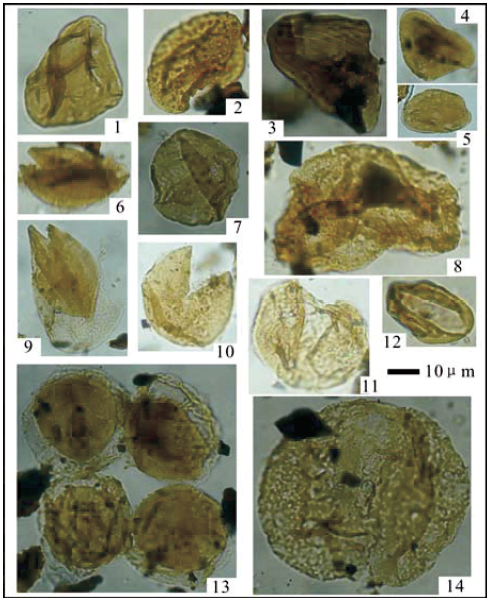
 Typical palynological fossils from the Shahezi Formation in the Shahezi Coal Mine. (1), Cyathidites; (2), Baculatisporites; (3), Cicatricosisporites; (4), Granulatisporites; (5), Polypodiaceaesporites; (6), Schizosporis; (7), Inaperturopllenites; (8), Pinuspollenites; (9), Concentrisporites; (10), Taxodiaceaepollenites; (11), Abietineaepollenites; (12), Cycadopites; (13), Perinopollenites; (14), Paleoconiferus. Wang et al. (2018).

The samples did not contain any Angiosperm pollen, which is found in the Yingcheng Formation, nor warm-climate adapted Fern spores such as Ruffordia and Coniopteris, which dominate the Yingcheng Formation, allowing differentiation of the two formations.

See also...

https://sciencythoughts.blogspot.com/2014/07/the-origin-of-lake-vattern.htmlhttps://sciencythoughts.blogspot.com/2014/05/pollen-and-spores-from-early-cretaceous.html
https://sciencythoughts.blogspot.com/2013/12/angiosperm-like-pollen-from-middle.htmlhttps://sciencythoughts.blogspot.com/2012/06/pollen-from-potshards-and-what-it-can.html
Follow Sciency Thoughts on Facebook.

Friday, 21 November 2014

Reconstructing extinct Arachnid orders in three-dimensions.


There are currently sixteen recognized orders of Arachnids, twelve of which are extant (have living relatives) and four extinct, the Trigonotarbids (Trigonotarbida), Phalangiotarbids (Phalangiotarbida), Haptopodids (Haptopoda) and Uraraneids (Uraraneida).  Members of two of these groups, the Triganotarbids and Uraraneids, have previously been reconstructed as three-dimensional computer models based upon computerised tomography (CT) scans.

In a paper published in the journal Peer J on 13 November 2014, RussellGarwood of the School of Earth, Atmospheric and Environmental Sciences and The Manchester X-ray Imaging Facility at The University of Manchester and JasonDunlop of the Museum für Naturkunde at the Leibniz Institute for Research onEvolution and Biodiversity at the Humboldt University Berlin present three-dimensional reconstructions of members of the two remaining extinct Arachnid orders, the Phalangiotarbids and Haptopodids.

Garwood and Dunlop obtained specimens of the single known Haptopodid species, Plesiosiro madeleyi, from the Natural History Museum in London, and of two Phalangiotarbid species, Goniotarbus tuberculatas and Goniotarbus angulatus from the Lapworth Museum in Birmingham and the Natural History Museum in London. All specimens originally came from the Coseley Lagerstätte, near Dudley in Staffordshire, England, making them about 315 million years old (Late Carboniferous). The Birmingham specimen (Goniotarbus tuberculatas) was found to be too poorly preserved for the project, so only the Natural History Museum specimens (Plesiosiro madeleyiand Goniotarbus angulatus) were used.

The reconstruction of the Haptopodid Plesiosiro madeleyi conformed largely to previous reconstructions made using conventional techniques. It was not possible to reconstruct the tips of the limbs, due to a crack that ran through the nodule in which it was preserved, and the specimen was slightly distorted due to lateral compression. Some joints could not be resolved due to ridges on the exoskeleton. The median (middle) eyes appeared to be set in a depression, which is unusual in Arachnids, where such eyes are normally raised, and Garwood and Dunlop suggest this may be an artefact of preservation.

Digital visualisations of the Haptopod Plesiosiro madeleyi. (A) Dorsal view showing opisthosomal segmentation and prosomal shield architecture. (B) Lateral view of the anterior ventral prosoma, nearest limbs and lateral prosoma removed, showing the nature of haptopod chelicerae. (C) Ventral view, showing ventral segmentation, and divided sternum. (D) Haptopod walking leg. Abbreviations: 1–10, opisthosomal segment number; as, anterior sclerite; ch, chelicerae; cx, coxa; fa, fang; fe, femur; L1–L4, walking legs 1–4; me, media eyes; mt, metatarsus; pa, paturon; pp, pedipalps; ps, pofsteriorsclerite; pt, patella; ta, tarsus; ti, tibia; tr, trochanter.Scale bars 3mm in (A) and (D), 1 mm in (B) and (C). Garwood & Dunlop (2014).

The Phalangiotarbid Goniotarbus angulatus was found to have a broad prosoma-opisthosoma boundary. The eye arrangement could not be fully resolved, which reflects earlier work on the group where between two and six eyes were reported. The chelicerae (mouthparts) cannot be resolved and are thought to be very small. The pedipalps (foremost limbs, not usually used for locomotion) are also small.

Digital visualisations of Phalangiotarbid Goniotarbus angulatus. (E) First left walking leg, showing typical segmentation. (F) Lateral view of the anterior ventral prosoma, showing the small pedipalps, median ridge, and possible chelicerae—below the resolution of the scan. (G) Fourth right walking leg. (H) Dorsal view showing median eyes and dorsal opisthosomal segmentation. (I) Ventral view showing opisthosomal segmentation and coxo-sternal region. Abbreviations: 1–10, opisthosomal segment number; as, anterior sclerite; ch, chelicerae; cx, coxa; fa, fang; fe, femur; L1–L4, walking legs 1–4; me, media eyes; mt, metatarsus; pa, paturon; pp, pedipalps; ps, pofsteriorsclerite; pt, patella; ta, tarsus; ti, tibia; tr, trochanter.Scale bars 3mm in (F - I), 1 mm in (E). Garwood & Dunlop (2014).

Garwood and Dunlop also hoped that these three dimensional reconstructions would help to resolve our understanding of phylogenic relationships between Arachnid groups. In recent years attempts to build a family tree for the group upon genetic analysis have proved problematic, while those based upon anatomy have proved consistent. However the addition of these formerly poorly known groups led to a severe rearrangement of computer generated family trees for the Arachnids, suggesting that these are not as robust as currently assumed.

See also…

In 2011 Paul Seldon of the College of Life Sciences at Capital Normal University in Beijing, the Paleontological Institute and Department of Geology at the University of Kansas and the Natural History Museum in London, along with ChungKun Shih and Dong Ren, also of the College of Life Sciences at Capital Normal University, described a large female Spider, from the Middle Jurassic Daohugou Biota of Inner Mongolia...

Scorpions are thought to have been among the earliest Animals to colonize the land, with specimens known...

Mygalomorph Spiders (Tarantulas and related species) are considered to be one of the most ancient groups of Spiders. They have two pairs of book lungs (many other Spiders have lost a pair) and downward pointing, rather than opposable fangs, again considered to be a primitive state in Spiders. Many species of Mygalomorph attain large sizes, all have flattened, disk-shaped bodies (rather than the more globular bodies of most other Spiders), and most are ambush predators.


Follow Sciency Thoughts on Facebook.

Wednesday, 30 May 2012

The enigmatic Carboniferous Arthropod Camptophyllia.

The Carboniferous Arthropod Camptophyllia is known from coal measure deposits in northern England. It is always preserved as a dorsal exoskeleton about 25 mm in length, made up of 10 segments, each segment being split into five plates; three apparently dorsal and two apparently lateral. Since its discovery in the 1920s it has been assigned to a number of different Arthropod groups, but none with any degree of confidence.

Line drawing of Camptophyllia. Gill (1924).

In a paper published in the journal Palaeontologica Electronica in April 2012, Russell Garwood of the Manchester X-ray Imaging Facility at the School of Materials at The University of Manchester and Mark Sutton of the Department of Earth Science and Engineering at Imperial College London present the results of a high-resolution X-ray micro-tomography study of Camptophyllia, which attempted to gain a better insight into the structure and affinities of the animal.

Examples of Camptophyllia from museum and private collections. (1) 44 mm specimen from the Tyne Coalfield, Natural History Museum, London. (2) 28 mm specimen from Crawcrook, near
Ryton-On-Tyne, Durham, Natural History Museum, London. (3) Counterpoint to (2), 30 mm. (4) 39 mm specimen from the Tyne Coalfield, Natural History Museum, London. (5) 42 mm specimen from Crock Hey, private collection of Stephen Livesley. (6) 35 mm Specimen from Crock Hey, private collection of Sean Sale. (7) 18 mm specimen from Coseley Colliery, Natural History Museum. (8) 13 mm specimen from Coseley Colliery, Natural History Museum, London. (9) 20 mm specimen from Coseley Colliery, Natural History Museum, London. (10) 20 mm specimen from Coseley Colliery, Natural History Museum, London. Garwood & Sutton (2012).

Garwood and Sutton carried out high-resolution X-ray micro-tomography studies of six specimens from the Natural History Museum in London and from the private collections of Stephen Livesley and Sean Sale. These were not, however able to resolve any features of the underside of Camptophyllia on any specimen. They concluded that this was unlikely to be a coincidence, and that therefore the undersides had not been preserved for a common reason. Arthropods shed their outer shells periodically as they grow, making it possible that the Camptophyllia specimens are in fact shed carapaces. However no known Arthropod sheds its dorsal carapace intact in this fashion without any other part of the exoskeleton, making this scenario unlikely. For this reason Garwood and Sutton favor the alternative possibility, that the dorsal shell of Camptophyllia was significantly mineralized, but that the underside was not; a pattern found in several Arthropod groups. Unfortunately this in no way helps to resolve the problem of Camptophyllia's taxonomic position.

Garwood and Sutton have placed two animations made from high-resolution X-ray micro-tomographs of specimens online. (Animation 1. Animation 2.).

Even thought they were unable to resolve the taxonomic position of Camptophyllia Garwood and Sutton were able to make some deductions from their study. A heavily mineralized dorsal skeleton, combined with soft underparts, is often associated with an ability to roll up into a ball for defensive reasons, something which would seem to be possible from Camptophyllia's bodyplan. None of the preserved specimens show any sign of eyes or similar structures, suggesting that Camptophyllia lived in an environment where vision was not a useful sense. The fossils have previously been identified as coming from shallow lacustrine (lake) environment, which could quite possibly have been murky or cloudy. In addition Camptophyllia has a 'snowshoe' shape, which is often associated with animals that live on soft sediments, and need to avoid sinking in such.

See also An Eocene False Scorpion from Baltic amberTwo new species of True Bug from the Mesozoic of ChinaAn Assassin Bug from the Palaeocene of Spitsbergen IslandA fossil termite from the Late Oligocene of northern Ethiopia and Preserved Trilobite digestive tracts from the Middle Cambrian of Utah.

Follow Sciency Thoughts on Facebook.