Showing posts with label Okinawa Islands. Show all posts

Sunday, 7 June 2020

Enchiridium daidai: A giant Polycladid Flatworm from southwest Japan.

Polycladids are a diverse group of marine Flatworms (Bilateran Worms which lack a body cavity) found from litoral (tidal) environments to deep sea hydrothermal vents, though they are most numerous and diverse around Coral Reefs. They typically range from about 3-20 mm in length, with a flattened oval bodyshape, and often have paired tentacles at their front ends. Members of the Family Prosthiostomidae are characterised by (i) an elongated body with a ventral sucker after the female gonopore, (ii) a plicate tubular pharynx, and (iii) paired prostatic ducts, each of which extends from a spherical prostatic vesicle and enters the penis or the ejaculatory duct independently, instead of uniting to each other before the entrance. The Prosthiostomidae is composed of five genera: Enchiridium, Enterogonimus, Euprosthiostomum, Lurymare, and Prosthiostomum. The genus Enchiridium is distinguished from other Prosthiostomids by having a muscle sheath (or bulb) that encloses just the two prostatic vesicles among other male reproductive organs; i.e., the seminal vesicle and the male atrium are not enclosed by the muscle sheath.

In a paper published in the journal ZooKeys on 12 March 2020, Aoi Tsuyuki of the Graduate School of Science at Hokkaido University and Hiroshi Kajihara of the Faculty of Science at Hokkaido University, describe a new species of Enchiridium from Kagoshima and Okinawa.

Three Polyclad specimens were collected subtidally from under rocks on the coast of Bonomisaki in Kagoshima Prefecture and the coast of Nago on Okinawa Island, southwestern Japan. Worms were anaesthetised in seawater containing menthol before fixation. The relaxed worms were photographed with a Nikon D5600 digital camera with external strobe lighting provided by a pair of Morris Hikaru Komachi Di flash units. For DNA extraction, a posterior piece of the body was removed and stored in 99.5% ethanol. The rest of the body was fixed in Bouin’s solution for 24 hours and preserved in 70% ethanol for long-term storage.

Map showing distribution of Enchiridium daidai: point (A) off the coast of Bonomisaki, Kagoshima (type locality); point (B) Nago, Okinawa Island. Tsuyuki & Kajohara (2020).

The new species is named Enchiridium daidai, where 'daidai' means 'orange', in reference to a thin marginal orange line surrounding the entire dorsal fringe. The species is described from three specimens, all collected by Aoi Tsuyuki. One was collected at 13–14 m depth off the coast of Bonomisaki in Kagoshima Prefecture, and the other two were both collected at 5 m depth at Nago on Okinawa Island.

Enchiridium daidai, photograph taken in life and eyespots observed in fixed state after being cleared in xylene. Entire animal, dorsal view (left) and ventral view (right). Abbreviations: fg, female gonopore; mg, male gonopore; op, oral pore; ph, pharynx; su, sucker. Scale bar is 10 mm. Tsuyuki & Kajohara (2020).

The body of Enchiridium daidai is elongate, tapered posteriorly, 28–77 mm long (77 mm in the holotype) and 4.6–14 mm maximum width (14 mm in the holotype) in the living state; the anterior margin is rounded; the mid-point of the posterior margin is acute. Tentacles are absent. The dorsal surface is smooth, translucent, and fringed with a thin marginal orange line. The ventral surface is translucent, without colour pattern. A pair of cerebral-eyespot clusters is present, each consisting of 20–52 eyespots (left 20 and right 23 in holotype); each cluster is of an antero-posteriorly elongated spindle shape. Marginal-eyespot clusters form a single marginal band, extending to position of mouth (about anterior one-eighth of the body length) along margins on both sides; marginal eyespots are abundant along the anterior margin, diminishing posteriorly. Ventral eyespots are absent. The intestine is highly branched, spreading all over body. A plicated pharynx is tubular in shape, about one-fifth of the body length, and located in the anterior one-third of the body. The oral pore is situated at the anterior end of the pharynx, behind the brain. The Male gonopore and female gonopore are closely set, both situated behind the posterior end of pharynx. The male copulatory apparatus consists of a large seminal vesicle, a pair of prostatic vesicles, and an armed penis papilla. The antero-posterior length of  the seminal vesicle is more than twice as long as the diameter of each prostatic vesicle. Spermiducal vesicles form a single row on each side of the midline, separately entering into seminal vesicle. An ejaculatory duct with a thick muscular layer, enters the penis papilla. Prostatic ducts with muscular layer are connected to the ejaculatory duct separately at the proximal end of penis papilla. A pair of spherical prostatic vesicles is coated within thin non-nucleated muscular wall, arranged anterodorsally to the ejaculatory duct. A common muscular sheath encloses the two prostatic vesicles. The seminal vesicle is oval, coated with a thick muscular wall, narrowing anteriorly and forming the ejaculatory duct; the latter almost immediately penetrating the common muscular sheath. The penis papilla is armed with a pointed tubular stylet, enclosed in a penis pouch, and protrudes into the male atrium. The male atrium is elongated anteriorly, and lined with a ciliated, muscularised epithelium. The female reproductive system is immediately posterior to the male reproductive system. Cement glands are numerous, concentrated around the vagina and release their contents into a cement pouch. The vagina curves anteriorly, leading to two narrow lateral branches of uteri. Each branch of uteri turns laterally and then runs backwards. The Lang’s vesicle is absent. A sucker is set on the body centre.

The specimens from Kagoshima and Okinawa differed in body size. The holotype from Kagoshima was 77 mm long and 15 mm wide, whereas the paratype specimens from Okinawa were 28–37 mm long and 4.6–7.4 mm wide. In spite of the noticeable difference in body size, specimens from Kagoshima and Okinawa, all having reached sexual maturity, were identified as conspecific. They shared the following morphological characteristics: (i) a body dorsally fringed with a thin orange line, (ii) a marginal-eyespot band extending to the position of the mouth (about anterior one-eighth of the body), (iii) two prostatic vesicles covered by a common muscle sheath, and (iv) common muscle sheath penetrated by ejaculatory duct. In addition the proportion of nucleotide sites at which two sequences being compared were different was very low, indicative of being the same species.

Difference in mature body size among Enchiridium daidai. (A) ICHUM 5993 (holotype), from Kagoshima, (B) ICHUM 5995 (paratype), from Okinawa, (C) ICHUM 5994 (paratype), from Okinawa. Scale bar 10 mm. Tsuyuki & Kajohara (2020).

Reaching 77 mm in body length, Enchiridium daidai is the largest species in the genus, superseding Enchiridium punctatum (about 40 mm in body length). Indeed, Enchiridium daidai is the second largest species in the Prosthiostomidae after Prosthiostomum cyclops, which reaches 90 mm. Among about 80 species of Prosthiostomids, only Enchiridium daidai and Prosthiostomum cyclops are known to exceed 70 mm in body length, while most of the other species are less than 30 mm long. Therefore, Tsuyuki & Kajohira's new species is considered to be unusually big in body size for a Prosthiostomid.

See also...

https://sciencythoughts.blogspot.com/2020/03/temnocephala-ivandarioi-new-species-of.htmlhttps://sciencythoughts.blogspot.com/2019/01/difroehlichia-elenae-new-species-of.html
https://sciencythoughts.blogspot.com/2018/01/xenoturbella-japonica-new-species-of.htmlhttps://sciencythoughts.blogspot.com/2016/08/cratera-viridimaculata-new-species-of.html
https://sciencythoughts.blogspot.com/2015/03/a-trematode-flatworm-from-intestines-of.htmlhttps://sciencythoughts.blogspot.com/2014/07/a-new-species-of-liver-fluke-from.html


Follow Sciency Thoughts on Facebook.

Wednesday, 3 June 2020

Cyaegharctus kitamurai: A new species of Marine Tardigrade from a submarine cave in the Ryukyu Archipelago, Japan.

Tardigrades, or Water Bears, are a distinctive group of small (usually less than 1 mm) invertebrates related to Arthropods, Nematodes and Velvet Worms. They have a simple segmented body with four pairs of limbs, and are remarkably resilient to environmental stress, being able to withstand extremely high and low temperatures, complete desiccation and even exposure to vacuum. Marine Tardigrades, specifically Arthrotardigrades, exhibit remarkable morphological diversity. The Styraconyxidae is one of the Arthrotardigrade families and it is currently comprised of 38 species and subspecies placed withingten genera: Angursa (eight species), Bathyechiniscus (one species), Lepoarctus (one species), Paratanarctus (one species), Pleocola (one species), Raiarctus (five species), Rhomboarctus (three species), Styraconyx (15 species and subspecies), Tetrakentron (one species), and Tholoarctus (three species and subspecies). In addition to these ten genera, an undescribed genus related to Styraconyx and Tetrakentron has been reported from a submarine cave in Japan, although no formal description of that species has ever been made and two voucher micrographs of a specimen used for a molecular phylogenetic study have been published.

In a paper published in the journal Zoosystematics and Evolution on 23 March 2020, Shinta Fujimoto of the Research Center for Marine Biology at Tohoku University, and Naoto Jimi of the Japanese National Institute of Polar Research formerly describe this submarine cave-dwelling Tardigrade.


Specimens were collected from Daidokutsu, a submarine cave off Iejima Island in the Okinawa Islands, part of the Ryukyu Archipelago, Japan, by Koshin Yasumura and Shinta Fujimoto in 2013 and 2019. For extraction of meiofauna, the cave sediment samples were stirred with tap water and the supernatants were concentrated using a 30 μm opening mesh net to separate coarse sediment and to wash away seawater. Subsequently, the meiofauna and fine sediment were separated using LUDOX HS-40 colloidal silica and a 32 μm opening mesh net. The type material was sorted under a stereomicroscope and fixed in 2–4% buffered formaldehyde. Specimens for light microscopy were mounted in distilled water for brief observation and mounted in glycerol. Differential interference contrast microscopy was conducted using an Olympus BX53 and phase contrast microscopy was conducted using an Olympus BX41. One specimen for scanning electron microscopy was post-fixed in 2% OsO₄ for 2 hours, dehydrated through a series of ethanol and acetone washes, critical point dried, osmium coated, and observed using a JEOL JSM-7001F Schottky Emission Scanning Electron Microscope. Type material was deposited in the Zoological Collection of Kyoto University. Adobe Illustrator CS6 and Photoshop CS6 were used to prepare figures and to obtain morphometric data.

The species is placed in a new genus, named Cyaegharctus, which is a combination of 'Cyäegha-', a deity of darkness and caves in the writings of Eddy Bertin, and '-arctus', a Latinised Greek word meaning 'bear', commonly used as a suffix when naming Tardigrades, and given the specific name kitamurai, in honour of Akihisa Kitamura of Shizuoka University, who has been studying Daidokutsu Cave and its Bivalve assemblage. The species is described from four specimens, three adult females and a juvenile, all collected from Daidokutsu Cave.

The holotype of Cyaegharctus kitamurai (when describing a new species one specimen is designated the holotype; all future specimens determined to belong to the same species as this holotype therefore bellong to the species) is an adult female with a dorso-ventrally flattened body 202 μm in length and 117 μm wide at level of leg III. The cephalic region (head) has an unpaired median cirrus (tendril), paired internal cirri, paired external cirri, paired lateral cirri, paired primary clavae (clublike structures), paired secondary clavae and an antero-ventral directed mouth. Paired spine-like cirri (38 μm) on cirrophores (stalks) arise from round lateral processes at level between legs III and IV. A rosette-like gonopore (genital opening) is 9 μm anterior to anus. There are four pairs of legs, each with an usual leg sensory organ on the dorsal side of the femur’s proximal portion, a pocket organ on the dorsal side of femur’s distal margin and four digits terminating in claws. 

Drawings of Cyaegharctus kitamurai, holotype KUZ Z2624. (A) Habitus (ventral view). (B) Leg IV pocket organ. Abbrreviations: an, anus; bt, buccal tube; ca, cavity; cE, cirrus E; db, dense body; ec, external cirrus; go, female gonopore; ic, internal cirrus; lc, lateral cirrus; mc, median cirrus; pc, primary clava; pl, placoid; op, opening; sc, secondary clava; soₗ, ₗᵥ legs I and IV sensory organs; sr, seminal receptacles; ss, stylet support; st, stylet. Fujimoto & Jimi (2020).

Fixation of specimens using formaldehyde seems to have introduced an artefact in the cuticle, i.e. the detached (or loose) outer epicuticle. When the specimens were sorted in distilled water before fixation, the outer epicuticle did not look loose at 63× magnification (all four specimens) and also at 400× magnification (only observed for KUZ Z2627) as it would appear in Tholoarctus. Although these are only brief observations and Fujimoto and Jimi did not conduct any experiment to test this artefact, they consider the outer epicuticle’s detached state as an artefact and excluded this character state from the diagnoses of this genus and species.

The adult female paratype KUZ Z2625 revealed the presence of a cuticular ring surrounding the rosette gonopore and the presence of the spine-like leg III sensory organs with no subdivisions. In the adult female paratype KUZ Z2626, the legs I–III sensory organs, claws and peduncles were orientated better than the holotype and the other paratype for observation. However, the pocket organs were not recognised for this specimen, probably due to the excessively-squeezed state.

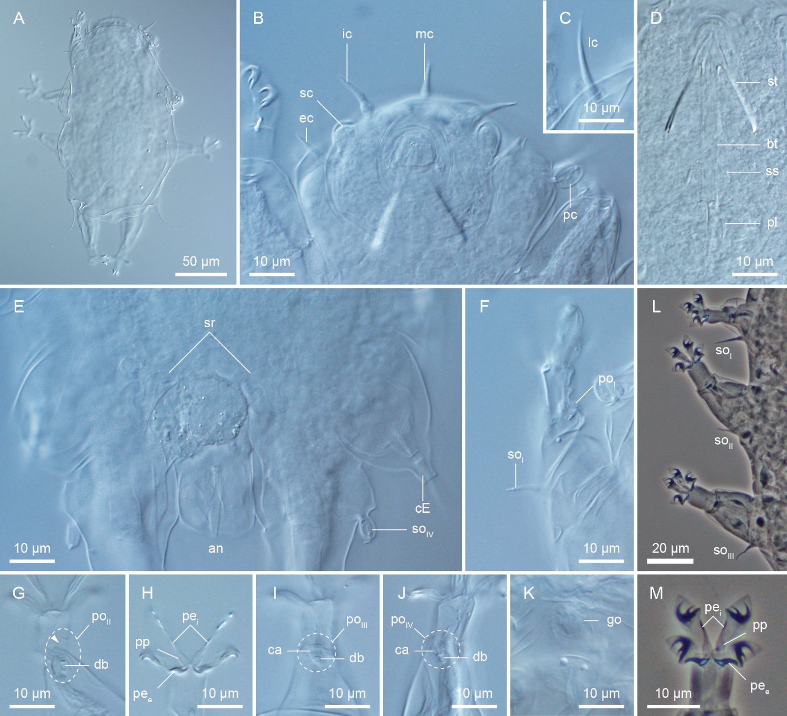
DIC and PhC micrographs of Cyaegharctus kitamurai, adult female. (A) Habitus (dorsal view), (B) cephalic region (ventral view), (C) lateral cirrus. (D) buccal apparatus, (E) caudal region (ventral view) (epicuticle pillars visible), (F) leg I sensory organ and pocket organ, (G) leg II pocket organ (arrowhead indicates protruding portion), (H) leg II digits and claws, (I) leg III pocket organ, (J) leg IV pocket organ, (K) female gonopore, (L) legs I–III sensory organs, (M) leg III digits and claws. (A)–(J) holotype KUZ Z2624, (K) paratype KUZ Z2625, (L), (M) paratype KUZ Z2626. Abbreviations: an, anus; bt, buccal tube; ca, cavity; cE, cirrus E; db, dense body; ec, external cirrus; go, gonopore; ic, internal cirrus; lc, lateral cirrus; mc, median cirrus; pc, primary clava; peᵢ,ₑ, peduncles of internal and external digits; pl, placoid; poₗ–ₗᵥ, legs I–IV pocket organs; pp, proximal pad; sc, secondary clava; soₗ–ₗᵥ, legs I–IV sensory organs; sr, seminal receptacles; ss, stylet support; st, stylet. Fujimoto & Jimi (2020).

The scanning electron micrographs of a four claw juvenile confirmed the results of light microscopy and also provided further detail. However, this scanning electron micrograph specimen also seems to have its outer epicuticle detached. If the outer epicuticle is attached to the underlying layer, a pattern is recognised on the surface of the body due to the pillar layer, but no such indentations were found, suggestive of the detached state of the outer cuticle. The proximal part of each leg has an inflated appearance not recognised in light microscopy. The view of the cephalic region revealed the three-dimensional morphology and the arrangement of the cephalic appendages and also confirmed the presence of terminal pores on the cephalic cirri and the primary clavae. The conical shape of the secondary clavae seems not as evident as in light microscopy probably due to the overlying outer epicuticle. The large anus is not on the ventral surface and rather direct posteriorly. The leg sensory organs were recognised and those of legs I and IV revealed to have terminal pores. The pocket organs were recognised on all legs, however, with slightly different degrees of protruded appearances. The protruded state might be an artefact caused during specimen preparation since specimens on microscope slides do not always have these appearances or the pocket organs are capable of moving. In the latter case, since no muscles seem to be attached to the structures, a passive movement is likely. Regarding claw/digit morphology, the peduncles (internal structures) were not recognised but the three hooks of the internal claws and the single-pointed external claws were recognised.

Scanning electron micrographs of Cyaegharctus kitamurai, four-claw juvenile paratype KUZ Z2627. (A) Habitus (lateral view), (B) cephalic region (frontal view), (C) cephalic region (ventral view), (D) anus, (E) leg I sensory organ, (F) leg II sensory organ, (G) leg III sensory organ, (H) leg IV sensory organ, (I) leg IV pocket organ, (J) leg III digits and claws. Abbreviations: ah, accessory hook; an, anus; cE, cirrus E; ec, external cirrus; fe, femur; ic, internal cirrus; lc, lateral cirrus; mc, median cirrus; pc, primary clava; ph, primary hook; poₗ, ₗᵥ, legs III and IV pocket organs; pp, proximal pad; sc, secondary clava; sh, secondary hook; soₗ–ₗᵥ, legs I–IV sensory organs; ti, tibia. Fujimoto & Jimi (2020).

Fujimoto and Jimi consider the dense body inside the pocket organs of Cyaegharctus kitamurai to be related to the van der Land’s body, often situated at the base of the primary clavae and leg IV sensory organs and suggest that the pocket organs are chemoreceptors. However, only one previous study, in 1981, investigated the function and ultrastructure of Arthrotardigrade sensory organs by transmission electron microscopy and, with our poor knowledge on Arthrotardigrade sensory organs, this remains a matter of speculation. Another possibility for the new structure is a secretory organ. However, no gland was recognised in its vicinity and there is no evidence supporting this hypothesis. To understand the true functions and evolutionary significance of the pocket organs, comparative ultrastructure studies of Arthrotardigrades (including Cyaegharctus kitamurai) are necessary.

See also...

https://sciencythoughts.blogspot.com/2018/11/macrobiotus-hannae-new-species-of.htmlhttps://sciencythoughts.blogspot.com/2018/09/bryodelphax-arenosus-new-species-of.html
https://sciencythoughts.blogspot.com/2018/03/macrobiotus-shonaicus-new-species-of.htmlhttps://sciencythoughts.blogspot.com/2017/09/bryodelphax-kristenseni-new-species-of.html
https://sciencythoughts.blogspot.com/2017/08/estimating-possibility-of-all-life.htmlhttps://sciencythoughts.blogspot.com/2017/06/macrobiotus-polypiformis-new-species-of.html
Follow Sciency Thoughts on Facebook.