Showing posts with label Okinawa. Show all posts

Wednesday, 17 June 2020

Pontopolycope orientalis: A new species of Polycopid Ostracod from Nagannu Island, Japan.

Ostracods are small bivalved Crustaceans that live in various aquatic environments including freshwater and marine habitats ranging from the supralittoral zone to the hadal zone. A number of ostracods are also known to be symbiotic with Invertebrates and Vertebrates. The Polycopid Ostracod genus Pontopolycope is characterized by a fully developed rostrum and concavity immediately ventral to the rostrum on the anterior margin of the carapace. This is a special characteristic of the family Polycopidae. To date, all four living congeners have been described from the Atlantic: Pontopolycope rostrata, Pontopolycope dentata, Pontopolycope mylax, and Pontopolycope storthynx.

In a paper published in the journal Zoological Studies on 20 April 2020, Hayato Tanaka of the Tokyo Sea Life Park, and Yusuke Kondo and Susumu Ohtsuka of the Setouchi Field Science Center of Hiroshima University, describe the first living representative of the genus Pontopolycope from the Indo-Pacific region.

Sediment samples were obtained from the sandy bottom at 52 m depth off the Nagannu Island, Okinawa Prefecture, southwestern Japan, on May 21, 2016, by using a dredge (mouth 50 cm wide × 15 cm high; mesh size 5 mm) towed along the bottom twice by the TRV Toyoshio-maru (Hiroshima University). Sediments were stirred in seawater, and the supernatant was filtered with a small plankton net (mesh size 0.1 mm). All living ostracod specimens were extracted from the remaining deposits under a binocular stereomicroscope. 

Map showing the sampling site. (A) Japan; (B) sampling locality. Tanaka et al. (2020).

The new species is named Pontopolycope orientalis, where 'orientalis' is Latin for 'in the east', in reference to the fact that this is the living species of the genus Pontopolycope in the Indo-Pacific region. The species is described from ten specimens, six males and four females.

The carapace of Pontopolycope orientalis is circular in lateral view, with developed rostrum and concavity immediately ventral to the rostrum of anterior margin. The posterior end is situated slightly near to the ventral side. The surface of the carapace is covered with shallow pits. Along anterior to posterior margin of the left valve and anterior to ventral margin of the right valve are a series of serrations. There is a pore system consisting of a shallow depression and a small dome with pore a canal. The adductor muscle scar consists of three closely spaced scars. A marginal infold with groove runs around the internal marginal zone of  the postero-dorsal, anterior and ventral margins in the right valve; there is a corresponding ridge in the left valve. The hinge structure consists of a simple groove in the right valve and of a corresponding bar in the left valve.

Pontopolycope orientalis, scanning electron microscope images. (A), (B), (G), paratype male (NSMT-Cr 27374); (C), (D), (H), paratype male (NSMT-Cr 27375); (E), (F), paratype female (NSMT-Cr 27379). (A), (E), external lateral view of right valve; (B), (F), external lateral view of left valve; (C), internal lateral view of right valve; (D) internal lateral view of left valve; (G) external surface of left valve; (H) adductor muscle scar, internal view of right valve. Scale bars: (A)–(F) 100 μm; (G) 15 μm; (H) 30 μm. Tanaka et al. (2020).

See also...

https://sciencythoughts.blogspot.com/2018/11/spiricopia-aurita-cylindroleberidid.htmlhttps://sciencythoughts.blogspot.com/2018/05/rosaliella-svalbardensis-new-species-of.html
https://sciencythoughts.blogspot.com/2017/09/cypria-lacrima-new-species-of-candonid.htmlhttps://sciencythoughts.blogspot.com/2014/11/a-new-species-of-ostracod-from.html
https://sciencythoughts.blogspot.com/2014/09/a-new-species-of-freshwater-ostracod.htmlhttps://sciencythoughts.blogspot.com/2014/04/three-dimensional-soft-tissue.html


Follow Sciency Thoughts on Facebook.

Sunday, 7 June 2020

Enchiridium daidai: A giant Polycladid Flatworm from southwest Japan.

Polycladids are a diverse group of marine Flatworms (Bilateran Worms which lack a body cavity) found from litoral (tidal) environments to deep sea hydrothermal vents, though they are most numerous and diverse around Coral Reefs. They typically range from about 3-20 mm in length, with a flattened oval bodyshape, and often have paired tentacles at their front ends. Members of the Family Prosthiostomidae are characterised by (i) an elongated body with a ventral sucker after the female gonopore, (ii) a plicate tubular pharynx, and (iii) paired prostatic ducts, each of which extends from a spherical prostatic vesicle and enters the penis or the ejaculatory duct independently, instead of uniting to each other before the entrance. The Prosthiostomidae is composed of five genera: Enchiridium, Enterogonimus, Euprosthiostomum, Lurymare, and Prosthiostomum. The genus Enchiridium is distinguished from other Prosthiostomids by having a muscle sheath (or bulb) that encloses just the two prostatic vesicles among other male reproductive organs; i.e., the seminal vesicle and the male atrium are not enclosed by the muscle sheath.

In a paper published in the journal ZooKeys on 12 March 2020, Aoi Tsuyuki of the Graduate School of Science at Hokkaido University and Hiroshi Kajihara of the Faculty of Science at Hokkaido University, describe a new species of Enchiridium from Kagoshima and Okinawa.

Three Polyclad specimens were collected subtidally from under rocks on the coast of Bonomisaki in Kagoshima Prefecture and the coast of Nago on Okinawa Island, southwestern Japan. Worms were anaesthetised in seawater containing menthol before fixation. The relaxed worms were photographed with a Nikon D5600 digital camera with external strobe lighting provided by a pair of Morris Hikaru Komachi Di flash units. For DNA extraction, a posterior piece of the body was removed and stored in 99.5% ethanol. The rest of the body was fixed in Bouin’s solution for 24 hours and preserved in 70% ethanol for long-term storage.

Map showing distribution of Enchiridium daidai: point (A) off the coast of Bonomisaki, Kagoshima (type locality); point (B) Nago, Okinawa Island. Tsuyuki & Kajohara (2020).

The new species is named Enchiridium daidai, where 'daidai' means 'orange', in reference to a thin marginal orange line surrounding the entire dorsal fringe. The species is described from three specimens, all collected by Aoi Tsuyuki. One was collected at 13–14 m depth off the coast of Bonomisaki in Kagoshima Prefecture, and the other two were both collected at 5 m depth at Nago on Okinawa Island.

Enchiridium daidai, photograph taken in life and eyespots observed in fixed state after being cleared in xylene. Entire animal, dorsal view (left) and ventral view (right). Abbreviations: fg, female gonopore; mg, male gonopore; op, oral pore; ph, pharynx; su, sucker. Scale bar is 10 mm. Tsuyuki & Kajohara (2020).

The body of Enchiridium daidai is elongate, tapered posteriorly, 28–77 mm long (77 mm in the holotype) and 4.6–14 mm maximum width (14 mm in the holotype) in the living state; the anterior margin is rounded; the mid-point of the posterior margin is acute. Tentacles are absent. The dorsal surface is smooth, translucent, and fringed with a thin marginal orange line. The ventral surface is translucent, without colour pattern. A pair of cerebral-eyespot clusters is present, each consisting of 20–52 eyespots (left 20 and right 23 in holotype); each cluster is of an antero-posteriorly elongated spindle shape. Marginal-eyespot clusters form a single marginal band, extending to position of mouth (about anterior one-eighth of the body length) along margins on both sides; marginal eyespots are abundant along the anterior margin, diminishing posteriorly. Ventral eyespots are absent. The intestine is highly branched, spreading all over body. A plicated pharynx is tubular in shape, about one-fifth of the body length, and located in the anterior one-third of the body. The oral pore is situated at the anterior end of the pharynx, behind the brain. The Male gonopore and female gonopore are closely set, both situated behind the posterior end of pharynx. The male copulatory apparatus consists of a large seminal vesicle, a pair of prostatic vesicles, and an armed penis papilla. The antero-posterior length of  the seminal vesicle is more than twice as long as the diameter of each prostatic vesicle. Spermiducal vesicles form a single row on each side of the midline, separately entering into seminal vesicle. An ejaculatory duct with a thick muscular layer, enters the penis papilla. Prostatic ducts with muscular layer are connected to the ejaculatory duct separately at the proximal end of penis papilla. A pair of spherical prostatic vesicles is coated within thin non-nucleated muscular wall, arranged anterodorsally to the ejaculatory duct. A common muscular sheath encloses the two prostatic vesicles. The seminal vesicle is oval, coated with a thick muscular wall, narrowing anteriorly and forming the ejaculatory duct; the latter almost immediately penetrating the common muscular sheath. The penis papilla is armed with a pointed tubular stylet, enclosed in a penis pouch, and protrudes into the male atrium. The male atrium is elongated anteriorly, and lined with a ciliated, muscularised epithelium. The female reproductive system is immediately posterior to the male reproductive system. Cement glands are numerous, concentrated around the vagina and release their contents into a cement pouch. The vagina curves anteriorly, leading to two narrow lateral branches of uteri. Each branch of uteri turns laterally and then runs backwards. The Lang’s vesicle is absent. A sucker is set on the body centre.

The specimens from Kagoshima and Okinawa differed in body size. The holotype from Kagoshima was 77 mm long and 15 mm wide, whereas the paratype specimens from Okinawa were 28–37 mm long and 4.6–7.4 mm wide. In spite of the noticeable difference in body size, specimens from Kagoshima and Okinawa, all having reached sexual maturity, were identified as conspecific. They shared the following morphological characteristics: (i) a body dorsally fringed with a thin orange line, (ii) a marginal-eyespot band extending to the position of the mouth (about anterior one-eighth of the body), (iii) two prostatic vesicles covered by a common muscle sheath, and (iv) common muscle sheath penetrated by ejaculatory duct. In addition the proportion of nucleotide sites at which two sequences being compared were different was very low, indicative of being the same species.

Difference in mature body size among Enchiridium daidai. (A) ICHUM 5993 (holotype), from Kagoshima, (B) ICHUM 5995 (paratype), from Okinawa, (C) ICHUM 5994 (paratype), from Okinawa. Scale bar 10 mm. Tsuyuki & Kajohara (2020).

Reaching 77 mm in body length, Enchiridium daidai is the largest species in the genus, superseding Enchiridium punctatum (about 40 mm in body length). Indeed, Enchiridium daidai is the second largest species in the Prosthiostomidae after Prosthiostomum cyclops, which reaches 90 mm. Among about 80 species of Prosthiostomids, only Enchiridium daidai and Prosthiostomum cyclops are known to exceed 70 mm in body length, while most of the other species are less than 30 mm long. Therefore, Tsuyuki & Kajohira's new species is considered to be unusually big in body size for a Prosthiostomid.

See also...

https://sciencythoughts.blogspot.com/2020/03/temnocephala-ivandarioi-new-species-of.htmlhttps://sciencythoughts.blogspot.com/2019/01/difroehlichia-elenae-new-species-of.html
https://sciencythoughts.blogspot.com/2018/01/xenoturbella-japonica-new-species-of.htmlhttps://sciencythoughts.blogspot.com/2016/08/cratera-viridimaculata-new-species-of.html
https://sciencythoughts.blogspot.com/2015/03/a-trematode-flatworm-from-intestines-of.htmlhttps://sciencythoughts.blogspot.com/2014/07/a-new-species-of-liver-fluke-from.html


Follow Sciency Thoughts on Facebook.

Friday, 11 January 2019

Hana hanagasa and Hana hanataba: Two new species of Stoloniferous Octocorals from the northwest Pacific.

Octocorals are colonial Corals, Anthozoa, lacking the extensive mineralised skeletons of Stony Corals. Each polyp of the colony has only eight tentacles, giving the group its name, though these often have numerous side branches, giving them a feathery appearance. Gorgonians are a group of Octocaorals that tend to form fan shaped colonies in shallow environments with strong currents. The Stolonifera is a group within the Octocorallia comprising Corals with polyps which are not set within a common coenenchymal tissue, but rather arise from a branching, ribbon-like stolon which spreads over the substrate, typically another Coral or Sponge. These Corals are less well studied than other Soft Corals, and relationships within the group are poorly understood.

In a paper published in the journal ZooKeys on 15 October 2018, Yee Wah Lau of the Molecular Invertebrate Systematics and Ecology Laboratory at the University of the Ryukyus, Frank Stokvis and Leendert van Ofwegen of the Naturalis Biodiversity Center, and James Davis Reimer, also of the the Molecular Invertebrate Systematics and Ecology Laboratory, and of the Tropical Biosphere Research Center at the University of the Ryukyus, describe two new species of Stoloniferous Octocorals from the northwest Pacific Ocean.

The first species is placed in a new genus, named Hana, which means ‘flower’ in Japanese, and given the specific name hanagasa, in reference to a flowered head-dress worn during traditional dances in Okinawa, in both cases this refers to the polyps of the species, which are somewhat floral in appearance. This species typically forms colonies of 50-100 polyps arising from ribbon-like stolons about 0.5 mm in diameter. Each polyp lives within a calyce 2.5-3.0 mm in height, into which they can completely withdraw. The polyps are spaced irregularly on the stolen, with the gap between them ranging from about 0.3 mm to about 2.5 mm. The species was found living on hard Coral rock on the northwest coast of Okinawa Island and southeast coast of Iheya Island, both in the Ryukyu Island chain.

Hana hanagasa from Okinawa. Lau et al. (2018). 

The second species is also placed in the genus Hana, and given the specific name hanataba, meaning a bouquet in Japanese, again in reference to the floral appearance of the polyps. This species typically forms colonies of 30-100 polyps, again rising dwelling within calyxes 2.5-3.0 mm high and arising from a branching stolon 0.5 mm in width. It was found dwelling off the southeast coast of Palau and off the Taiwanese atoll of Dongsha.

Hana hanataba from Ngemelis Island, Palau. Lau et al. (2018). 

See also...

https://sciencythoughts.blogspot.com/2018/04/adelogorgia-osculabunda-adelogorgia.htmlhttps://sciencythoughts.blogspot.com/2017/06/sinularia-mesophotica-new-species-of.html
https://sciencythoughts.blogspot.com/2017/04/flagelligorgia-gracilis-new-species-of.htmlhttps://sciencythoughts.blogspot.com/2017/02/muricea-subtilis-new-species-of.html
https://sciencythoughts.blogspot.com/2014/12/a-new-species-of-soft-coral-from.html
Follow Sciency Thoughts on Facebook.

Wednesday, 28 February 2018

Antipathozoanthus obscurus, Antipathozoanthus remengesaui, & Antipathozoanthus cavernus: Three new species of Parazoanthid Soft Coral from the Indo-Pacific region.

Parazoanthid Corals are colony forming Soft Corals noted for their tendency to grow on other benthic filter feeding animals, typically Sponges but sometimes Corals or other organisms, a form of facultative parasitism in which the Parazoanthid benefits from currents generated by the larger organism, while at the same time covering part of its surface, reducing the host’s ability to feed itself. Parazoanthids usually lack symbiotic Algae, and inhabit dark environments such as marine caves and deep sea cold seeps, where there are few predators, but also little food and limited natural currents, and thereby rely on their host’s ability to generate a current to survive.

In a paper published in the journal ZooKeys on 29 December 2017, Hiroki Kise of the Molecular Invertebrate Systematics and Ecology Laboratory at the University of the Ryukyus, and the Palau International Coral Reef Center, Takuma Fujii, also of the Molecular Invertebrate Systematics and Ecology Laboratory at the University of the Ryukyus, and of the Research Center for Island Studies Amami Station of Kagoshima University, Giovanni Diego Masucci and Piera Biondi, again of the Molecular Invertebrate Systematics and Ecology Laboratory at the University of the Ryukyus, and James Davis Reimer, once again of the Molecular Invertebrate Systematics and Ecology Laboratory at the University of the Ryukyus, and the Palau International Coral Reef Center, and of the Tropical Biosphere Research Center of the University of the Ryukyus, describe three new species of Parazoanthid Soft Coral from the Indo-Pacific region. All are placed in the genus Antipathozoanthus, which gets its name from its habit of overgrowing Antipatharian (Black) Corals.

The first new species described is given the name Antipathozoanthus obscurus, meaning ‘dark’ in reference to the dark environments in which it was found. The species was found in caves and crevices on reefs at depths of between 3 and 15 m, and, unusually, grew on a rocky, non-living substrate, where it was possibly able to survive due to strong currents generated by tidal action, which may have carried planktonic food into the caves. Polyps of this species reach 5-10 mm in height, with an oral disk that opens to a similar size in diameter, and have between 26 and 32 brown or orange tentacles. These polyps are connected by stolons which form a mesh-like network. The whole is covered by an encrustation of sand and other particles up to 8 mm in size. The species was found in the Okinawa Islands of Japan, and around Al Wajh Shaybarah on the Red Sea Coast of Saudi Arabia, which suggests a far wider distribution, probably encompassing the entire Indian Ocean and Western Pacific, and possibly further afield. 

Specimen of Antipathozoanthus obscurus. Collected from Cape Bise, Motobu, Okinawajima Island, Japan (26°42'34.4"N, 127°52'49.2"E) at a depth of 5 m by James Davis Reimer. Kise et al. (2017).

The second species described is named Antipathozoanthus remengesaui, in honour of Tommy Esang Remengesau, Jr., the current president of the Republic of Palau, for his support of marine research and conservation in Palau. This species was found growing on colonies of Corals of the genus Antipathes growing around the entrances to caves at depths of between 9 and 40 m, at Kagoshima (Japan) and Palau in the Pacific Ocean, in the Maldives in the Indian Ocean and around Al Wajh Shaybarah on the Red Sea Coast of Saudi Arabia. Polyps of this species are white in colour and reach 3-8 mm in height, with oral disks up to 8 mm in diameter, surrounded by 40-42 pinkish, translucent tentacles up to 4 mm in length. These polyps are solitary, or only weakly connected by a poorly developed coenenchyma. This species also tends to develop a coating of sand particles, though these tend to be smaller, at 1-3 mm.

Antipathozoanthus remengesaui, colony connected by poorly developed coenenchyma with white polyps on Antipathes sp., collected from Blue Hole, Palau (7°8'29.4"N, 134°13'23.3"E) at a depth of 23 m by James Davis Reimer. Kise et al. (2017).

The final species described is named Antipathozoanthus cavernus, in reference to its habit of living in caves. This species was found growing on colonies of the Coral Myripathes around cave entrances and on steep slopes at depths of between 19 and 39 m in Kagoshima and the Maldives. Polyps of this species reach 3-10 mm in height and have an orange oral disk 4-15 mm in diameter, surrounded by 32-40 translucent tentacles up to 5 mm in length. The polyps are connected by a highly developed coenenchyma, and again the colony is coated in sand and other particles 1-8 mm in size. 

Antipathozoanthus cavernus polyps connected by highly developed coenenchyme with orange ring around oral disk, collected from Siaes Tunnel, Palau (7°18'54.8"N, 134°13'13.3"E) at a depth of 39 m by James Davis Reimer. Kise et al. (2017).

See also...

http://sciencythoughts.blogspot.co.uk/2018/01/charting-long-term-coral-decline-in.htmlhttp://sciencythoughts.blogspot.co.uk/2018/01/porites-australiensis-exceptionally.html
http://sciencythoughts.blogspot.co.uk/2017/12/terpios-hoshinota-tracking-progress-of.htmlhttp://sciencythoughts.blogspot.co.uk/2017/09/looking-for-animals-in-wengan-biota.html
http://sciencythoughts.blogspot.co.uk/2017/04/flagelligorgia-gracilis-new-species-of.htmlhttp://sciencythoughts.blogspot.co.uk/2016/06/sclerangia-floridana-new-species-of.html
Follow Sciency Thoughts on Facebook.

Wednesday, 3 January 2018

Porites australiensis: An exceptionally large Coral microatoll from Okinawa, Japan.

Stony Corals of the genus Porites are found on Reefs around the world, where they form large flat topped microatolls, rising above the surface of the reef. They are often used  by palaeoclimatologists to study ancient climates, since they tend to preserve oxygen isotope levels in the same proportion as the water they lived in, a proxy for the temperature of the sea. Due to this fossil Porites colonies have been widely sought out, and some remarkable specimens found, with the current largest known specimen coming from China and measuring about 9 m across.

In a paper published in the journal Coral Reefs on 3 October 2017, Ichiro Takeuchi of the Graduate School of Agriculture at Ehime University, and Hideyuki Yamashiro of the Tropical Biosphere Research Center of the University of the Ryukyus, describe the discovery of an exceptionally large Porites microatoll from the Okinawa Islands of Japan.

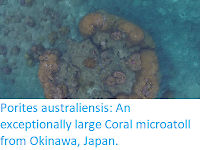
The colony was found about 150 m off the south coast of the island of Sesoko (which is itself about 500 m off the west coast of Okinawa Island) during an aerial drone survey of the area in March-April 2017. The atoll stands in water which is about 1.2-3.0 m deep at low tide, with a tidal range of about 2.3 m. The colony is a specimen of Porites australiensis 11.1 m across, making it the largest recorded Porites colony known, living or fossil, which given an estimated growth rate of 2.6–10.8 mm per year, makes the colony between 500 and 2100 years old.

Large Porites microatoll at Sesoko Island, Okinawa, Japan. Aerial photo taken from an altitude of 11 m at near low tide. The top of the microatoll is near the sea surface. Takeuch & Yamashiro (2017).

See also...

http://sciencythoughts.blogspot.co.uk/2017/12/terpios-hoshinota-tracking-progress-of.htmlhttp://sciencythoughts.blogspot.co.uk/2017/06/sinularia-mesophotica-new-species-of.html
http://sciencythoughts.blogspot.co.uk/2017/04/flagelligorgia-gracilis-new-species-of.htmlhttp://sciencythoughts.blogspot.co.uk/2017/02/muricea-subtilis-new-species-of.html
http://sciencythoughts.blogspot.co.uk/2016/06/sclerangia-floridana-new-species-of.htmlhttp://sciencythoughts.blogspot.co.uk/2016/04/assessing-impact-of-land-reclamation-in.html
Follow Sciency Thoughts on Facebook.

Saturday, 4 February 2017

Testing the strength of the Coconut Crab.

Coconut Crabs, Birgus latro, are the largest terrestrial Crustacean species, and indeed largest terrestrial Arthropod of any description. It is found across much of the Indian and Pacific Oceans, living on most landmasses where Coconuts are found (though in some areas they have been exterminated to protect commercial Coconut crops). They are descended from Hermit Crabs, and younger Coconut Crabs still use borrowed Snail shells for protection, but the adult Crabs have developed tough calcareous exoskeletons. Like other Decapod Crustaceans, Coconut Crabs have large chelae (claws) which are their main method of food manipulation and defence. Studies of other Decapods have been shown to be capable of exerting a considerably greater pinching force with their claws than the bite of similarly sized Vertebrates, making the pinching force of the Coconut Crab potentially very formidable.

In a paper published in the journal PLoS One on 23 November 2016, Shin-ichiro Oka, Taketeru Tomita and Kei Miyamoto of the Okinawa Churashima Foundation, describe the results of an experiment designed to test the strength of the claws of the Coconut Crab.

Oka et al. collected 29 Coconut Crabs from Ocean Expo Park on Northern Okinawa Island, ranging in size from 16.2 mm in length and 33 g in weight to 64.5 mm in length and 2120 g in weight and measured their pinching force with a device used to measure crushing force when an animal bites down on a stainless steel stick-shaped sensor, and in addition took measurements of their claws.

Measurement of the pinching force and claw morphology in the Coconut Crabs. (a) The force was measured with the SK-MBF-01F device (SkyScience Co. Tokyo, Japan) and related sensors and (b) demonstration of the method by which pinching force was measured. (c) Claw measurements of the Coconut Crab used in this study. The placement of the sensor used for pressure measurement is highlighted in green. The measurements used for claw length (CL), claw height (CH), and claw width (CW) are also indicated. L1: in-lever length from the fulcrum to the apodemes insertion; LBAE: out-lever length from the fulcrum to the tubercle (the contact point with the device sensor). Oka et al. (2016).

Oka et al. found that the pinching force of the claws related directly to the size of the Crabs, with the smallest Crab exerting a pinch of 29.4 Newtons, and the largest 1765.2 Newtons. However even the largest Crabs used in the study were considerably smaller than the maximum size for the species, about 4 kg. Assuming that the strength of larger Crabs continues to scale up in the same way, Oka et al. calculate that they could exert a pinch-force of 3300 Newtons, greater than the pinch-force measured from any other Crustacean, and higher than the bite-force recorded from any terrestrial predator except Alligators.

See also...

http://sciencythoughts.blogspot.co.uk/2016/12/iphiculus-eliasi-new-species-of.htmlhttp://sciencythoughts.blogspot.co.uk/2016/11/geosesarma-batak-geosesarma-tagbanua.html
http://sciencythoughts.blogspot.co.uk/2016/08/tiwaripotamon-pluviosum-new-species-of.htmlhttp://sciencythoughts.blogspot.co.uk/2016/05/decapod-crustaceans-from-tarioba-shell.html
http://sciencythoughts.blogspot.co.uk/2015/11/sundathelphusa-brachyphallus-new.htmlhttp://sciencythoughts.blogspot.co.uk/2015/04/fizesereneia-panda-new-species-of-gall.html
Follow Sciency Thoughts on Facebook.

Friday, 25 March 2016

Teinotarsina aurantiaca: A new species of Clear-winged Moth from Okinawa.

Clear-winged Moths, Sensiidae, are found across much of the globe, being particulalry numerous in the tropics. They lack the wing scales typical of Lepidopterans (Butterflies and Moths), instead having clear wings, combined with body-shapes and colouration resembling that of Wasps or Hornets. This is considered to be an example of Batesian Mimicry (a harmless organism evolving to resemble a harmful one) which enables the Moths to be active during daylight hours without suffering heavy predation. The larvae of Clear-winged Moths bore into wood or plant roots, with some species being considered agricultural pests.

In a paper published in the journal ZooKeys on 7 March 2016, Sadahisa Yagi and Toshiya Hirowatari of the Entomological Laboratory at Kyushu University, and Yutaka Arita of the Zoological laboratory at Meijo University, describe a new species of Clear-winged Moth from Okinawa Island, Japan.

The new species is placed in the genus Teinotarsina, and given the specific name aurantiaca, meaning 'orange'. Like other members of the genus Teinotarsina it is long-legged, and resembles an Ichneumon Wasp, but unlike any other member of the genus it has extensive orange markings. The species is described from a single male specimen found dead by a roadside. Nothing is known about the life habbits of the living Moth, and the females and larvae are unknown.

Teinotarsina aurantiaca, male specimen. Yagi et al. (2016).

The orange colouration of Teinotarsina aurantiaca is unique within the genus, but many Wasps on Okinawa do exhibit orange or red colouration not seen in close relatives on nearby landmasses, an example of Müllerian mimicry (harmful organisms coming to resemble one-another, to present would-be predators with a comon warning signal), a pattern which Teinotarsina aurantiaca also appears to have adopted.

See also...

http://sciencythoughts.blogspot.co.uk/2015/12/micropterix-gaudiella-new-species-of.htmlMicropterix gaudiella: A new species of Micropterigid Moth from the Italian Alps.       The Micropterigidae are considered to be the most primitive family of living Lepidoptera (Butterflies and Moths), retaining a number of archaic traits, such as functioning mandibles as adults (which are...
http://sciencythoughts.blogspot.co.uk/2015/02/a-new-species-of-grass-moth-from-tibet.htmlA new species of Grass Moth from Tibet.  Grass Moths, Crambidae, are generally small and inconspicuous, particularly when in a resting posture, with rolled wings on a Grass stem, though they are a variable group and some species are...
http://sciencythoughts.blogspot.co.uk/2015/01/a-new-species-of-giant-swallowtail.htmlA new species of Giant Swallowtail Butterfly from North and Central America.          Swallowtails, Papilionidae, are among the most conspicuous of American Butterflies, due to their large size and conspicuous colouration. The group has been extensively studied by taxonomists and...
Follow Sciency Thoughts on Facebook.