Sea Anemones, Actiniaria, are one of the least known Invertebrate groups in the fossil record, despite being abundant in most marine ecosystems today and holding a phylogenetic position which suggests that they are likely to have appeared early in the history of Animal life. Putative Anemones have been reported from both Ediacaran and Cambrian faunas, but the evidence for the placement of these fossils is at best weak. Post Cambrian, only a single fossil Anemone has been described, Palaeoanemona marcusi, from the Carboniferous of Argentina.
In a paper published in the journal Papers in Palaeontology on 8 March 2023, Roy Plotnick of the Department of Earth & Environmental Sciences at the University of Illinois at Chicago, Graham Young of the Manitoba Museum, and James Hagadorn of the Department of Earth Sciences at the Denver Museum of Nature & Science, re-describe Essexella asherae, an abundant fossil from the Carboniferous Mazon Creek Lagerstӓtte of Illinois, which has previously been considered to be a Scyphozoan Jellyfish, as a well preserved and abundant fossil Sea Anemone, and that this Anemone is likely linked to the trace fossil Conostichus, which is widespread in the Palaeozoic fossil record.
Numerous terrestrial Plant and Animal fossils, referred to as the Braidwood Biota, have been collected from the Francis Creek Shale Member of the Carbondale Formation at coal mines to the southeast of Morris, Illinois, since the mid-nineteenth century, with the strip mines east of the Mazon River (or Mazon Creek) being particularly productive. In the 1960s a marine fauna apparently coeval with the Braidwood Biota was described from the Peabody Coal Company Pit No 11, which lies slightly to the south of the Braidwood Biota sites. This marine fauna was named the Essex Fauna, with the two together having become known as the Mazon Creek Lagerstӓtte.
The Essex Fauna contains a high proportion of soft-bodied organisms, something quite unusual in later-Palaeozoic fossil deposits, making the site of great potential interest, yet it remains relatively understudied compared to the more famous Cambrian sites, such as the Burgess Shale. Despite this lack of formal study, hundreds of thousands of Essex Fauna specimens have been collected, predominantly from nodules found on spoil heaps by amateur collectors, and can be found in both museums and private collections, and in the past decade a number of studies into these organisms have been published.
The Mazon Creek deposits were laid down in a tropical deltaic environment, about 5° south of the equator, in which a coastal swamp was repeatedly drowned by rising sealevels, then prograded back over the drowned deposits as more sediment was deposited on the delta. The fossils of the Braidwood Biota is thought to contain both organisms that lived in the coastal swamps and those that lived further inland and which were carried to the coast during flood events, while the Essex Fauna is interpreted as having lived in the delta estuary, where the river input and frequent flood events caused the salinity to be lower that fully marine environments and prone to fluctuations. This fauna therefore lacks fully marine organisms incapable of dealing with low or variable salinity, such as Brachiopods or Crinoids, which tend to dominate other Late Palaeozoic marine faunas.
The fossils of the Mazon Creek Lagerstӓtte are typically preserved within siderite (iron carbonate) nodules, and appear to have been buried rapidly in an environment with abundant iron but limited sulphate, facilitating a Methanogen-dominated microbiota, in which Bacteria facilitated the catalytic development of siderite and pyrite (iron disulphide) around decomposing carcasses, with siderite growth quickly coming to dominate the growing nodule.
The most abundant fossils within the Essex Fauna are sack-like soft-bodied organisms which collectors refer to as 'blobs'. These are so abundant that they are frequently discarded, and can be purchased locally for a few dollars. Tens of thousands of these 'blobs' are available for study in the collections of various museums.
These 'blobs' were first studied in the 1970s, when it was assumed that they were Scyphozoan Jellyfish, being formally described as such by palaeontologist Merril Foster in 1979, and given the name Essexella asherae. These Jellyfish were apparently odd Animals, with a bell and tentacles similar to that of modern Jellyfish, but with their tentacles enclosed within a 'skirt' which hung down from the bell. This is unlike anything seen in modern Scyphozoans, and its function is difficult to explain, as it would apparently have interfered with both feeding and locomotion. This skirt was sometimes covered by a pattern of regularly arranged pustules or ridges, though the purpose of these was also difficult to explain, as is their presence in only a portion of the specimens. Some of these Jellyfish had a tiny Gastropod, Strobeus sp., preserved upon their skirts, which was compared to the modern Gastropod Janthina sp., which live on and consume planktonic Cnidarians.

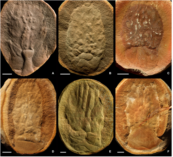
General morphology of Essexella asherae, showing the principal architectural elements. Unless otherwise indicated, all specimens illustrated here and in other figures are from the former Pit 11, Peabody Coal Co., near Godley and Braceville, Illinois, USA,Francis Creek Shale Member of the Carbondale Formation, Moscovian. (A) FMNH PE 92306; the inferred protracted tentacles (EF) andthe division into a smooth folded ‘disc’ (EA) area, inferred to be a physa and a topographically complex column (ED), interpreted as the scapus of an Anemone; note overall high relief. (B) FMNH PE 87065; the division into a smooth lower (EA) and topographically complex upper (ED; column) units with the top of the column being truncated at the region of the inferred oral disc: these are,respectively, the ‘accessory lobe’ (bell) and ‘skirt’ of Foster (1979). (C) FMNH PE 92305; this large specimen illustrates the high relief of Essexella specimens relative to other Mazon Creek animals and the overall topographic complexity; banded structures emerge from the top of the collapsed disc. The white film is kaolinite. (D) FMNH PE 39893, paratype; showing examples of the small Gastropod Strobeus sp. (S) near the centre line of a conical column. The column area is otherwise smooth, while the disc region (physa) has a triangular outline and folds. (E) FMNH PE 87330; close-up of Strobeus on a column, with kaolinite halo. (F) ROMIP64584; compare the truncated top edge of the column to the specimen in (B), the brown film is sphalerite, and the relief is lower compared with the specimens in(A) and (C). (G) ROMIP64584; close-up of specimen in (F), showing digitate extensions, inferred as retracted tentacles (EF). (H) FMNHPE 30711; specimen showing six elongate banded structures (EB), inferred to be contracted longitudinal muscles, running from the top of the disc to the top of the column. (I) FMNH PE 84617; two individuals in a single concretion, examples of multiple specimens in a concretion are rare and are usually small individuals; the specimen on the right shows external column ornamentation (EE) with pointed tips, while the specimen of the left has a collapsed outline. Abbreviations: S, Strobeus; Architectural elements: EA, element A (disc); EB, element B (elongate banded structures); ED, element D (column); EE, element E (column ornamentation); EF, element F (distal digitate structures). All scale bars represent 10 mm. Plotnick et al. (2023).
Specimens of Essexella asherae are typically preserved within nodules, parallel to the bedding plane. When split they break into two parts, with the upper being thicker and the lower thinner, which suggests the lower part formed as an impression of the original decaying Animal, and the upper part formed as a mold after this Animal had decayed away and the space it left was infilled by sediment. The preservation is variable, with some specimens showing exquisite detail while others can only tentatively be assigned to the taxon at all. Some of the specimens are surrounded by halos of precipitated pyrite, kaolinite, or sphalerite.
Almost all of the fossils associated with the Essex Fauna, including the vast majority of the Essexella asherae specimens, come from Peabody Coal Company Pit No 11, although some specimens are known from waste piles at the Sun Spot Mine, to the north of Astoria in Fulton County, Illinois, and from an outcrop of shale at the base of the Verdigris Formation near Windsor in Henry County, Missouri.
Previous interpretations of Essexella asherae have seen these fossils as representing a Medusa (Jellyfish) lying on its side, with a smooth, bowl-shaped bell and a long skirt surrounding a series of tentacles, all of equal length. These fossils appear to be of entirely soft bodied organisms, with a body divided into two sections, and a roughly-circular margin at one end, but have no other diagnostic features which would place them firmly within the Scyphozoa, such as oral-arms, tentacles, lappets, gonads, or radiate symmetry. Furthermore, no other Jellyfish, living or fossil, has ever been found which has a skirt surrounding its tentacles, In addition, specimens of Essexella asherae never show signs of having been dragged over sediments, something common in modern and fossil stranded Jellyfish.
These observations cause Plotnick et al. to reject the hypothesis that Essexella asherae is a Scyphozoan Jellyfish, and instead to hypothesize that these fossils represent Cnidarian polyps, predominantly preserved in a splayed lateral view, and, more specifically, Actiniarian Sea Anemones. These Anemones appear to have had an infaunal or semi-infaunal lifestyle, and to have been responsible for the trace fossils Conostichus and Bergaueria.
Plotnick et al. examined thousands of specimens of Essexella asherae in the collections of the Field Museum, Yale Pea-body Museum, Royal Ontario Museum, Manitoba Museum and the University of Illinois at Chicago, as well as several private collections. Fossils were photographed under normal and cross-polarized light, and where there was significant vertical-relief, image stacking was used to keep all of the image in focus. Trace fossils were photographed in diffuse light. Only specimens from institutional collections were used in formal descriptions of the new interpretation, to ensure that these remain available for future researchers.
Specimens of Essexella asherae vary considerably in size, with the smallest observed being 35 mm high and 27 mm wide, while the largest specimens are about 130 mm high and 70 mm wide. Most specimens are flattened and show no vertical relief, but some have considerable three-dimensional preservation. Notably, this is considerably more pronounced than is seen in any other soft-bodied organism from the Mazon Creek Biota, including Sea Cucumbers, Shrimps, and the famous Tully Monster, Tullimonstrum gregarium. This implies that Essexella asherae had a particularly firm body for a 'soft bodied' organism, possibly with a dense mesoglea and tough periderm.
The body is divided into two sections, a smooth circular, semicircular, or sometimes almost triangular region, and a larger barrel shaped region with greater relief, with an indented or convex margin where it attaches to the smaller body segment, and a truncated margin at the opposite end. This barrel-shaped body portion is quite variable in shape, and was apparently capable of expansion and contraction in life. Small tuberculate or digitate extensions can sometimes be seen extending beyond the truncated margin of the barrel-shaped region.
Plotnick et al. observed six recurrent elements in specimens of Essexella asherae, upon which their new diagnosis is based. (A) A smooth circular to semicircular, triangulate, or crescentic disc-like structure hat may exhibit evidence of folding, creasing, and/or contraction., and that bears faint net-like surface details in exceptional specimens. (B) Packets of elongate striated to occasionally banded structures that emanate from the interpreted interior edge of the disc. This may be twisted en masse, but can also exhibit Y-shaped alternations. (C) Short, flexible digitate structures emanating from the interpreted exterior edge of the disc. (D) A basket–bag or barrel-shaped mass hat may be ornamented to smooth, and can contain views of the elongate packets arranged such that the mass rises upward from the disc and has a straight/flat or gently curving top that is often associated with external ornamentation of the column. (E) Variable external ornamentation of the column, which may be diamond-shaped or ovate, elongate and striated (occasionally) with pointed tips, diffuse or weakly beaded, or smooth. (F) Rare short, thin, often-overlapping finer-scale structures that appear to emanate from the distal end of the column, These are of variable abundance and length and have tips that are rounded or pointed. Some examples have sediment interspersed between the short fine structures.

Specimens illustrating architectural elements and their variable expression. Element A (EA), the disc, is interpreted as equivalent to the physa in modern Anemones. Element B (EB), elongate banded and striated structures, are inferred to be contracted mesenterial retractor and parietobasilar muscles. Element D (ED), the column, is referred to the scapus of modern Anemones. (A) FMNH PE 92303, (counterpart); specimen preserves only the outline of the column (ED), including the truncated top margin; the disc element (EA) is folded and creased and is nearly as large as the column. (B) ROMIP66245; the disc element (EA) is proportionately large, and folded and creased. (C) FMNH PE 9806; the column preserves evidence of weakly beaded ornamentation, whereas the disc is smooth. (D) ROMIP66246; the surface of the disc (EA) has faint net-like surface details. (E) FMNH PE 39847; nearly circular disc element, with elongate banded structures (EB), presumed to be longitudinal retractor muscles, extending from the top of the disc to the frontal margin and twisted towards the left. (F) FMNH PE 85928; element B running from the top of the disc to the frontal margin. (G) FMNH PE 85901; element B running laterally from the basal disc to the side of the column, they are inferred to be parietobasilar muscles. (H) FMNH PE 36861, paratype; well-preserved element B (retractor muscles) attached to the top of the disc. (I) ROMIP64586; specimen showing evidence of both parietobasilar (diagonal) and retractor (longitudinal) muscles. (J) FMNH PE 46836; Francis Creek Shale, Coal Co. N Mine (Pond A), collected by Gordon Baird, well-preserved diagonal contracted parietobasilar muscles. Abbreviations: EA, element A; EB, element B; ED, element D. All scale bars represent 10 mm. Plotnick et al. (2023).
At is the case with all fossils, the preserved form of Essexella asherae reflects its form during life, changes that took place during and after death, the results of decay before and during burial, and chemical changes that took place during the formation of the nodules within which the specimens are enclosed. Thus in different specimens, different layers of tissue have been exposed by the collapse of the organisms during decay and compaction, the actions of scavenging Animals, and the way in which the nodules split open when they were cracked by fossil collectors. This creates taphonomic variations in the fossils, in which different combinations of features from the original organisms can be seen. These range from simple lateral views of the organism's exterior surface morphology, to complex combinations of internal and external features with distorted body margins. In addition, a few specimens are preserved in oral or aboral (from above or below) views rather than lateral.

Specimens illustrating architectural elements C, D and E and their variable expression. Element C (EC) are rarely preserved short, flexible digitate structures at the upper edge of the disc. Possibly tenaculi or verrucae. Element D (ED) is the entire column. Element E is the ornamentation of the column, which may be verrucae, tenaculi or tubercles; shown in (C)–(H). (A) FMNH PE 60678, holotype; element C is highlighted. (B) ROMIP64594; sediment separates element C from the column (element D). (C)–(F) illustrate variability in the relative sizes and shapes of the preserved disc and the column and the preservation of the external ornamentation of the column: (C) ROMIP53728, tapered column with weakly beaded surface ornamentation; (D) ROMIP64598, globose column with diffuse surface ornamentation; (E) ROMIP48547, column of relatively reduced size with smooth surface; (F) ROMIP64589, surface of globose column showing an overprint of the surface ornamentation and underlying element B (muscles). (G) FMNH PE 29351; ovate ornamentation. (H) FMNH PE 49830; striated ornamentation with pointed tips .Abbreviations: EC, element C; ED, element D (column). All scale bars represent 10 mm. Plotnick et al. (2023).
Plotnick et al. divided specimens of Essexella asherae into four taphonomic groups, to help understand how variations in preservation express themselves as different morphologies, reflecting the same original species. Type (I) specimens bear crisply preserved,ornamented, scale- or diamond-shaped examples of element E as preserving the original external surface of the organism and the original form. These specimens appear to best represent one view of the organism’s original morphology, and have columns that are frequently elongate with their widest part some what above the base. Each specimen also has a smooth to slightly folded basal disc, which may have a digitate distal margin and F elements ‘peeking’ out from the flat top of the disc. The disc is usually as wide or almost as wide as the column but is distinctly smaller in overall size. Type (II) specimens are typically characterized by a column ornamented by variable, often beaded to tuberculate examples of element E; these elements are significantly less distinct and crisp than the ornament in Type (I) specimens. The column may be somewhat flattened, with an ovoid outline and a flat or gently curved top from which small digitate structures (element F) may protrude. In some cases the column may appear deflated, allowing the outline of the underlying striated structures (element B) to be discerned, or the external layers are partly or completely removed to show the striated structure; this style of preservation demonstrates the three-dimensional relationship of features that were originally internal and external. The disc is frequently creased or wrinkled and may be narrower or wider than its associated column. The relative sizes of the two major components are variable; the disc is usually much smaller than the column, but in some examples the column is distinctly shrunken whereas the disc may appear inflated. Type (III) specimens either have columns with obscure pustulose ornament or have surfaces that are smooth to weakly longitudinally striated. The column may be somewhat wrinkled and can have indistinct margins; it often has a broadly conical shape, tapering from base to tip. The distal tip of the column may have small bulbous structures (element F), although in many instances its features are indistinct, or it may appear to have partly disintegrated. The disc is often heavily creased or wrinkled, appears partly inflated, is frequently wider than the column, and in some instances may be as long as or larger than the column. The majority of specimens that bear the Gastropod Strobeus belong to Type (III). It is likely that most of the tens of thousands of Essexella known to be present in various collections could be assigned to Type (III); many of the previously described ‘curtains’ and ‘blobs’ would fall into this variant. It would be possible to define another taphonomic variant that has even poorer and less distinct preservation than variant III, but this would not have practical value because such specimens would have so little recognizable morphology that they could not be definitively assigned to Essexella, or to any other Mazon Creek taxon. Plotnick et al. retain the term ‘blobs’for these specimens. Type (IV) specimens may be variably affected by decomposition and diagenesis; this type is used for comparatively small specimens preserved with only an inferred oral/aboral view of the disc or the column, which cannot therefore be assigned to types (I)-(III). Plotnick et al. subdivide this group into two divisions. Type (IVa) has a quadrate central pattern or concave or subcircular central area with a surrounding smooth ring; it resembles the closed oral disc of an Anemone. In some instances, small rounded or pointed protrusions may represent expressions of element F, the structures that protrude from the distal end of the column. These resemble the contracted tentacles of an anemone. In some instances Type (IVa) examples may be oriented obliquely so that the ‘mouth’ is toward one edge of the fossil. Type (IVb) specimens have a smooth central area and a radiating or reticulate outer ring, often raised on a mound. Plotnick et al. interpret this feature as representing the bottom of the pedal disc, with the remainder of the animal compressed above it; in some instances, a considerable portion of the upper part of the animal can be seen splayed around the base of the disc. The central portion of this disc is often offset from the central mass of the fossil. This taphonomic variant includes specimens previously referred to as the Scyphozoan Reticulomedusa greenei, which Plotnick et al. regard as a junior synonym of Essexella.

Specimens illustrating architectural element F (EF) and its variable expression. This rarely preserved element is interpreted as the tentacles surrounding the oral disc. (A) FMNH PE 50262, with elongate EFs. (B) FMNH PE 29351. (C)–(D) sediment (S) between EFs showing that they were not laterally contiguous: (C) FMNH PE 92302; (D) ROMIP64589. (E) ROMIP66246, collapsed and irregular EFs. (F) ROMIP64586; EFs have a slender thorn-like form. All scale bars represent 10 mm. Plotnick et al. (2023).
Many of the Essexella asherae specimens show signs of having been scavenged after death, Simple, unbranching trace fossils often cut across the specimens, suggesting that scavenging Animals had burrowed through their bodies. In modern environments carrion is known to serve as a nutritional hot spot which can promote local biodiversity, and this has also been demonstrated in the Cambrian Sirius Passet Lagerstӓtte, and possibly in some Ediacaran deposits.

Specimens illustrating taphonomic variant (I). This variant is interpreted as best preserving the external morphology and tentacles (element F). (A) FMNH PE 29351; specimen with creased disc and column with crisp ovate ornament. (B) FMNH PE 49830; column has striated ornament with pointed tips. (C) FMNH PE 84609; column has striated ornament and possible overprint of underlying muscles. (D) FMNH PE 84614; column has weakly expressed ornament and a truncated tip. (E) FMNH PE 48698; column has collapsed in the middle; notice the colour change in that area. (F) FMNH PE 60678, holotype; note the faint trace of element C (i.e. short, flexible digitate structures) at the base of the column. All scale bars represent 10 mm. Plotnick et al. (2023).
The Soleniscid Gastropod Strobeus is frequently found associated with Essexella asherae. When the latter was interpretted as a Scyphozoan Medusa, it was suggested that Strobeus might be a floating neustonic predator similar to the modern Janthina, which predates floating Cnidarians, but this clearly does not fit with the interpretation of Essexella asherae as a bottom-dwelling Anemone. Furthermore, there are no predation marks on any Essexella asherae specimens likely to have been caused by a Gastropod hunter, and Strobeus is considerably smaller than Essexella asherae, whereas Janthina is large compared to its prey.

Specimens illustrating taphonomic variant (II). External ornamentation, if present, is less distinct than in taphonomic variant I, often with superimposition of external and internal morphological features or total loss of external morphology. Note the variable relative sizes of the column and disc but the commonality of the overall outline. (A) FMNH PE 29041; specimen with disc collapsed on the sides. (B) FMNH PE 87099; extensively creased disc area, weakly triangulate ornament on the column. (C) ROMIP64593; creased and proportionally large disc. (D) ROMIP64597; note the small relative size of the disc area. (E) FMNH PE 40097; except on the lower right, the external morphology has been stripped from the column, revealing the underlying striated muscle structures. (F) ROMIP66246; digitate structures at the top, underlying muscle structures are visible within the partly collapsed column. All scale bars represent 10 mm. Plotnick et al. (2023).
Alternative interpretations of the presence of Strobeus on Essexella asherae are that the Gastropods were scavenging on dead Anemones, or that they were parasitic, living on the Anemones while they were alive. Modern scavenging Gastropods such as Nassariids (Dog Whelks) or Buccinids (True Whelks) are generally found in aggregations upon carrion, which is found opportunistically. These are generally environmentally tolerant species, able to cope with fluctuations in salinity and oxygen levels, conditions predicted for the offshore Mazon Creek environment where these fossils originated. The alternative suggestion is that had a lifestyle similar to that of modern Wentletraps of the genus Epitonium, which are parasitic or kleptoparasitic on Sea Anemones.

Specimens illustrating taphonomic variant (III). Examples illustrate total or near total loss of external ornamentation, and obscure internal structures. Some are associated with the Gastropod Strobeus, preserved medially near the highest parts of a prone column. The disc is often broader than the column and heavily wrinkled. (A) FMNH PE 87330; note the cluster of three Strobeus and the broad disc area. (B) FMNH PE 87054; medial specimen of Strobeus surrounded by kaolinite. (C) ROMIP64592; creased disc and collapsed column. (D) FMNH PE 9806; heavily creased disc and collapsed, flattened column with ‘smeared’ tip. (E) ROMIP64601; few details are visible; note the digitate outline. All scale bars represent 10 mm Plotnick et al. (2023).
Plotnick et al. believe that Strobeus is more likely to have been a scavenger than a parasite. They are almost always found in clusters on the midline of the columns of Essexella asherae, which would have been the highest point on the carcass and the last part to be buried by sediment. A parasite, which was attached before the Anemone died and remained in position after the host died, would have no such preferred orientation on a toppled Anemone. Furthermore, these Snails are generally found in clusters, which is behaviour seen in modern Gastropod scavengers, and are rare on specimens showing little or no decay, implying that they were late-stage colonisers of decaying organisms, possibly capable of surviving environmental stresses which killed the Anemones.
The study and classification of Actiniarian Sea Anemones is based entirely on the soft tissues of these Animals, i.e. features usually beyond the reach of palaeontologists. Anemones generally have hexamerous symmetry, which can be observed in the distribution of mesenteries and tentacles and often the body wall of the column. The column of the Anemone can often be divided into sections, with the portion directly above the basal disk, known as the scapus, generally being the largest. The scapus, and quite often the entire column, may be covered by a thick periderm, which may have external decoration such as tubercles. The basal disc in burrowing Sea Anemones is often modified to form a bulb-like phyla.

Specimens illustrating taphonomic variant (IV), which previously have been assigned to Reticulomedusa greenei (itself assigned to Scyphozoa). Plotnick et al. recognize these rare and small individuals as representing the oral or aboral ends of Essexella, preserved vertically. Examples with a quadrate central pattern or concave or subcircular central area with a surrounding smooth ring (A)–(F) are interpreted as the mouth and oral disc, whereas specimens with a radiate or reticulate outer ring (G)–(L) are the bottom of the pedal disc or physa. (A) FMNH PE 92301; mouth and oral disc; note the inner quadrate region. (B) FMNH PE 13949; mouth and oral disc,the arrow indicates possible preserved tentacles or tentacle bases. (C) FMNH PE 45157; mouth and oral disc; note the quadrate form. (D) ROMIP47991; mouth and oral disc, arrows indicate possible preserved tentacles. (E) FMNH PE 82157; mouth and oral disc; arrows indicate possible preserved tentacles; specimen donated by Jack Wittry. (F) FMNH PE 93016; mouth and oral disc; specimen donated by Rob Coleman. (G) FMNH PE 29408, holotype of Reticulomedusa greenei; aboral surface (base of disc), showing a characteristic reticulate pattern surrounding a smooth central area. (H) FMNH PE 56975; aboral surface (base of disc) with a smooth central part surrounded by a reticulate structure. (I) FMNH PE 87194; aboral surface surrounded by a compressed remnant of the remainder of the Animal. (J)–(K), FMNH PE 93015 part and counterpart; aboral surface (base of disc) with the compressed remnant of the remainder of the Animal surrounding it and extending obliquely toward the top of each photograph; specimen donated by Rob Coleman. (L) FMNHPE 33629; aboral surface (base of disc) preserved in oblique view. All scale bars represent 10 mm. Plotnick et al. (2023).
Internally, the Anemone contains a series of paired radial mesenteries (membranes) which extend inwards from the column wall and are attached to the base and oral disc, and may be attached to the actinopharynx (perfect mesenteries) or not (imperfect mesenteries). The longitudinal retractor muscles run along the entire length of one side of the mesenteries. These can be used to shorten the column and retract the oral disc. On the other side of the mesenteries are the parietobasilar muscles, which run obliquely about half way up the column wall, and are used to shorten and widen the column during retraction. Circular muscles run around the margin of the column; in some species the circular muscles at the top of the column can form a sphincter which protects the retracted tentacles and oral disk. Many Sea Anemones also have radial endodermal basilar muscles, which runalong the insertion of the mesenteries into the basal disc, and which can be used to attach to or detach from the substrate, and sometimes to move about.

Specimens showing evidence of post-mortem burrowing. (A) FMNH PE 60211; note the different scales of burrows in the disc and the column. (B) FMNH PE 49340; large burrows radiate across a collapsed and blob-like specimen. Both scale bars represent 10 mm. Plotnick et al. (2023).
Most modern Sea Anemones live attached to hard surfaces, but both extant sub-orders of the Actiniaria contain burrowing species. These can be divided into two morphotypes (both found in both sub-orders), with one form being long and slender with a pedal disc expanded to form an ampullaceous (ampulla-shaped) phyla, while the other morphotype is less Worm-like in form and lacks a phyla, but can expand and contract the pedal disc.

Examples of modern epifaunal and burrowing Anemones. (A) Unidentified burrowing Anemone, Castle Rocks, Cape Peninsula, South Africa (Peter Southwood/Wikimedia Commons). (B) YPM IZ080459; burrowing Anemone Haloclava producta (Enthemonae, Haloclavidae), preserved specimen, note the rows of verrucae, and the base expanded to form a physa (Eric Lazo-Wasem/Yale Peabody Museum). (C) YPM IZ 009387.CN; epifaunal Anemone Actinauge verrilli, preserved specimen from continental shelf, approximately 160 km SSW of Martha’s Vineyard, Massachusetts, USA. (D)–(F) Burrowing Anemone, Paranthus rapiformis (Sea Onion; Enthemonae, Actinostelidae); live individual showing expansion and contraction of the column and of the pedal disc; (E) and (F) show the base of the pedal disc (specimen provided by the Woods Hole Oceanographic Institution). (G) Unidentified partially contracted Anemone, Guam (Ryo Sato/Wikimedia Commons). (H) Epifaunal Anemone, Anthopleura elegantissima, Isla Santa Cruz, California, USA, showing extended tentacles, mouth and oral disc (Ed Bierman/Wikimedia Commons). (I) Anthopleura elegantissima?, Puget Sound, Washington, USA, with tentacles partly retracted (Demon Days 64/Wikimedia Commons). (J) Anthopleura elegantissima, Puget Sound, Washington, USA, exposed at low tide with tentacles retracted and oral disc covered with sediment (Wikimedia Commons). All scale bars represent 5 mm. Plotnick et al. (2023). All extant burrowing Sea Anemones are able to use their hydrostatic skeletons to expand and contract their aboral ends, either a physa or a pedal disk, as well as being able to change their bodyforms significantly by retracting different muscle groups. The surfaces of these Anemones are frequently covered by rows of tubercles or similar ornamentation.
Due to their soft-bodied nature, and more-or-less complete absence from the fossil record, the Taphonomy (decay processes) of Sea Anemones has not been extensively studied. However, some recent studies looking at the taphonomy of extinct soft-bodied organisms from Ediacaran and Cambrian biotas have included Sea Anemones in experimental data. These studies found that the tentacles of Sea Anemones decay rapidly after death, followed by the outer dermal layer and internal organs, with the interior muscles and pedal disk being the most decay-resistant tissues. This means that the decay of the outer dermal layer tends to expose the internal musculature, which eventually breaks down to form structureless masses similar to many Mazon Creek 'blobs'.
This fits well with the interpretation of Essexella asherae as an Anemone, with the fossils typically showing a mixture of external morphology and exposed internal musculature, as well as the resilient pedal disc. The variation seen in the fossils would be driven by the predicted decay path, combined with irregular events such as modification of the body by escaping purification gasses and the action of burrowing Animals, and halted at the point where the Anemone is buried and encased within a concretion.
Plotnick et al. reconstruct Essexella asherae as a relatively tough and firm-bodied Anemone, capable of retaining its mass and shape some way into the decay process. The body is divided into a smooth disc and a columnar region with greater surface relief, with the disc generally smaller than the column.
Many specimens show evidence of folding, creasing, and shrinkage of the smooth disc. Plotnick et al. interpret this disc as the pedal region of the Anemone, which was probably capable of expansion and contraction, forming an organ similar to the phyla of modern burrowing Anemones. In life this would probably have formed a fluid-filled sack, used to anchor the Anemone in soft sediment, with the collapse of this sack after death forming the folds and creases observed.
The column of the body is typically barrel-shaped, indented where it meets the pedal disc and truncated at the opposite end. Plotnick et al. suggests that the barrel of Essexella asherae would have been the scapus of the living Anenome, with the truncated area representing the location of the oral disk. The variable preservation of the specimens mean that a variety of structures may be visible here, sometimes superimposed on the same specimen. The packets of elongated striated or banded structures, which originate from the pedal disk and run longitudinally to the oral disc, and which appear to be about six in number, are interpreted by Plotnick et al. as being longitudinal retractor muscles. In some cases the position of these features hint at the presence of an underlying actinopharynx and gastrovascular cavity. Bands of about six banded structures can also be seen extending from the interior edge of the pedal disc diagonally to the margin of the scapus, which Plotnick et al. interpret as contracted parietobasilar muscles.

Stylized reconstruction of Essexella asherae. (A) External view of the expanded anemone, showing division into column (scapus) and expanded pedal disc (physa), reconstructed external sculpture and inferred tentacles. (B) cutaway view of the expanded Anemone, showing mesenteries with longitudinal contractor (right) and diagonal parietobasilar (left) muscles; the physa is hollow and muscular, with a small central aperture connecting to the interior of the column. (C) Partially contracted Anemone, with withdrawn tentacles and narrowed oral disc. The nature of the structures emerging from the top of the physa is speculative. Marjorie Leggitt in Plotnick et al. (2023). The apparent six-fold symmetry of the mesenteries and other strucutures supports the placement of Essexella asherae within the Hexacorallia (the wider group that includes the Actiniarian Sea Anenomes, as well as several types of Corals). The surface of the column of many specimens is covered by diamond-shaped or scale-like structures, while the pedal disc appears to have elongate structures protruding from its upper edge, which Plotnick et al. consider to be elongate verrucae or tubercles.
The truncated end of some specimens have short, tube-like structures visible, which Plotnick et al. interpret at the tips of retracted tentacles. The rare specimens preserved in oral or aboral view are consistent with an interpretation of the fossils as being Actiniarians. These show the retracted mouth to be quadrate to subcircular in shape, with rare specimens showing this to be surrounded by a ring of tentacles.

Possible actinopharynx, mouth and microbial structures. (A) FMNH PE 92304; specimen with distinct striated longitudinal retractor muscles (element B; EB); the elevated axial portion may show the form of the underlying actinopharynx and gastric cavity; associated structures running aborally from them may represent a preserved actinopharynx. (B)–(C) ROMIP64590; unique specimen with iron oxide staining and rounded structures at the oral end, interpreted as the mouth (M) region. (D) ROMIP66281; showing a flattened to con-cave oral end with staining and texture different from those of the column. (E)–(F) FMNH PE 93017, partially contracted example with con-cave iron-stained region on the oral end interpreted as the mouth (M), and subtle surface sculpture on the column. (G) ROMIP48547,detail of column of the counterpart, with filamentous to clotted texture. Scale bars represent: 10 mm (A)–(F); 1 mm (G). Plotnick et al. (2023).
Fossil Sea Anemones are incredibly rare, with the Essex Fauna of the Mazon Creek Lagerstӓtte representing the only deposit in which these are a frequent find. Trace fossils attributed to burrowing Sea Anemones, however, are relatively common throughout the Phanerozoic, with five ichnogenera of plug-shaped burrows thought to have been formed by these Animals.
The ichnogenus Conostichus comprises conical, downward pointing burrows between 15 and 145 mm in diameter. These trace fossils are common in Pennsylvanian (Late Carboniferous) deposits across North America. The apex of the cones of Conostichus have a 12-fold symmetry, which is also seen in specimens of Essexella asherae seen in oral view, and which is thought to reflect the internal septal divisions of the living Anemone. Internally these traces resemble a series of stacked cones. Although most abundant in the Pennsylvanian of North America, Conostichus ranges from the Cambrian to the Early Permian, with one possible Cretaceous example. These traces are most common in fine-grained siliciclastic sediments, but are also found in carbonates, and always in shallow environments which were either fully marine or of variable salinity. These burrows have been considered to be the dwelling of an unknown filter feeding Animal, with the stacking of the cones formed by the Animal moving upwards as sediment is deposited. These burrows were first proposed to have been created by an Actiniarian by Charles Kent Chamberlain in 1971, who envisioned a burrowing Sea Anemone stouter than modern forms, with a large, bulbous phyla and a barrel-shaped upper body. Chamberlain's proposed Anemone matches closely with Plotnick et al.'s reconstruction of Essexella asherae.

Examples of the trace fossil Conostichus. (A), (B) and (D) are specimens originally described by Joseph Devera, from near Old Town, Illinois, in 1989. Tradewater Formation (Lower Pennsylvanian); uncatalogued specimens in the collections of the Illinois Natural History Survey; specimens have been coated in varnish to arrest pyrite decay; photos courtesy of Jared Thomas, Prairie Research Institute. (C) FMNH PE 92300; bottom view of a Conostichus, from Tunnel Hill, Illinois, Tradewater Formation. All scale bars represent 10 mm. Plotnick et al. (2023). Plotnick et al. suggest that Essexella asherae or a very similar organism were responsible for the Conostichus traces, on the basis of the overall similarity in size and shape between the fossil and the ichnofossil, the apparently hexamerous symmetry of both, the correspondence in form between the interpreted basal disc of Essexella and the lower apex of Conostichus, as well as that of other plug-shaped forms such as Bergaueria and Conichnus, and the environmental and stratigraphic overlap between the taxon and the ichnotaxon. Plotnick et al. further report the known existence of a specimen of Essexella asherae within a Conostichus trace, although this is in a private collection, and not available for study.
Neither Essexella asherae nor the Conostichus burrows closely resemble slender modern burrowing Sea Anemones, being stouter and having a greatly expanded pedal disc. Essexella asherae appears to have been morphologically intermediate between the two modern groups of burrowing Anemones, with an enlarged pedal disc area which was able to expand and contract during burrowing.
Essexella asherae is extremely common within the Mazon Creek deposits, apparently having been the dominant macroscopic benthic organism in the shallow-marine palaeocommunity. This is in keeping with the behaviour of modern burrowing Anemones, which often form high abundance communities; for example the small Anthopleura hermaphroditica has been shown to reach densities of around 30 000 individuals per m³ on intertidal mudflats in Chile. The only detailed study of the distribution of Mazon Creek fossils found that Essexella asherae was found throughout the marine environment, but becomes more abundant further from shore, which was interpreted at the time as being indicative of a plancktonic Jellyfish being carried into the estuary and stranded, bu which Plotnick et al. suggest indicates that the species was a bottom-dwelling species, favouring muddy environments and able to cope with changes in salinity, similar to the modern estuary-dwelling Nematostella vectensis.
Plotnick et al. observe that a considerable size range is present within the Essexella asherae fossils, suggesting that if they were being buried by seasonal climatic events, many specimens were clearly able to survive several such events and relocate themselves upwards in the process. Alternatively, the Anemones may have periodically been killed off by another event, possibly an input of fresh water beyond their ability to survive, and then buried by more gradual (or variable) sedimentation, something which is supported by the variation in decomposition seen in the specimens. This range of decomposition is also seen in burrowing Sea Cucumbers from the Mazon Creek Biota.
Sea Anemones are a highly successful group, found in almost all marine environments, and showing a remarkable degree of diversity for an Animal with such a simple bodyplan. This success has lead to a high degree of convergence within the group, with different groups of Anemones both gaining and losing features to adapt to similar environments. This means that phylogenetic analysis of the group using morphology is notoriously difficult, something made harder by the almost complete absence of a fossil record.
Some highly speculative fossil Sea Anemones have been reported from the Ediacaran, plus a small number of possible species from the Cambrian Kuanchuanpu and Chengjiang formations of China. The only post-Cambrian fossil Sea Anemones described are Essexella asherae from the Mazon Creek Lagerstӓtte and Palaeoanemona marcusi from the Carboniferous Leoncito Formation of Argentina, both of which have been reconstructed as burrowing species.
The hexamerous radial symmetry seen in Essexella asherae is good evidence that the species should be placed within the Hexacorallia, and the absence of a calcified skeleton suggests that it should further be placed within the Actiniaria (or possibly the Corallimorpharia), and the morphological similarity to modern burrowing Sea Anemones, combined with a known specimen found inside a burrow, strongly suggests that it was a burrowing species, although no features which would be deemed useful in further classifying a modern Actiniarian are preserved.
Molecular genetic studies of the Actiniaria suggest that the group should be split into two suborders, the Anenthemonae and the Enthemonae, both of which contain burrowing species. These suborders can be further differentiated by the arrangement of their mesenteries. Morphologically, Essexella asherae is closer to the Anenthemonae than the Enthemonae, but not sufficiently so for this diagnosis to be made with any degree of confidence. Molecular clock studies have suggested that the Actinarians divided from other Hexacorallians as early as 470 million years ago (Ordovician), making it quite possible that Essexella asherae lived before the group split into the Anenthemonae and the Enthemonae.
See also...
Online courses in Palaeontology.
Follow Sciency Thoughts on Facebook.
Follow Sciency Thoughts on Twitter.