After the publication of its discovery from the famous Hell Creek Formation in 1905, the carnivorous dinosaur Tyrannosaurus rex was met with intense scientific interest and public popularity, which persists to the present day. Numerous hypotheses concerning Tyrannosaurus rex biology and behavior result from decades of research primarily focused on skeletal morphology and biomechanics. Only within the past 15 years has bone histology been applied to investigate the aspects of Tyrannosaurus rex life history inaccessible from gross examinations, addressing questions concerning ontogenetic age, growth rate, skeletal maturity, and sexual maturity. In 2004, two teams independently assessed the growth dynamics of Tyrannosaurus rex using osteohistology. Their results suggest that T. rex had an accelerated growth rate compared with other Tyrannosaurids and achieved adult size in approximately two decades.The teams focused on growth curves, rather than on detailed analyses or interpretations of bone tissue microstructures. However, osteohistology is critical for establishing a baseline against which skeletal maturity and growth changes in cortical morphology related to life events in this taxon can be tested. Identifying the timing of growth acceleration and empirically quantifying juvenile Tyrannosaurus rex growth rates are of special importance because the juvenile growth record is lost in older individuals because of bone remodeling and resorption.
In a
paper published in the journal
Science Advances on 1 January 2020, Holly Woodward of the
Department of Anatomy and Cell Biology, at the
Oklahoma State University Center for Health Sciences, Katie Tremaine of the
Department of Earth Science and the
Museum of the Rockies at
Montana State University,
Scott Williams, also of the Museum of the Rockies,
Lindsay Zanno of
Paleontology at the
North Carolina Museum of Natural Sciences, and the
Department of Biological Sciences at
North Carolina State University,
John Horner of
Chapman University, and
Nathan Myhrvold of
Intellectual Ventures, present the results of the bone microstructure of the femur and tibia of two Tyrannosaur skeletons of controversial taxonomic status recovered from the Hell Creek Formation and now in the collection of the
Burpee Museum of Natural History.
These specimens are BMRP 2002.4.1, a largely complete specimen composed of nearly the entire skull and substantial postcranial material, and BMRP 2006.4.4, a more fragmentary specimen. Respectively, Woodward et al. estimate these specimens to be 54 and 59% the body length of FMNH PR 2081 (popularly known as 'Sue'), one of the largest known Tyrannosaurus rex specimens. The ontogenetic age of BMRP 2002.4.1 was previously reported as 11 years based on fibula osteohistology. However, because the fibula grows more slowly than the weight-bearing femur and tibia, it does not reflect annual increases in body size or relative skeletal maturity as accurately. Woodward et al. used femur and tibia data to (i) provide detailed comparative intra- and interskeletal histological descriptions, (ii) quantify the ontogenetic age and relative skeletal maturity of these specimens, and (iii) allow empirical observation of annual growth rate, with emphasis on variability during the life history of Tyrannosaurs.

Hind limb elements of BMRP 2006.4.4. Left femur in (A) lateral, (B) cranial, (C) medial, and (D) caudal views. Proximal portion of left tibia in (E) lateral and (F) proximal views. Abbreviations: ac, accessory lateral condyle; cc, cnemial crest; ctf, crista tibiofibularis; fc, fibular crest; fh, femoral head; ft, fourth trochanter; g, greater trochanter; if, intercondylar fossa; L2, lobe on lesser trochanter; lc, lateral condyle; lt, lesser trochanter; mc, medial condyle; mdc, mesiodistal crest; pf, popliteal fossa; ts, trochanteric shelf. Scale bar 5 cm. (F) not to scale. Woodward et al. (2020).
Moreover, by histologically quantifying the ontogenetic age of BMRP 2002.4.1 and BMRP 2006.4.4 and inferring skeletal maturity, Woodward et al. present new data that can be used to evaluate competing taxonomic hypotheses regarding these and other mid-sized Tyrannosaur specimens discovered in the Hell Creek Formation, specifically whether BMRP 2002.4.1 (and by proxy other specimens) represents an adult 'pygmy' genus of Tyrannosaurid, 'Nanotyrannus'.
In general, the femur and tibia cortical bones of BMRP 2002.4.1 and BMRP 2006.4.4 can be classified as a woven parallel complex. Vascularity and osteocyte lacuna density are uniformly high throughout. In the femora, the primary and secondary osteons surrounding vascular canals are frequently isotropic in the transverse section and anisotropic in the longitudinal section. Also in the transverse section, femur primary tissue exhibits moderate anisotropy regionally and weak anisotropy locally, corresponding to a loose arrangement of mineralized fibers in parallel.

Femur histology of Tyrannosaurid specimens BMRP 2002.4.1 and BMRP 2006.4.4. (A) Mid-cortex of the transverse thin section of BMRP 2002.4.1. Plane-polarised light emphasizes osteocyte lacuna density and variability in shape within the laminae, as well as longitudinal primary osteons. In circularly polarised light, there is a weak preferred fiber arrangement parallel to the transverse plane of section reflected by regional birefringence. Many primary osteons have uniformly isotropic fibers with rounded osteocyte lacunae. (B) Mid-cortex of the transverse thin section of BMRP 2006.4.4. Osteocyte lacuna density and variability in shape within the laminae are evident in plane-polarised light. Circularly polarized light reveals varying birefringence associated with bone fiber orientation, but there is a weak preferred fiber arrangement parallel to the transverse plane of section reflected by regional birefringence. Many primary osteons are composed of uniformly isotropic fibers with rounded osteocyte lacunae. (C) Longitudinal section of the mid-cortex of BMRP 2006.4.4. Vascular canals appear as near-vertical, thin, dark columns. As in the transverse section, the primary laminae between primary osteons contain variably arranged osteocyte lacunae. In circularly polarized light, the laminae are weakly isotropic (I), corresponding to the poorly organized parallel orientation of fibers in the transverse plane. The laterally compressed osteocyte lacunae in primary osteons are embedded within a uniformly birefringent (anisotropic) matrix in circularly polarised light, indicating that the primary osteon lamellae are longitudinally oriented parallel-fibered bone. (D) On the posteromedial side of the transverse section of BMRP 2006.4.4, there is a parallel-fibered annulus located at the periosteal surface (thickness indicated with blue line). Photographed in circularly polarized light. (E) In the transverse section on the posterolateral side, the annulus shown in (D) (blue lines) is overlain by highly isotropic woven-fibered laminae. Abbreviations: AN, anisotropic Matrix; CCL, circularly polarised light; I, weakly isotropic laminae; PPL, plane-polarised light; PO, primary osteons. Woodward et al. (2020).
In the tibia transverse section of BMRP 2002.4.1, longitudinal primary osteons are isotropic in circularly polarized light, but fibers of primary osteons encircling laminar, circular, and plexiform vascular canals are anisotropic. In contrast, primary osteons in the tibia of BMRP 2006.4.4 are frequently isotropic regardless of vascular canal orientation. Because of its proximal sampling location, the cortical shape of the tibia from BMRP 2006.4.4 in transverse section differs from that of BMRP 2002.4.1 and incorporates the fibular crest on the lateral side. Highly vascularized reticular woven tissue is present on the anterior and anterolateral periosteal surfaces. In both individuals, the thickest tibial cortex is located anteriorly.

Tibia histology of Tyrannosaurid specimens BMRP 2002.4.1 and BMRP 2006.4.4. (A) Transverse mid-cortex thin section of BMRP 2002.4.1. Longitudinal primary osteons are evident, and plane-polarised light emphasizes osteocyte lacuna density and variability in shape within laminae. Circularly polarized light reveals varying birefringence associated with bone fiber orientation, but with a weak arrangement of fibers parallel to the transverse plane of section. Many primary osteons are composed of highly isotropic fibers with rounded osteocyte lacunae. (B) Longitudinal thin section of the mid-cortex of BMRP 2002.4.1. Vascular canals appear as near-vertical, dark columns. Adjacent to the vascular canals, the primary osteons contain laterally compressed osteocyte lacunae. Circularly polarised light demonstrates that the laterally compressed osteocyte lacunae of primary osteons are embedded within a uniformly birefringent matrix (anisotropic), indicating that the lamellae of primary osteons are parallel-fibered bone. Osteocyte lacunae orientation varies in the thin laminae between primary osteons. In circularly polarized light, the laminae are weakly isotropic, corresponding to the weak arrangement of parallel fibers in transverse section. (C) In transverse thin section, the periosteal surface of BMRP 2006.4.4 on the anterior side consists of reticular primary osteons within laminae of highly isotropic, woven tissue. (D) Within the anterior and anteromedial innermost cortex of BMRP 2006.4.4, in transverse thin section, six closely spaced lines of arrested growth are visible interstitially. Blue lines highlight the lines of arrested growth trajectories. Abbreviations: AN, anisotropic Matrix; CCL, circularly polarised light; I, weakly isotropic laminae; PPL, plane-polarised light; PO, primary osteons. Woodward et al. (2020).
Of special note, within the medullary cavity of the femur and tibia of BMRP 2006.4.4, isotropic, vascularized, primary tissue is separated from the cortex by a lamellar endosteal layer. These features are morphologically consistent with medullary bone; however, additional studies on the systemic nature of this tissue throughout BMRP 2006.4.4 and biochemical tests on this tissue are necessary to test this hypothesis.

Fragmentary femur transverse thin section of BMRP 2002.4.1. (A) Mid- to outer cortex in circularly polarized light, with the periosteal surface at the bottom left of the image. The rich vascular network shown here is comprised of longitudinal vascular canals within a woven-parallel complex of primary tissue. Many primary osteons display isotropic lamellae encircling the vascular canals. (B) Magnified region of femur cortex in circularly polarised light. Regional anisotropy gives way to varying birefringence and osteocyte lacuna shape within the laminae surrounding primary osteons suggesting weakly woven or poorly organized parallel-fibered tissue. Regions of uniform isotropy within primary osteons in (B) appear 'bubbly' in (C) with plane polarised light because the fiber bundles are arranged perpendicular to the plane of section. Abbreviations: CPL, circularly polarised light; PPL, plane polarised light. Woodward et al. (2020).
Cyclical growth marks, resembling tree rings in transverse thin section, were observed in the femora and tibiae of both BMRP specimens. Studies on extant vertebrates demonstrate that cyclical growth marks result from brief interruptions in osteogenesis, occurring with annual periodicity and typically coinciding with the nadir. The annual pauses in bone apposition are recorded as cyclical growth marks in cortical microstructure as either pronounced lines of arrested growth or diffuse annulus rings. On the basis of counting cyclical growth marks, BMRP 2002.4.1 was at least 13 years old at death (13 cyclical growth marks in the femur and 10 cyclical growth marks in the tibia), and BMRP 2006.4.4 was at least 15 years old at death (15 cyclical growth marks in the femur and 13 to 18 cyclical growth marks in the tibia). Typically, vertebrate long bone cortices will exhibit widely spaced cyclical growth marks within the cortex when young, corresponding to high annual osteogenesis. In subadults, cyclical growth marks become more closely spaced as osteogenesis decreases approaching adult size. In contrast to these frequently observed patterns, the spacing of CGMs was unexpectedly variable throughout the femur and tibia cortices of both BMRP specimens.

Femur and tibia histology overview of Tyrannosaurid specimens BMRP 2002.4.1 and BMRP 2006.4.4. (A) Top: the fragmentary femur of BMRP 2002.4.1 was transversely sectioned (blue line). Scale bar is 10 cm. Bottom: the resulting thin section is incomplete. Scale bar is 1 cm. (B) Top: the right tibia of BMRP 2002.4.1 was transversely sectioned (blue line). Scale bar is 10 cm. Left: the resulting complete transverse thin section. The embedded material was also sectioned longitudinally along a lateral (L)–medial (M) transect (blue line) to produce a longitudinal thin section. Scale bar is 1 cm. Right: the resulting longitudinal thin section. (C) Top: the left femur of BMRP 2006.4.4 was transversely sectioned (blue line). Scale bar is 10 cm. Left: the resulting complete transverse thin section. The embedded material was also sectioned longitudinally along an anterolateral (AL) – posteromedial (PM) transect (blue line) to produce a longitudinal thin section. Scale bar, 1 cm. Right: the resulting longitudinal thin section. (D) Top: the partial left tibia of BMRP 2006.4.4 was transversely sectioned (blue line). Scale bar is 10 cm. Bottom: the resulting complete transverse thin section, which incorporates the lateral crest. Scale bar is 1 cm. Woodward et al. (2020).
In the femur of BMRP 2006.4.4, there is an annulus at the periosteal surface on the medial side, but when followed posteriorly, the annulus is within the outer cortex, while fibrolamellar tissue makes up the cortex of the periosteal surface. Within the innermost cortex on the anterolateral side, six lines of arrested growth are closely spaced. Because of resorption from the medullary drift, these lines of arrested growth are absent within the innermost cortex of the posterior and lateral sides.

Transverse thin section histology of the right tibia of BMRP 2002.4.1. (A) Transverse and longitudinal thin sections were produced from the right tibia of BMRP 2002.4.1. (B) In circularly polarised light the innermost cortex of the anteromedial side shows a primary cortex with longitudinal and laminar vascularity as well as a scattering of secondary osteons. The cementing lines bounding secondary osteons are highly birefringent, but secondary osteon lamellae are isotropic. (C) Vascularity becomes circular to plexiform on the lateral side, shown in plane polarised light. (D) Anterolaterally, primary tissue gives way to a column of Haversian systems radiating from inner to outer cortex, likely associated with tendon insertion. Viewed in circularly polarised light, these secondary osteons are also isotropic. (E) Magnified view of anterolateral isotropic secondary osteons. When viewed in plane polarised light (F), the same isotropic regions of the secondary osteons appear 'bubbly', likely due to the longitudinally parallel arrangement of mineralised fibers. (G) Relative zonal spacing between cyclical growth marks (highlighted in blue and numbered from earliest to latest) is greater on the medial side than (H) the lateral side, accounting for the thicker cortex posteromedially. Increased zonal spacing posteromedially corresponds to more apposition occurring annually on the medial side relative to the lateral side prior to death. (G) and (H) shown in plane polarised light. Woodward et al. (2020).
It has previously been demonstrated that inaccurate bone microstructure interpretations are possible if the mineralized tissue is observed in only a single plane; specifically, the more slowly formed parallel-fibered mineral arrangement could be mistaken for the rapidly deposited woven-fibered mineral arrangement, which has direct bearing on growth rate interpretations. Therefore, the femur of BMRP 2006.4.4 was longitudinally sectioned in an anterolateral-posteromedial plane, and the tibia of BMRP 2002.4.1 was sectioned in a medial-lateral plane to accurately assess tissue organization and associated relative growth rates. In the femur of BMRP 2006.4.4, vascular canals are arranged parallel to the plane of section and to the shaft of the long bone. Adjacent to the vascular canals, bone fibers are highly anisotropic in circularly polarised light and contain osteocyte lacunae with long axes arranged parallel to the vascular canals and plane of section. Tissue of the laminae between primary osteons varies locally in degree of isotropy, with corresponding variable shape in osteocyte lacunae. On the medial side of the longitudinal section through the tibia of BMRP 2002.4.1, vascular canals are arranged obliquely with numerous communications. From the mid- to the outer cortex, vascular canals are more uniformly parallel to the bone shaft, with fewer transverse Volkmann’s canals. Adjacent to vascular canals, fibers of the primary osteons are anisotropic in circularly polarised light with longitudinally flattened osteocyte lacunae. Fibers within the primary laminae vary locally in isotropy and osteocyte lacuna orientation. The lateral cortex is thinner than the medial cortex, and vascular canals are more closely spaced with fewer communicating canals.

Transverse thin section of the left tibia of BMRP 2006.4.4. (A) Only the proximal end of the left tibia was preserved, so that the transverse thin section incorporates the fibular condyle. (B) The continuous lamellar endosteal layer is thickest on the posteromedial side (blue arrow), and contains radial vascular canals. The cortex on the posteromedial side is primarily laminar, with scattered radial canals (visible in lower right of image). Plane polarised light. (C) On the anterolateral side, the majority of primary tissue is laminar, circular, and plexiform, but vascularity in three zones becomes exclusively reticular. Circularly polarised light. (D) Following the reticular zones of tissue to the anterior side, the majority of the cortex within those zones is replaced by secondary osteons. Circularly polarised light. (E) An annulus (blue arrow) is visible at the periosteal surface on the posterolateral side, here shown in Plane polarised light (top) and Circularly polarised light (bottom). (F) Histology of the lateral fibular condyle. Primary tissue on the anterolateral side was growing radially, and primary osteons in this region are parallel to the transverse plane of section. The center of the fibular condyle consists of large erosion rooms bounded by lamellar endosteal layers. Woodward et al. (2020).
Comparison of BMRP 2002.4.1 and BMRP 2006.4.4 bone fiber organization in the transverse and longitudinal sections using circularly polarised light confirms that primary tissue is generally poorly organized parallel fibered to weakly woven. Dense osteocyte lacunae and poor bone fiber organization, in combination with a rich vascular network of reticular, laminar, and plexiform primary osteons, are characteristics that empirically correspond to elevated osteogenesis ranging from 5 to 90 μm/day. Nonetheless, the frequency of longitudinal vascularity, as well as regionally prevalent poorly organized parallel fiber bundles within the transverse sections, suggests that annual growth rates were nearer the lower bound The BMRP individuals did, however, experience occasional periods of faster growth indicated by bands of regionally isotropic woven laminae with reticular vascularity.

Longitudinal thin section of BMRP 2002.4.1 tibia. (A) The tibia of BMRP 2002.4.1 was cut longitudinally along a medial – lateral transect (blue line) resulting in a longitudinal thin section (lower right). (B) Throughout the cortex, osteocyte lacunae are so dense they cause the thin section to appear fibrous. Magnification of the inner cortex on the medial side shows randomly organized vascular canals with frequent Volkmann’s canals. (C) Within the mid- to outer cortex, vascular canals are vertically oriented proximal to distal, with fewer Volkmann’s canals. (D) On the lateral side, osteocyte lacuna density is somewhat less than the medial side. Vascular canals are vertically arranged in longitudinal thin section throughout the lateral cortex. Woodward et al. (2020).
In both BMRP specimens, the majority of primary osteons as well as some secondary osteons were isotropic in the transverse section. Corresponding anisotropy in longitudinal examination confirms that the fiber bundles within osteons are longitudinally arranged. Studies on long bone response to loading show that longitudinal collagen fiber orientation within secondary osteons is commonly found in habitually tension-loaded regions, which may also apply to primary osteon collagen fiber orientation. As such, future studies on tyrannosaurid locomotion biomechanics may benefit from incorporation of osteohistology.

Transverse and longitudinal thin sections were produced from the left femur of BMRP 2006.4.4. (A) The complete transverse thin section of the femur. A longitudinal thin section was also made along an anterolateral – posteromedial transect, which captures the thickest region of cortex. (B) A lamellar endosteal layer (top of image) separates the innermost cortex from the medullary cavity, except where it was broken off post-burial. This anteromedial view of the inner cortex shows a sparse scattering of secondary osteons and longitudinal to laminar vascularity. Image shown in circularly polarised light, with endosteal surface on the upper left. (C) There is a column of secondary osteons radiating from inner to outer cortex on the posteromedial and posterior sides, likely associated with tendon attachment. The annulus at the periosteal surface beginning on the medial side continues on the posterior side and is visible at the surface in the upper right of the circularly polarised light image (arrow). A column of secondary osteons is also present on the lateral and anterior sides of the cortex. (D) The annulus visible at the periosteal surface medially and posteriorly is within the outer cortex (arrow) on the posterolateral side, followed by longitudinal and reticular primary osteons within a regionally isotropic matrix. Within the outer cortex, there are several bands of tissue alternating between laminar primary osteons and a combination of longitudinal to reticular primary osteons. Each band is separated by a cyclical growth mark. Image shown in circularly polarised light. (E) The anterolateral side shown in plane polarised light consists of longitudinal and laminar vascular canals in primary tissue, with dense osteocyte lacunae and no annulus at the periosteal surface. Woodward et al. (2020).
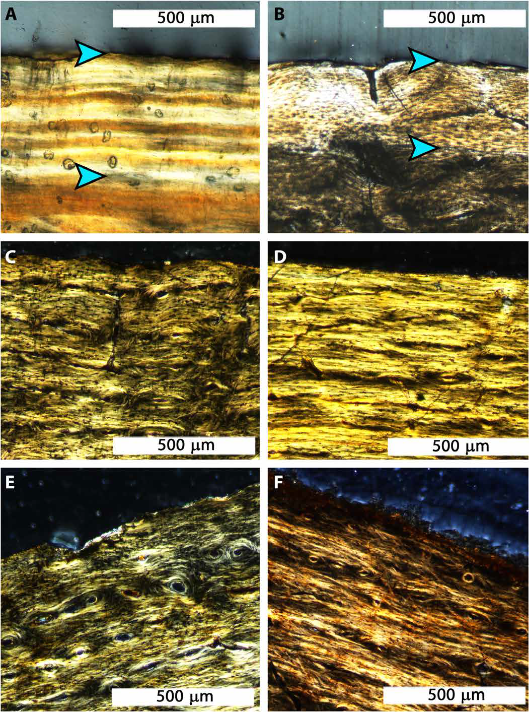
Rather than exhibiting an external fundamental system, a woven-parallel complex extends to the periosteal surface in both Tyrannosaurid specimens. Thus, histology supports morphological observations that BMRP 2002.4.1 and BMRP 2006.4.4 were skeletally immature individuals at death. In lieu of epiphyseal fusion, which most Reptile taxa lack, an EFS is the only way to conclusively confirm attainment of asymptotic adult body length from the long bones of a Vertebrate. When present, the external fundamental system occupies the periosteal surface as either closely spaced lines of arrested growth (separated by micrometers) or as a thick, primarily avascular annulus. Cyclical growth marks close to the periosteal surface can sometimes be mistaken for an external fundamental system. In the case of BMRP 2006.4.4, an annulus is present at the periosteal surface of both the femur and tibia, but when the annulus is followed around the cortex, in both cases it becomes embedded within the outer cortex and superseded by woven primary tissue. The proximity of the annulus to the periosteal surface instead suggests that BMRP 2006.4.4 died soon after growth resumed following the annual hiatus and that cortical osteogenesis was directional.

The presence of an external fundamental system at the periosteal surface of a long bone indicates skeletal maturity, while the absence of an external fundamental system indicates that the bone is still growing at the time of death. (A) An external fundamental system composed of tightly stacked birefringent lines of arrested growth (between blue arrowheads) at the periosteal surface of an Alligator, Alligator mississippiensis. (B) The external fundamental system (between blue arrowheads) in an Ostrich, Struthio camelus, is made of nearly avascular, birefringent parallel-fibered to lamellar primary tissue. (C) No external fundamental system is present at the periosteal surface of the femur of BMRP 2002.4.1, (D) the tibia of BMRP 2002.4.1, (E) the femur of BMRP 2006.4.4, or (F) the tibia of BMRP 2006.4.4. All panels are shown in transverse thin section, with circularly polarised light. Woodward et al. (2020).
On the basis of femur cyclical growth mark count, BMRP 2002.4.1 was over 13 years old at death, which is 2 years older than the original estimate based on fibula cyclical growth mark count. The slightly larger BMRP 2006.4.4 was over 15 years old. The number of cyclical growth marks missing due to medullary expansion is unknown, precluding an exact age at death for BMRP 2002.4.1 and BMRP 2006.4.4. Although the number of missing cyclical growth marks could be predicted on the basis of innermost zonal thicknesses and a process of retrocalculation, the variable spacing between cyclical growth marks observed in BMRP 2002.4.1 and BMRP 2006.4.4 and other Tyrannosaurs renders the technique unreliable in this case, and it was not attempted.
Within the innermost cortex of BMRP 2006.4.4, there is a tight stacking of six cyclical growth marks. Because the cyclical growth marks remain parallel about the cortex and do not merge, they either represent a single hiatus in which growth repeatedly ceased and resumed (totaling 13 years of growth) or up to 6 years where relatively little growth occurred annually (totaling up to 18 years of growth). This tight stacking of six cyclical growth marks is not observed in the femur of BMRP 2006.4.4, which preserves 15 cyclical growth marks. The cyclical growth mark count from the partial tibia of BMRP 2006.4.4 is questionable because the proximal sampling location away from midshaft incorporates the fibular crest, introducing associated regions of remodeling and directional growth affecting apposition interpretations. Because of this and their absence in the femur, the observed grouping of six cyclical growth marks is conservatively interpreted as a single hiatus event. Similar instances of a single hiatus represented by narrowly spaced lines of arrested growth are reported in other Tyrannosauroids. If this grouping of cyclical growth marks instead represents 6 years of protracted growth, then BMRP 2006.4.4 demonstrates the extent to which these individuals could adjust growth rate based on resource availability, in this case prolonging the ontogenetic duration of BMRP 2006.4.4 as a mid-sized carnivore.
Bone tissue organization was similar across femora and tibiae, suggesting that both bones record annual increases in body size equally well. If the stacked cyclical growth marks of BMRP 2006.4.4 reflect a single hiatus, then each femur preserved more cyclical growth marks than the associated tibia. Previous studies demonstrated that intraskeletal inconsistencies in cyclical growth mark counts are due to variable rates of medullary cavity expansion or cortical drift across elements when sampled at midshaft. Therefore, our preliminary assessment of Tyrannosaurus rex intraskeletal histology suggests that the femur is more informative than the tibia, despite regions of cortical remodeling from tendinous entheses about the cortex. Additional intraskeletal histoanalyses of Tyrannosaurid specimens are necessary to test whether the femur is the preferred weight-bearing bone for simultaneous assessments of annual growth rates and skeletochronology.
In addition to ontogenetic zonal thickness variability within the cortex, zonal thickness also changed with respect to cortical orientation. That is, zones were often much thinner relative to one another on one side of the transverse section and much thicker on another side. This pattern is particularly noticeable in the tibia of BMRP 2002.4.1 (medial cortical zones are thickest) and the femur of BMRP 2006.4.4 (posteromedial cortical zones are thickest). This observation implies that directional cortical growth occurred over ontogeny and stresses the necessity of complete transverse sections for histological analysis: Obtaining a fragment or core for study from one orientation may result in erroneous interpretations of growth rate and skeletal maturity.
Interpretations of relative maturity in nonavian Dinosaurs often rely on reported trends in the thickness of cortical zones between cyclical growth marks from the inner to the outer cortex. Zone thickness is typically greatest within the innermost cortex, corresponding to rapid annual growth early in life. Zones become progressively thinner in the mid-to the outer cortex of older individuals, as annual growth rate decreases approaching asymptotic body length. These general trends provide the interpretive foundation for the two previous histology-based ontogenetic studies on Tyrannosaurus growth. The spacing of cyclical growth marks within the outer cortices of BMRP 2002.4.1 and BMRP 2006.4.4 is narrower than between some cyclical growth marks deeper within the cortices, which suggests that, although not adults, the specimens were approaching a body length asymptote at about one-half the body length of FMNH PR 2081. However, annual zonal thicknesses between cyclical growth marks deeper within the cortices of BMRP 2002.4.1 and BMRP 2006.4.4 are variable, and zones do not consistently progress from widely spaced within the inner cortex to more closely spaced in the outer cortex. Because of unpredictable spacing within the cortex, reduced zonal thickness near the periosteal surface is likely an unreliable indicator of skeletal maturity in BMRP 2002.4.1 and BMRP 2006.4.4. Variable zonal thicknesses are, thus, likely to be observed in ontogenetically older Tyrannosaurus rex individuals. To test this hypothesis, we examined femur and tibia thin sections from Tyrannosaurus rex specimens USNM PAL 555000, MOR 1125, MOR 1128, MOR 1198, and CCM V33.1.15. In all individuals, variability in annual zonal thicknesses was observed. In particular, compared to zone spacing within the mid-cortex, noticeably thinner zones are present within the innermost cortex of USNM PAL 555000 and MOR 1128. These results contradict the mathematically predictable zonal spacing in Tyrannosaurus rex long bones reported in earlier studies, which used some of the same specimens reassessed by Woodward et al.. The results further suggest not only that BMRP 2002.4.1 and BMRP 2006.4.4 had not yet entered the accelerated growth period proposed for this taxon but also that the accuracy of the generalised Tyrannosaurus rex body mass curve proposed in 2004 would be affected by undetected individual variation in annual growth.

Examples of variable cyclical growth mark (blue lines) spacing in Tyrannosaurids examined by Woodward et al. (A) The variability of cyclical growth mark spacing in the femur of BMRP 2002.4.1 and (B) the tibia of BMRP 2006.4.4 may imply that these individuals were approaching asymptotic body length. However, cyclical growth marks within the innermost cortices of much larger Tyrannosaurus rex specimens (C) USNM PAL 555000 and (D) MOR 1128 demonstrate that the cyclical growth mark spacing is not a reliable indicator of relative maturity status. All panels are shown in transverse thin section. Woodward et al. (2020).
Variable lines of arrested growth spacing is reported in Ornithomimids, Ornithopods, and other Tyrannosauroids, and may correlate with annual resource abundance. Woodward et al.'s data suggest that this trait also characterizes Tyrannosaurus rex: Because the level of bone tissue organisation within zones remained the same from the innermost cortex to the periosteal surface in the BMRP specimens, growth rates were within a similar range from year to year. To produce these extremes in annual bone apposition, the duration of the growth hiatus must have varied annually. On the basis of the larger Tyrannosaurus rex specimens examined here for comparison, the adjustment of annual growth hiatus duration in response to resource abundance is a physiological characteristic observed throughout Tyrannosaurus rex ontogeny. Regardless of cause, unpredictable cyclical growth mark spacing observed by Woodward et al. and in previous studies stresses caution when inferring relative maturity based on cortical lines of arrested growth spacing. The observation of closely spaced cyclical growth marks within the innermost cortices of larger Tyrannosaurus rex validates our interpretation that the thin zonal spacing observed in the outermost cortices of BMRP 2002.4.1 and BMRP 2006.4.4 are not reliable indicators of relative maturity when an external fundamental system is absent.
The bone microstructural interpretations discussed here not only provide insight into Tyrannosaurus rex ontogeny but also have bearing on discussions concerning CMNH 7541 and Nanotyrannus. CMNH 7541 consists of a small isolated skull 572 mm in length. Inferred to be sympatric with Tyrannosaurus rex, it was originally named Gorgosaurus lancensis. In 1988 CMNH 7541 was redescribed as an adult specimen of a new genus, Nanotyrannus. Using an extensive empirical dataset, Nanotyrannus was formally synonymised into Tyrannosaurus in 2004, supporting the interpretation of CMNH 7541 as a juvenile Tyrannosaurus rex. Presently, most Tyrannosaurid specialists consider CMNH 7541 and possible referred specimens to be juvenile Tyrannosaurus rex based on morphological skull features shared with those found in undisputed juvenile individuals of other Tyrannosaurid taxa. Nonetheless, several publications have since argued for the validity of Nanotyrannus based not only on morphological characters of the CMNH 7541 type skull but also on characters from the somewhat larger skull of BMRP 2002.4.1 (720 mm in length), which some researchers have assigned to Nanotyrannus based on shared morphological characters they consider adult autapomorphies of the taxon. Currently, BMRP 2002.4.1 is the only accessioned specimen with postcranial skeletal elements preserved that is specifically argued by proponents of Nanotyrannus as belonging to that genus. Because CMNH 7541 lacks the postcranial skeleton and proponents of Nanotyrannus refer BMRP 2002.4.1 to that taxon, the limb bone histology of BMRP 2002.4.1 (and additionally BMRP 2006.4.4) reveals the life history of CMNH 7541 by proxy.
Woodward et al. provide histological data that can be used to reject the hypothesis that Nanotyrannus was erected on the basis of a skeletally mature 'pygmy' individual, resulting in two remaining alternative hypotheses: (i) Nanotyrannus is a valid taxon, but the holotype and all currently referred specimens including BMRP 2002.4.1 and BMRP 2006.4.4 are immature, with no skeletally mature individuals yet known; and (ii) CMNH 7541, BMRP 2002.4.1, BMRP 2006.4.4, and other mid-sized Tyrannosaurid specimens collected from the HCF represent juvenile ontogenetic stages of Tyrannosaurus rex. Thus far, the femur and tibia of BMRP 2002.4.1 and BMRP 2006.4.4 are the only weightbearing bones of Upper Cretaceous Hell Creek Formation Tyrannosaurids described histologically from complete transverse sections, and these universally demonstrate features characteristic of actively growing juvenile Dinosaurs that had not yet entered an exponential phase of growth (as demonstrated by our new data identifying noticeably thinner zones within the innermost cortex of large-bodied Tyrannosaurus rex specimens such as USNM PAL 55500). On the basis of these data, the latter hypothesis is most parsimonious. Incorporating additional mid-sized Hell Creek Formation Tyrannosaurid specimens into this histology-based relative maturity assessment is necessary to further support or refute the parsimonious hypothesis.
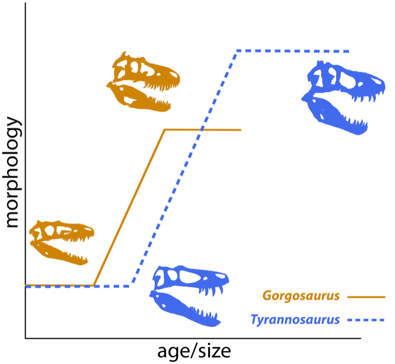
Synonymisation of Nanotyrannus with Tyrannosaurus rex means that rather than two sympatric tyrannosaurid taxa within faunal assemblages of the Hell Creek Formation, only one valid Tyrannosaur species, Tyrannosaurus rex, is currently recognised. As an adult, Tyrannosaurus rex occupied the large-sized carnivore niche in the latest Cretaceous Hell Creek Formation ecosystem, achieving an average adult body mass of about 9502 kg by 20 years of age. BMRP 2002.4.1 and BMRP 2006.4.4, at over 13 and over 15 years of age, respectively, were only half the length of an adult Tyrannosaurus rex. An earlier study obtained an averaged body mass estimate of 954 kg for BMRP 2002.4.1, which falls within the mid-sized Dinosaur body mass range of 50 to 1000 kg. Woodward et al.'s histological confirmation of BMRP 2002.4.1 and BMRP 2006.4.4 as mid-sized juveniles is therefore congruent with a hypothesized delayed onset of exponential growth in Tyrannosaurus rex relative to the ontogenetic timing of exponential growth in other Tyrannosaurids. Because Tyrannosaurus rex attained its great size late in ontogeny, many aspects of its biology likely differed between juvenile and adult individuals, leading to hypotheses that it used ontogenetic niche partitioning, where prey size is a function of body size. This feeding strategy is observed today in the extant Archosaur Alligator mississippiensis, which occupies different carnivore niches before and after achieving skeletal maturity. It has recently been demonstrated that although able to puncture bone, latestage juvenile Tyrannosaurus rex could not yet crush bone or engage in osteophagy, and therefore engaged in a feeding strategy distinct from adults.
Woodward et al.'s histological assessment of BMRP 2002.4.1 and BMRP 2006.4.4 provides data critical to understanding juvenile Tyrannosaurus rex biology and ecology, and additional evidence that there were no sympatric Tyrannosaurids in the Hell Creek Formation. Furthermore, they hypothesise that ontogenetic niche partitioning, coupled with an ability to adjust annual growth hiatus duration to track resource abundance, made Tyrannosaurus rex one of the most successful nonavian Theropods.
See also...