Commonly envisaged as a prelude to the Cambrian Explosion, the terminal interval of the Ediacaran Period (roughly 550–539 million years ago) chronicles several monumental events during the evolutionary dawn of Animal life. Among the most significant are the emergences of biomineralisation and active motility, which demarcate this interval from the rest of the Ediacaran Period. Toward the Period’s conclusion, the first Metazoan mass extinction event encompassed the downfall of the archetypal Ediacaran Biota. Their demise, however, was coincident with an ecological shift in which organisms such as
Cloudina and other occupants of this novel tube-building morphotype become increasingly populous. Collectively, these 'Cloudinomorphs' (to avoid conflating unresolved phylogenetic relationships with shared morphologies) were small, sessile, and epibenthic, but they appeared with several key adaptations that may have enhanced their chances for ecological success. These attributes include: (i) the advent of macroscopic biomineralization in the form of shelly external tubes, potentially serving as an impediment to predation; (ii) the establishment of gregarious habits that may signal the onset of Metazoan ecosystem engineering behaviors; and (iii) the development of enhanced larval dispersal mechanisms, and presumably both sexual and asexual reproductive habits, versus stolon-like reproductive modes of some members of the enigmatic softbodied 'Ediacara Biota'. These compounded ecological innovations may have helped to place the Cloudinomorphs as central players in ushering in a phase of fundamental ecosystem reform and increased trophic complexity. Although its cause is equivocal at present, the changing of the ecological guard from largely sedentary Ediacara-type communities to much more dynamic syn-'Cambrian Explosion' ecosystems was well underway in the terminal Ediacaran. Indeed, as recently proposed, this interval possibly displays an even larger step-change in organismal and ecological complexity than at the Ediacaran–Cambrian boundary itself. Nonetheless, the most crucial task that remains is to untangle the potential relationships between the organisms of the Ediacaran Period and those well-defined as metazoans in the Cambrian Period. The Cloudinomorphs are one of the few groups known to span the Ediacaran–Cambrian boundary and thus understanding their phylogenetic position is key to unraveling the evolutionary and ecological relationships between the seemingly disparate biomes of the Ediacaran and Cambrian Periods.
In a
paper published in the journal
Nature Communications on 10 January 2020,
James Schiffbauer and
Tara Selly of the
Department of Geological Sciences and
X-ray Microanalysis Core at the
University of Missouri,
Sarah Jacquet, also of the Department of Geological Sciences at the University of Missouri,
Rachel Merz of the
Biology Department at
Swarthmore College,
Lyle Nelson of the
Department of Earth and Planetary Sciences at
Johns Hopkins University, Michael Strange of the
Department of Geoscience at the
University of Nevada, Las Vegas,
Yaoping Cai of the Shaanxi Key Laboratory of Early Life and Environment,
State Key Laboratory of Continental Dynamics, and
Department of Geology at
Northwest University, and
Emily Smith, also of the Department of Earth and Planetary Sciences at Johns Hopkins University, provide a detailed report of internal soft-tissue preservation within Cloudinomorph fossils from the Wood Canyon Formation of Nye County, Nevada, and, moreover, one of the earliest reports of preserved internal anatomical structures in the fossil record. On the basis of the morphology and interpreted physiology of this soft-tissue structure, Schiffbauer
et al. suggest that this feature holds significant potential to shed new light on the phylogenetic placement of the Cloudinomorphs.
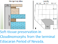
Generalized stratigraphy of the Montgomery Mountains site. Ediacaran–Cambrian boundary denoted by the presence of Treptichnus pedum. Cloudinomorphs recovered from silty-shale below first dolostone marker bed in the lower member of the Wood Canyon Formation. Nye County indicated on map, with yellow star marking approximate sample locality. ZQ, Zabriskie Quartzite; Stirling Qtz, Stirling Quartzite. Schiffbaur et al. (2020).
The phylogenetic position of the Cloudinomorphs has yet remained unresolved, albeit not without effort. Previous attempts have used the only available information (to date) from the fossil record; their external tubes. Such features, specifically demonstrated by
Cloudina, that have been employed to help constrain their phylogeny include (but are not limited to): (i) nested funnel-in-funnel tube construction; (ii) smooth inner tube wall lumen; (iii) presence of daughter-tube branching; iv) ovate tube cross-sections; (v) bulbous shape of the closed posterior bases; (vi) absence of basal attachment structures; (vii) calcareous composition in mineralized representatives; and (viii) microgranular tube wall ultrastructures. There are several caveats that should be considered, however. First and foremost, some of these features are not uniformly representative across all of the Cloudinomorphs, which should serve as a caution toward future attempts to resolve relationships within this morphotypic group. Moreover, at least some of these alleged diagnostic features (or lack thereof) may be taphonomic noise rather than primary biological signal. For instance, although a homogenous microgranular tube ultrastructure is commonly reported for
Cloudina, lamellar construction has also been observed, raising the question as to the influence of diagenetic recrystallisation on retention of primary ultrastructure, or, for that matter, original composition. It thus follows that the degree of tube wall biomineralisation in addition to the original biomineral chemistry has been met with differing interpretations, likely compounded by varying preservational and diagenetic histories between localities. The absence of substrate attachment structures may be a consequence of displacement and transport during storm events, a common mode of deposition of Cloudinomorphs that yields fragmental tubes in detrital hash resembling biohermal or reefal buildups. Alternatively, if the attachment structures were originally soft tissue, they may have been taphonomically lost, in which case the absence of evidence should not be construed as evidence for absence. Ovate crosssections may result from compression of a modestly flexible tube during sediment compaction, which is almost certainly the case for tubes of some Cloudinomorph taxa that are interpreted to have been originally organic. As such, and as should be the case with all enigmatic fossils, attempts at phylogenetic assessment would be best suited to focus on taphonomically robust features or those that can be best determined to be biologically and taxonomically informative.
In conjunction with the influences of a complex and wide array of taphonomic histories, placement of the Cloudinomorphs is further confounded when we consider the diversity of modern tube-building organisms and their assumed convergence of tubedwelling habits. Most agree that the Cloudinomorphs are at least of 'lower; (phylogenetically earlier branching) Diploblastic Metazoan-grade organisation. However, differences in the value with which the aforementioned characteristics are weighted in comparison with polyphyletic modern tube-builders can yield a broad assortment of plausible affinities, ranging from Chlorophytes (Green Algae) to Triploblastic Metazoans (the group which includes all Animals other than Sponges, Ctenaphores, Cnidarians, Placazoans, and Flatworms). Although more antiquated interpretations have included presumably Poriferan-grade Archaeocyathids, recent discussion urged not to discount a Macroalgal affinity, owing to comparable annulated tubular morphologies observed in modern calcareous Dasyclad Algae. Extinct Microconchid Lophophorates have also been offered as a possible analog on the basis of tube structure and shape. Similarly, some PterobranchHemichordates produce dichotomous organic-walled tubes with reasonably comparable morphologies, and thus may also warrant consideration. Other authors have instead refused to wedge the Cloudinomorphs into any extant or extinct group, proposing otherwise that they occupy their own
incertae sedis stem-metazoan family, the Cloudinidae. Satisfying the perceived majority of their exterior tube characteristics, however, most researchers currently fall into either Anthozoan Cnidarian or Polychaete Annelid camps, but further distinction has been hindered by the absence of preserved soft tissues.
The Wood Canyon fossil assemblage is dominated by Cloudinomorphic forms These fossils, as well as others from nearby units, have been taxonomically compared with the well-studied tubular fauna of the Gaojiashan Lagerstätte, South China and, more recently, to lesser-known Cloudinomorphs from the East European Platform. Systematic investigation of the Wood Canyon Cloudinomorph fossils has thus far formally described two new species,
Saarina hagadorni and
Costatubus bibendi, as the most abundant in this locality. Taphonomically, the Wood Canyon and Gaojiashan assemblages are highly comparable, with fossils from both units predominantly exhibiting three-dimensional pyritization. However, whereas the majority of Cloudinomorph tubes from the Gaojiashan are completely pyritised (e.g., the full tube volume is filled by pyrite mineralisation), those from Nevada show pyritised external tube walls retaining three-dimensionality but without pervasive pyrite infilling. As a result, the Nevadan Cloudinomorphs offer a unique potential for capturing resolvable soft tissues, and x-ray tomographic microscopy provides an ideal method for non-invasive exploration of internal fossil features.

Wood Canyon Cloudinomorphs of the Montgomery Mountains site. (a) Holotype of Saarina hagadorni, sample USNM-E1636_009_B13. (b) Paratype of Saarina hagadorni, sample USNM-WCF_005_01. (c) Holotype of Costatubus bibendi, sample USNM-MS_DS_12. Samples reposited at the Smithsonian Institution. All scale bars are 1 mm. Schiffbaur et al. (2020).
Unlike some of the Cloudinomorphs that built more robust shelly tubes, the exterior tubes of the described Wood Canyon Cloudinomorphs are inferred to have been organic in original composition from indications of plastic deformation much like the Gaojiashan taxon
Conotubus and East European representatives of
Saarina. Generic and specific taxonomic identification of the Nevadan tubular fossils containing soft tissues is unfortunately muddied by a lack of substantive exterior tube detail, likely resulting from chemical limitation during preservation. The soft-tissue-bearing tubular fossils exhibit exterior tube diameters (approximately 2–4 mm) that generally fall within the observed range for the two described Wood Canyon Cloudinomorph genera (maximum diameters 3.92mm and 6.36mm for
Saarina hagadorni and
Costatubus bibendi, respectively), albeit greater than the median diameter for either genus (median diameters 0.74mm and 1.09 mm for
Saarina hagadorni and
Costatubus bibendi, respectively). Schiffbaur
et al. interpret the annulation of the tubes observed both optically and by x-ray tomographic microscopy as a vestige of a 'funnel-in-funnel' tube construction, which supports the hypothesis of their Cloudinomorphic affinities.
Soft tissue-bearing Cloudinomorphs with schematic interpretation. 3D volume render from x-ray tomographic data shown in left image per frame (red-to-orange coloration indicates high density internal regions within exterior tube), with interpretive diagram in right image per frame. Examples here show (a) medial position and consistency (sample USNM-N1601_FL_018), (b) partial degradation/fragmentation (sample USNM-E1630_006), and (c) kinking and folding (sample USNM-N1601_FL_017). Soft tissue in sketches highlighted in red. Samples reposited at the Smithsonian Institution. All scale bars are 2 mm, sketches by Stacy Turpin Cheavens. Schiffbaur et al. (2020).
From three-dimensional reconstructions of x-ray tomographic data, internal structures were revealed within the external tubes from a smallsubset of the analysed specimens (roughly 11%; 4 of 35 analysed specimens), which Schiffbaur
et al. interpret as preserved soft tissues. The soft-tissue feature manifests as a sub-millimetric to millimetric diameter, centrally positioned cylinder that largely follows the curvature of the sagittal external tube length. In three of four cases, the cylindrical feature is mostly continuous through nearly the full length of the external tube, and only fragmented taphonomically. One of these specimens shows significant kinking and sinuous bending of the internal cylinder relative to its external tube. The other specimen shows instead an incomplete internal cylinder broken at a fragmented section of the external tube and also assumed to be unpreserved at the apical/posterior end of the external tube. When viewing the x-ray tomographic data transversely to the tube length, the internal cylinder rests adjacent to the lower (with respect to bedding) internal surface of the tube wall.
3D reconstruction full movie of sample USNM-WCF_001. Movie progression: (1) sequential 2D tomograph slices moving through the host rock and fossil, bright regions indicate pyrite tube wall and gut. (2) Layered dissolve of host rock, revealing Avizo-segmented data. Red color = tube wall and disseminated pyrite. (3) Removal of disseminated and non-continuous pyrite around the tube wall structure. (4) Rotation and reveal of gut structure (gold/orange). Hints of tube wall funnel/transverse annulation structure can be observed during rotation. (5) Dissolution of tube wall for full gut reveal. Schiffbaur et al. (2020).
To better explore the transverse morphology and preservation of the internal cylinders, a portion of the fragmented specimen was selected for destructive preparation (via manual serial grinding) and subsequent scanning electron microscopic analyses. The sectioned soft-tissue cylinder was observed to be either infilled by sediment or fully mineralised, and verified to be pyritic in composition. The external tube was additionally confirmed to have been pyritised (mostly weathered to iron oxyhydroxides), within a fine-grained siliciclastic host rock matrix. In cross-sectional view, the external tube can be complete, but appears more robustly pyritised at the bottom edge, and tenuous at the upper edge, with respect to bedding. Where the interior tube directly abuts the exterior tube, the exterior tube may be very thin, but this appears to be a localized phenomenon and is not apparent in all of the x-ray tomographic- or scanning electron microscope-observed transverse cross-sections. The exterior tube shows marginal lateral compression. In portions where the internal cylinder is broadly sediment-filled, it displays ovate cross-sections comparable in shape to the compressed external tube. In some of these sediment-filled portions, pyrite does exist within the interior of the tube, potentially replicating an organic template. Portions of the internal cylinder that are fully mineralized, in contrast, show circular, uncompressed cross-sections. Where the internal cylinder is sediment-filled, pyrite mineralisation appears to extend both inward (towards the cylinder interior) and outward (into the lumen of the external tube) from a discernable cylinder wall.

Optical imaging and x-ray tomography of Cloudinomorph pyritised tube and soft tissue. (a) Light image of entire specimen (sample USNM-WCF_001) in planview, specimen partially obscured at rock surface. (b) Corresponding 3D volume render, showing soft tissue (orange) and tube wall (gray); boxes (d), (e) are marked in both (a), and (b) to help guide slight differences in orientation. (c) Close-up view of labeled box in (a), highlighting funnel rims (arrows) on external tube. (d) Close-up view of labeled boxes in (a), (b), 3D volume render showing partial soft tissue and funnel rims (arrows); (d) largely overlaps with (c), but includes also host rock encased portion of the fossil. (e) Partial soft tissue from labeled boxes in (a), (b), (f). Cross-sectional view of e showing relative position of soft tissue that has settled to the bottom of the external tube wall. Sample reposited at the Smithsonian Institution. All scale bars 2 mm. Schiffbaur et al. (2020).
Each case of soft-tissue preservation presents a balance between taphonomically constructive and destructive processes, wherein retention and replication of biological information necessitates that decay does not eradicate, and mineralisation does not overwrite, informative features. Impeding both decay and mineralization early in the taphonomic sequence of the Nevadan Cloudinomorphs created a 'goldilocks' scenario in which soft tissues may be distinguishably preserved, as opposed to their Gaojiashan contemporaries. Pyritisation proceeds because of a confluence of chemical and microbiological factors, including: (i) a limited source of organic material (usually the soft tissues of the deceased organism); (ii) focused degradation of that organic material by sulphate-reducing Bacteria; and (iii) anoxic pore waters rich in reduced iron along with available sulphate. While oxidizing the remnant organic material of the organism, sulphate-reducing Bacteria (in normal seawater pH) convert sulphate to bisulphide, which then serves as one of the building blocks of pyrite along with reduced iron as the other.
2D stacked tomograph, latitudinal cross-section slices through sample USNM-WCF_001. Movie progresses from upper part to lower part. Schiffbaur et al. (2020).
If any part of this process becomes chemically starved, fossil pyritisation will be halted. There are three paths that this can take, based on limitation of either organic matter, reduced iron, or sulphate. If Bacterial sulphate reduction proceeds uninhibited by sulphate availability, the organics of the decaying organism are likely to be entirely consumed. This process, limited only by the availability of organics, would leave no soft tissues to be preserved, and should result in authigenic, centripetal pyrite infilling. In the other two cases, pyritisation can cease relatively early in the taphonomic sequence once the burial environment becomes chemically limiting (assuming no replenishment). If the availability of reduced iron is limited, pyrite formation will discontinue, but further degradation of the organics by sulphate reducers could continue unrestricted. Where sulphate concentration is instead limited, decay by sulphate-reducing Bacteria would cease once the sulfate supply is expended. In turn, with no further generation of bisulphide, pyrite formation would be subsequently suspended once the available bisulphide is exhausted. Regardless which pathway is realized in the Wood Canyon burial environment, the necessary ingredient to preserve these soft tissues, and have them remain perceivable, is to terminate pyritisation before overgrowth can obscure or homogenise the features.

Cross-sectional morphology of preserved Cloudinomorph soft tissue. Cross-sections revealed by serial grinding of specimen USNM-WCF_001; portion of the fossil chosen for grinding shown. (a)–(e) Light and SEM images matched with approximately equivalent μCT tomographic slices (differences in obliquity imposed during serial grinding). Far right in a shows tube and gut pyritisation via EDS elemental mapping. (f) Position of slices (a)–(e) shown on μCT tomographic slice through the transverse plane. All scale bars 2 mm. Schiffbaur et al. (2020).
In the Gaojiashan, pyritisation likely proceeded uninhibited by sulphate or reduced iron. Thus, even though the external tube morphology may be faithfully replicated in this assemblage, any internal structures were homogenised or obliterated by the combination of continued decay and mineralisation. Conversely, we infer that pyritisation of the Nevadan Cloudinomorphs was abbreviated early in the taphonomic sequence by sulphate or reduced iron limitation. To briefly summarise taphonomy in the Wood Canyon: (i) The initial burial event emplaced the Cloudinomorphs within the sulphate reduction zone of the sediment (oriented prone to bedding, whether or not this was their in-vivo position). (ii) Decay by sulphate-reducing Bacteria commenced, producing bisulphide that initiated pyrite mineralisation. (iii) In a significantly sulphate-restricted local environment (with no sulphate replenishment), we infer that the rate of Bacterial sulphate reduction may have also been diminished once sulphate concentrations dropped below rate-independent levels. With tempered Bacterially mediated decay, the earliest stages of mineralisation focused on the two most histologically suitable loci for pyrite nucleation, the robust organic walls of the exterior tube and the presumably more labile internal soft-tissue cylinder. Schiffbaur
et al. suggest that pyrite mineralisation of the external tube and internal cylinder occurred nearly simultaneously, as evidenced by the observed similarity in their compressed, ovate cross-sections from sediment compaction. (iv) Once structural integrity of supporting soft tissues was compromised through decay, the pyritizing soft tissues gravitationally settled to the imposed bottom of the external tube. Thus, both the ventral positioning of the internal cylinders within the recumbent external tubes and the distinction between bedding-respective dorsal and ventral coherency of exterior tube pyritisation (or perhaps ventral-inward pyrite infilling) serve as geopetal indicators. The gravitational slumping of the decaying soft tissue within the tube, as oriented recumbently, would have increased the distance for diffusion of bisulphide toward the upward-positioned wall of the exterior tube. If the reduced iron concentration was high in the burial setting, pyritisation would have therefore been focused more towards the decaying soft tissues, resulting in the observed preservational pattern. The kinked soft tissue observed in sample USNM-N1601_FL_017 may present a slightly different scenario, wherein the organism had died and slumped within its external tube prior to burial positioning or repositioning. And (v), either early sulphate exhaustion caused microbial decay by sulphate reducers to cease, or reduced iron was expended in the burial environment, thus halting continued pyritisation. The former chemical limitation may be more realistic. That is, if local sulphate concentrations instead remained sufficient to fuel continued (and less rate-restricted) bacterial sulphate reduction, it is probable that all of the soft tissues of the tube-dweller, including the internal cylindrical structure, would have been more rapidly exhausted. This taphonomic scenario likely would have yielded preservation of the exterior tube with more substantive detail, but leaving no soft tissues to be preserved. Schiffbaur
et al. suggest that this is likely the norm for the majority of the specimens recovered from the Wood Canyon Formation.
2D stacked tomograph, longitudinal cross-section slicesthrough sample USNM-WCF_001. Sample in the reverse orientation (180°) as that from above. Schiffbaur et al. (2020).
In order to provide an improved phylogenetic resolution on the Cloudinomorphs, Schiffbaur
et al. first consider which soft tissues are most likely to fossilise. Although they may be rare, there is no shortage of preserved internal soft-tissue structures reported from the fossil record. Fossilised internal soft tissues in the Ediacaran are limited to one possible occurrence of a muscular Cnidarian; on the other hand, Cambrian examples are much more numerous and diverse, including cardiovasculature, nervous and neurological tissues, musculature, and copious reports of digestive tracts. In Cambrian lagerstätten, guts are the most frequently preserved internal structures. Whereas fossil vasculature or nervous tissues are preserved as compressed or flattened features and musculature as bundled fibrous structures, fossil guts can reveal a broadly tubular nature where three-dimensionally preserved, and sometimes occur with the presence of associated digestive glands. Cambrian guts are typically preserved either as carbonaceous films, sediment infillings, or via phosphatisation, the latter of which is potentially reflective of the organism’s digestive physiology. However, there are limited (and perhaps contentious) examples of gut pyritisation as well as gut-content pyritisation. The consistent geopetal nature of the pyritised soft-tissue structures observed here supports the notion that they were originally centrally located structures in vivo, rather than adjacent to the exterior tube wall. At this stage, we can only speculate on the potential histological underpinnings that resulted in preferential pyritisation of these features. It is instead their cylindrical expression, propensity for preservation in Cambrian fossils, and consistent size, shape, and position within the external tube that most endorse a gut interpretation.

Additional detail of cross-sectional morphology. SEM backscattered electron micrographs (Z-contrast) of specimen USNM-WCF_001. Each row corresponds to a single slice at increasing magnifications from left to right, rows (a)–(c); dashed boxes in left and middle columns correspond to location for higher magnification images. Right-most frame in row (c) shows EDS elemental map of middle frame in row (c). Soft tissues in these slices are partially pyrite-infilled, increasingly so from (a) to (c), though distinct sediment grains can be observed. Note also distinct soft-tissue wall boundaries, indicated by black arrows in higher magnification views. White arrows in higher magnification views of rows (a), (b) indicate inferred direction of pyrite precipitation from soft-tissue wall, centripetally toward the interior and centrifugally from the exterior. Scale bars 200 μm for left-most column, and 100 μm for middle and right most columns. Schiffbaur et al. (2020).
Despite being soft tissues, the tendency for gut tracts to be preserved is likely amplified by several factors. Not only can portions of the digestive tract in some organisms be lined with decay-resistant cuticle, but guts are also segregated environments hosting their own microbiome and ions sourced from microbial metabolisms and ingested contents at the time of death. Guts can thus be isolated and accentuated taphonomic vessels, providing ideal conditions for self-contained mineralization. As observed by Schiffbaur
et al., the presence of centripetally precipitated pyrite inward from an apparent soft-tissue cylinder wall suggests that their preservation did indeed proceed from the interior. The next key challenge is to identify, within reasonable Cloudinomorph assignments and from both morphological and taphonomic perspectives, which soft tissue structures, whether guts or otherwise, could conceivably leave comparably preserved cylindrical structures. Schiffbaur
et al. detail the two primary but debated assignments for the Cloudinomorphs, Cnidarians and Annelids, and offer supplemental treatment on other possibilities (Hemichordates and Phoronids).

Proposed taphonomic sequence of the Wood Canyon Cloudinomorph soft tissues. (a) Cloudinomorph in hypothesised life position. External soft tissue hypothesised, modeled after Siboglinid Polychaete. (b) Burial by rapid sedimentation and initiation of decay. Sediment begins to enter tube cavity. (c) Burial compaction of the outer tube from weight of overlying sediment. Early pyritisation begins on interior surface of external tube and on both interior and exterior surface of soft-tissue cylinder. (d) Continued pyritization of exterior tube and soft-tissue cylinder. Inset of soft-tissue cylinder wall showing both inward and outward framboidal pyrite growth. (e) Remaining soft tissue decays, leaving pyritised exterior tube and interior soft-tissue cylinder. Gravitational settling of pyritised internal cylinder adjacent to lower external tube boundary. Stacy Turpin Cheavens in Schiffbaur et al. (2020).
Cnidarians, and more specifically Anthozoans, have probably received the most attention as a logical affinity for the Cloudinomorphs. Similarities reported between morphological characters of Anthozoans and Cloudina have served to propagate a cnidarian interpretation through the literature. On the other hand, Anthozoan internal anatomy is markedly disparate from the cylindrical structures observed by Schiffbaur
et al. Cnidarians, regardless of class affiliation, are defined in part by the possession of a sac-like gastrovascular coelenteron; this simple two-way digestive system has a single orifice for the intake of food and expulsion of waste. Within the Anthozoans, the upper portion (the pharynx) can be broadly tubular, opening into a larger, mesentery-lined, and grossly tubular gastrovascular cavity with numerous outpocketings defined by septa, unlike anything observed herein. These numerous septa, which can be calcitic and thus easily preservable, provide structural support of the tubular pharynx and gastrovascular cavity, but such structures are not observed in any Cloudinomorphs.

Diagrammatic comparison of candidate taxa for Cloudinomorph affiliation. Sections of the tubes and body walls are removed to illustrate gut tracts (red). (a) Anthozoan coelenteron showing upper, tubular pharynx and lower, sac-like gastrovascular cavity with mesentery structure. (b) Polychaete Annelid with straight through-gut path. Stacy Turpin Cheavens in Schiffbaur et al. (2020).
Another possibility that should be considered is that our preserved soft tissues could represent the entire soft-tissue body, rather than an internal feature, of tube-dwelling Hydrozoan polyps. Although generally rare and somewhat contentious in the fossil record, Hydroid fossils have been reported dating back to the Cambrian. Many Hydrozoans live in colonial habits joined by an interconnected network of canals and exterior skeletal branches, for instance, perhaps akin to such modern calcareous examples as
Millepora Fire Corals. The Cloudinomorph tube construction is strikingly different from the densely porous tubes of the Fire Corals, but a more important distinction may be found in the pattern of tube branching. If a colonial Hydrozoan assignment were fitting for the broader Cloudinomorphs, one may expect branching to be more common than observed. Although single-tube branching is known in
Cloudina and presumed to indicate asexual budding behavior, it has not been observed in most other comparable tubiform Cloudinomorphs, such as those reported here from Nevada and elsewhere. At last, no indications of tentacles are found in the soft tissues reported by Schiffbaur
et al., which have been considered diagnostic characters in a rigorous evaluation of putative fossil Hydrozoans. Although this may pose concern for such an interpretation here, rapid taphonomic loss of tentacles has been shown to be likely. Nevertheless, granting that features of Cloudinomorph external tubes have been deduced to be very generally Cnidarian as compared to other plausible affinities, the straight, sagittally continuous soft tissues, whether guts or not, are difficult to reconcile in favor of such an affinity.
The combination of straight, cylindrical soft tissues, and external tube structures may designate Polychaete Annelid Worms as the most fitting phylogenetic position for the Cloudinomorphs. Not only do Annelid through-guts express simple cylindrical morphologies, but the external tubes of the tube-building Annelids are also at least structurally comparable to the Cloudinomorphs, contrary to previous assertions. For instance, one of the features that has been used as a primary argument against a Polychaete affinity is the presence of closed posterior tube ends. Closed ends are known from some posteriorly complete Cloudinomorphs, notably
Cloudina and
Conotubus; although other Cloudinomorphs, like
Saarina, may have had only partially closed or constricted posterior tube ends. This feature may therefore not be ubiquitous within the Cloudinomorphs without clear evidence for a closed basal tube end across all members. Perhaps more importantly, the previous claim that closed bases are absent in modern tube-dwelling Polychaetes is unsupported by zoological literature. For example, Siboglinids are known to have closed bases and many other tube-dwelling Polychaetes possess dedicated anatomical structures (ciliated fecal grooves) or other behavioral strategies to keep waste from accumulating in a closed posterior end of the tube. A second unsubstantiated argument is that Polychaete tubes are not composed of nested funnels, but such a tube construction is in fact found in Siboglinids like
Oasisia. Finally, the mode of asexual reproduction by budding as inferred from branching in
Cloudina tubes is sometimes thought to be more indicative of a Cnidarian affinity. Tube-dwelling Serpulids among other Polychaetes, however, are known to undergo comparable clonal reproduction, though not all cloudinomorphs, including those reported here Schiffbaur
et al., show evidence of external tube branching. The point here is not to invalidate a valuable character evaluation of
Cloudina, but instead to offer caution to its applicability to the broader Cloudinomorphs and limited comparisons with modern tube-dwelling Polychaetes. One previous study effectually compares morphological characters of
Cloudina to broad-stroke Cnidarians, but makes a comparison with tube-dwelling Polychaetes, which much more narrowly focuses on three sessile, tube-dwelling families, Sabellids, Serpulids, and Cirratulids. The choice of these families clearly results from their calcareous tube-building habits in relation to the tubes of
Cloudina, but information provided by the fossil record seems incompatible with such comparisons. The records of Sabellids and Serpulids extend only into the Carboniferous and Triassic, respectively, and the Cirratulids have a much younger appearance in the Oligocene, thus casting doubt on the appropriateness of these families as acceptable comparators.
Scanning electron microscope image of exterior tube structure of Oasisia alvinae, a modern funnel-in-funnel tube-building Siboglinid Polychaete. Scale bar is 500 μm. Schiffbaur et al. (2020).
The overarching phylogenetic systematics of the ecologically diverse Annelids is complicated and controversial. They can be generally divided by life mode and feeding strategies into two reciprocal monophyletic major clades, the Errantia (free moving, predatory forms) and the Sedentaria (sessile, tube-dwelling forms), but they additionally include five basally branching lineages (Oweniidae, Magelonidae, Chaetopteridae, Amphinomidae, and Sipuncula). The lowest branching of these are tube-dwellers, the Oweniids and Magelonids. Together, these two families form a monophyletic sister
group to the other Annelids, the Palaeoannelida, followed by the basally branching, tube-building Chaetopterids.
Generalized phylogenetic scenario for divergence of stem-Lophotrochozoa and stem-Annelida with gut shape noted. Schiffbaur et al. (2020).
Outside of the three previously targeted Sedentarian families, placing the Cloudinomorphs within any other specific Polychaete designation may still impose a chronological gap, albeit likely more reconcilable, between the terminal Ediacaran and the earliest fossil record of readily identifiable Polychaete tubes. The earliest potential examples of Polychaete tubes previously reported are indeed Cambrian in age, including organophosphatic Chaetopterid tubes (
Hyolithellus) from Greenland and calcareous tubes of
Coleoides and
Ladatheca from Newfoundland and England. Although it is important to note that a record of Polychaete tubes is ostensibly absent from exceptional Cambrian lagerstätten, such deposits do provide several plausible tube-free Annelid fossils, such as (among others) stem-Annelids from the Sirius Passet; Sipunculids, remarkably similar to recent examples, with preserved gut tracts from the Maotianshan Shale, and numerous Polychaetes from the Burgess Shale, most of which preserve gut tracts. Furthermore, moderate taphonomic survival of Annelid gut tracts has been demonstrated by decay experiments with Polychaetes. These fossils ultimately suggest the divergence of at least the basal-most Annelid branches (the Palaeoannelids and Chaetopterids) within the Cambrian Period. Schiffbaur
et al. thus advocate an expanded investigation of the diversity of unresolved but comparable tubiform fossils across the Ediacaran–Cambrian transition in an effort to help potentially connect these records.
Although not a common interpretation for Cloudinomorphs, the tubicolous and vermiform Pterobranch Hemichordates do show some tubular similarities with organic-walled representatives of the morphoclade and thus have been previously considered1. The robust tubes of the Pterobranchs have left a considerable fossil record extending to the early Cambrian. They have additionally shown soft-tissue preservation, with a single example from the Chengjiang Lagerstätte. While no taphonomic details were reported, these soft tissues are presumed to have been pyritised but compressed (e.g., two-dimensionally pyritised). Commonly colonial, the Pterobranchs are stalked zooids with U-shaped guts that live within collared tubes. Although their digestive tract may not fit with the cylindrical morphology observed by Schiffbaur
et al., their contractile stalks, on the other hand, may be a feasible non-gut interpretation; comparable in shape, position within the external tube, and with lengths that can extend through the entirety of the external tube. Pterobranch stalks, however, are densely muscular structures with a ventral nerve, and thus are reasonably difficult to reconcile with the sediment-infilled portions of the cylinders as observed by Schiffbaur
et al..
Colonial Pterobranch Hemichordates; zooid on right illustrates U-shaped gut path. Contractile stalk shown in tube cut-out below zooid on right, and contracted zooid shown on left. Stacy Turpin Cheavens in Schiffbaur et al. (2020).
It may also be appropriate to consider the sister class to the Pterobranchs, Enteropneust Hemichordates. The Acorn Worms are not tube-builders in the modern-day; although, with a few Cambrian tubicolous representatives, perhaps this was a more common life mode early in their evolutionary history. For instance, the well-known Burgess Shale fossil,
Margaretia dorus, originally assigned to the Green Algae, has recently been shown to be a tubular dwelling structure of the vermiform Enteropneust
Oesia disjuncta. As opposed to the U-shaped gut of the Pterobranchs, the Acorn Worms have an anterior mouth and posterior anus connected by a straight through-gut, which has been preserved in Cambrian representatives. These are decidedly more comparable to the cylinders reported by Schiffbaur
et al., and the lack of hepatic sacs would suggest an affinity with modern Harrimaniid Worms, also similar to Cambrian representatives. Their external tubes, however, may pose the most significant obstacle for such an assignment of the Cloudinomorphs. The reported Cambrian tubes are solely organic in composition and their construction can be quite distinct, like the ornately perforated and anteriorly enclosed tube of
Oesia.
Stylized Cambrian Enteropneust, showing Margaretia-like tube structure and straight through-gut path and reduced hepatic sacs. Stacy Turpin Cheavens in Schiffbaur et al. (2020).
Limited Cambrian examples of possible Phoronids have been reported. Much like the broader group Cloudinomorphs, these Horseshoe Worm fossils exhibited both ‘soft shell' and mineralised tubes, with calcareous tubes suggested as ancestral, counter to previous inferences regarding possible ancestral relationships in the Cloudinomorphs. Phoronids may also serve as the most reasonable extant analogue to the extinct Worm-like Tentaculitids, including the Microconchids, which have been offered as a potential interpretation for the Cloudinomorphs. Possible tabulae in
Cloudina from Spain have been utilized to support the Microconchid reconstruction, but these structures are tenuous within a heavily recrystallised, sparry calcite-replaced specimen and thus may not be the most biologically informative of features. With regard to their internal anatomy, Phoronids and Microconchids, as other Lophophorates, should have a digestive tract that follows a U-shaped path with a superiorly positioned mouth and anus, again distinct from the morphology of the straight cylinders observed by Schiffbaur
et al..
Phoronid with deep U-shaped gut path. Stacy Turpin Cheavens in Schiffbaur et al. (2020).
While a straight through-gut has typically been considered homologous in Bilaterians, recent discussion on Lophotrochozoan anatomical organization and evolution proposes instead that, in sessile forms, U-shaped guts may be the basal groundplan. This claim has roots in the Cambrian fossil record, with fossil U-shaped guts documented for instance in stem-Rhynchonelliform Brachiopods and Orthothecimorph Hyoliths. The ‘U-tube theory’ could imply that the Cloudinomorphs, if stem-Lophotrochozoans, would be expected to follow suit. There are, however, several caveats that may argue against this idea. Perhaps the most important of these stipulations is that not all sessile tube-dwellers possess a U-shaped gut. For instance, some Cambrian organisms like the problematic
Hyolithellus have been inferred to possess a straight through-gut and are interpreted to be most likely Annelid-grade, potentially similar to Chaetopterid Polychaetes. If indeed guts, the soft-tissue structures observed here show no evidence of following a U-shaped path, which may call into question either the ‘U-tube theory’ on ancestral U-shaped guts or the suggestion of a basal Lophotrochozoan position for the Cloudinomorphs.
The structure and ingested contents of fossil guts hold significant potential to be behaviorally and ecologically informative. For instance, the preservation of digestive glandular structure and recognisable prey items in the gut contents of Cambrian Ecdysozoans have been used as verification of a predatory or scavenging life mode. These simple cylindrical Cloudinomorph soft tissues, however, are lacking any detail of differentiation or compartmentalization—which is not necessarily problematic for a Polychaete interpretation. Portions of the soft-tissue cylinder that are fully mineralised, as well as other sections that show sediment infill, can both be resolved with a gut interpretation. First, regions of pyrite infilling of the Cloudinomorph guts may tentatively represent mineralisation of ingested, non-descript, organic detritus, similar to gut-content/ cololite pyritisation observed in Cambrian Trilobites. Alternatively, these internal gut structures may represent pyritised internal gut folds like typhlosoles, which are known to occur in Annelids, though the taphonomic resolution and three-dimensional continuity of these features is unfortunately poor. Second, if the observed simple morphology is biologically faithful, in conjunction with their posited sessile habit, then we may be able to deduce that the Cloudinomorphs were likely detritivorous and presumably deposit-to-suspension feeders. The flexibility in feeding behaviors of modern-day tube-dwelling Polychaetes may provide insight on the presence of sediment encased within these fossil soft tissues. Specifically,
Owenia and several Spionids are among species that can switch between suspension feeding and deposit feeding behaviors depending on external conditions. These organisms are normally suspension feeders in higher current flow, taking food from the water column with their tentacular palps. However, when water current is low and suspended food is unavailable, they tend to employ surface deposit feeding by placing their palps on the surface of the substrate, during which sediment is commonly ingested. This is not meant to suggest that other tube-dwellers could not have behaved similarly, but it is actualistic evidence provided directly by potential modern analogs. The potential feeding flexibility of the Cloudinomorphs adds diversity in Ediacaran feeding modes, for example, building on recent suggestions of macroscopic suspension feeding by
Ernietta and scavenging by motile Bilaterians.
To our knowledge, the structures reported herein are not only the first recognisable soft tissues in Cloudinomorphs, but also the oldest guts yet described in the fossil record. As such, the Wood Canyon tubular fossil assemblage has provided a unique view into early animal anatomy. Nonetheless, for at least the cautions listed throughout the discussion above, Schiffbaur
et al. choose to refrain from shoehorning the Cloudinomorphs into any explicit Polychaete family. However, it is the sum of their parts, including the external tube structure, internal soft tissues, and presumed behavioral considerations, that may best denote placement amongst the Annelida as the most plausible. The accord of sequencing-based phylogenies and the available fossil record indicates that stem-Annelids, regardless of whether they exhibited a tube-dwelling habit or not, had diverged by at least the Early Cambrian; and thus a placement of the terminal Ediacaran Cloudinomorphs within basal branches of the Annelids is very likely not unreasonable. If these structures are indeed guts, they are the earliest in the record, fortify a terminal Ediacaran presence of Bilaterians, demarcate the divergence of the Lophotrochozoa, and, perhaps, help to build a phylogenetic bridge across the Ediacaran–Cambrian boundary to the diversity of Annelids known from post-Cambrian Explosion lagerstätten. Nevertheless, when taken together, the novelties provided by the Cloudinomorphs in the terminal Ediacaran, including the advent of macroscopic biomineralisation, the establishment of plausible ecosystem engineering behaviors, the enhancement of larval dispersal mechanisms and sexual and asexual reproductive habits, plausibly novel feeding strategies, and direct soft-tissue evidence of a through-gut, signpost an immense ecological leap towards the rapid Metazoan diversification that transpired geologically soon after.
See also...


%20(1)%20(1).png)