Showing posts with label Burgess Shale. Show all posts

Friday, 4 June 2021

Understanding the role of sediment-gravity flows in the formation of the Cambrian Burgess Shale Lagerstätte.

The fossil record has been used to reconstruct a history of the evolution of life on Earth, but itself preserves a rather incomplete record. Almost all fossils are of biomineralised or other hard tissues, with only rare sites, known as Konservat Lagerstätten, preserving soft tissues or the bodies of organisms which lack hard tissues. Estimates of the preservation potential of modern organisms suggest that 30% of marine megafuana, and 80% of megafauna overall, would leave no fossil record in normal deposits. Much of our understanding of the emergence of modern Animals comes from Cambrian Konservat Lagerstätten, such as the Burgess Shale, where it is estimated that 86% of the preserved fauna would not be preserved under normal conditions. This makes understanding the processes which led to the creation of these deposits particularlt important. It is often assumed that the Burgess Shale represents the near-faithful preservation of an intact biological community, and these fossils have been used to reconstruct food webs and community structures, but it is unclear whether the conditions which led to the preservation of these fossils did indeed preserve a high fidelity impression of a living community, or created biases which we have not detected and have worked into our understanding of how these ancient communities worked, and thus how modern communities developed from those early, Cambrian examples.

The preservation of soft tissues in fossils is rare, and thus not always as readily understood by palaeontologists as the preservation of more familiar hard tissues. Studies of the decay of modern organisms can help palaeontologists understand these preserved soft tissue fossils, but only a limited number of such studies have ever been undertaken. In particular, there are very few studies of the post-mortem transportation of soft bodied organisms, and how this effects the preservation process. 

The Burgess Shale Lagerstätte is considered to be one of the most important fossil faunas known, producing a large range of soft bodied organisms from an outer shelf Middle Cambrian environment. Understanding rhe mechanisms that led organisms to be preserved here is a key to understanding the biodiversity of the original ecosystem, as well as those of the forty plus other Cambrian sites around the world which show 'Burgess Shale-type preservation'. However, there has been a long-standing debate about the role of sediment transportation in the preservation of the Burgiss Shale Fauna, with the sediments that host the fossils originally diagnosed as having been produced in dilute turbidity currents, in which organisms were transported and then buried, then to have been in situ organisms which were buried by the turbidites. More recently, the deposits have been re-interpreted to be mud-rich slurry flows, an interpretation which, if right, would have profound implications for any soft bodied organism caught up in them.

In a paper published in the journal Communications Earth & Environment on 2 June 2021, Orla Bath Enright of the School of the Environment, Geography, and Geosciences at the University of Portsmouth, and the Institute of Earth Sciences at the University of Lausanne, Nicholas Minter, also of the School of the Environment, Geography, and Geosciences at the University of Portsmouth, Esther Sumner of the National Oceanography Centre at the University of Southampton, and Gabriela Mángano and Luis Buatois of the Department of Geological Sciences at the University of Saskatchewan, present the results of a study which used flume experiments to understand how transportation would impact the degradation of samples of the King Ragworm, Alitta virens, a modern marine species lacking hard tissues.

Field observations of the Burgess Shale exposure at Walcott Quary in British Colombia revealed that the deposits are made up of a series of silt and clay beds, with 'floating' quartz grains which average 100–500 μm in size, but can reach up to 1000 μm. These beds tend to have scoured bases, and structures such as parallel laminations, which are considered to be indicative of sediment transport. Bath Enright et al. interpret these structures as having been laid down by transitional cohesive flows, with both turbulent and laminar characteristics.

Bath Enright et al. next developed an index of degradation for Alitta virens, using specimens decayed for 0, 24, or 48 hours, and then subjected to conditions similar to those they interpret for the deposition of the Burgess Shale in a flume tank, for 25, 225, or 900 minutes. As a control measure, specimens were subjected to static decay for similar periods of time. Specimens that were partially decayed prior to transport produced a variety of results, ranging from whole, but shrivelled, to an unsupported gut with fluid escape and a general flattening of the body. The specimens tended to decay more rapidly at their posterior ends and mid-section; pre-decayed and then transported specimens tended to be more damaged towards their posterior ends. 

Next, Bath Enright et al. examined 197 specimens of Polychaete Worms from the Burgess Shale, 154 specimens of Burgessochaeta, and 43 specimens of Canadia. Very few of these showed any sign of degradation, and few of those that did showed little preference in where this occurred, although in these the preference was for the posterior to be more decayed.

 
Increasing states of Polychaete degradation. Alitta virens (right) and comparable states in the fossil, Burgessochaeta (left). (A) State 1-complete Polychaete, entire body segment intact (ROM–64913). (B) State 2-damage towards the mid-section and posterior transforms into tangled remains caused by the combination of transport and decay. The body remains intact as one segment (ROM–64916). (C) State 3-remains of the trunk and setae. The body structure has deteriorated significantly (ROM–64914). (D) State 4-remains of loose setae are attached to minute segments of cuticle and jaw elements only are recovered (ROM–64915). Bath Enright et al. (2021).

Based upon the analysis of the flume tank specimens, and comparison to specimens from Walcott Quarry, Bath Enright et al. conclude that the specimens of the Burgess Shale could have been transported in quasi-laminar flows for more than 20 km before being burried. This long distance preservation in quasi-laminar flows implied in this is in contrast to the situation seen in turbulent flows, which are more widely studied, and in which the amount of damage seen tends to increase with the distance of transport. However, Bath Enright et al. also note that this is not the case for specimens heavily decayed before transportation.

From these observations, Bath Enright et al. conclude that the specimens of the Burgess Shale were mostly un-decayed before being caught in the flows. Many of the Burgess Shale Polychaetes are compressed, but have no preferred orientation, and are typically found within the beds, rather than at their tops or bottoms. This suggests that they were transported within the flow, rather than being buried by it. At the end of Bath Enright et al.'s experiments, the sediments remained in a soupy state for some time, and living Worms were rapidly able to escape. Assuming the same applied at in the Burgess Shale, then any Worms remaining in the sediment must have been dead. Interpretation of the oxidation state of the Burgess Shale is complex, but it has been suggested that the oxic/anoxic boundary happened at the sediment surface, and the deposits are free of signs of burrowing or other bioturbation that would be expected if Worms were living in the sediment.


Schematic flow reconstruction for the Walcott Quarry in the Burgess Shale. (A) Schematic Representation in which the laminar plug extends towards the base of the flow and changes to a transitional plug regime. A turbulent cloud of sediment is suspended in the water column above the plug flow. The soft-bodied organisms (labelled 1, 2, and 3) have been picked up along the flow path, potentially kilometres apart from one another. (B) Bed A from the Greater Phyllopod Bed of the Walcott Quarry. (C) Graphic log showing Bed A; soft-bodied organisms (1, 2, and 3) from the flow type above (A) will become mixed in the deposit. (D) Thin-section scan from Bed A showing parallel laminae, erosive, scoured bases, and 'floating' quartz grains (Q). White arrows indicate transitional cohesive flow deposits. Bath Enright et al. (2021).

Burgess Shale-type lagerstätten are traditionally viewed as ‘windows’ into the biology and ecology of past life and are used to reconstruct in-life communities. However, Bath Enright et al.'s  study casts doubt on the validity of this, suggesting that the sediments present may have been transported very long distances, potentially collecting organisms from a variety of habitats before finally settling. In particular the Cambrian Chengjiang and Qingjiang biotas of China, and Upper Ordovician Beecher’s Trilobite Bed of the USA are widely interpreted to been emplaced by flows, and might therefore also have interpreted organisms from multiple environments. 

Konservat Lagerstätten provide us with a great deal of information about past life, due to their unique preservation of soft tissues and soft bodied organisms. These deposits tell us a great deal of information about the anatomy of long dead organisms, and have helped us to understand the origins of many living groups of organisms. However, Bath Enright et al. caution against attempts to reconstruct ancient biological communities based upon these deposits, without clear indication that the fossils have been preserved in their life-environment.

See also...


Online courses in Palaeontology. 

Follow Sciency Thoughts on Facebook.

Follow Sciency Thoughts on Twitter


Sunday, 20 December 2020

Gyaltsenglossus senis: A Hemichordate from the Middle Cambrian Burgess Shale.

Hemichordate relationships remain contentious due to conflicting molecular results and the high degree of morphological disparity between the two Hemichordate classes, Enteropneusta and Pterobranchia. Additionally, Hemichordates have a poor fossil record outside of the Cambrian, with the exception of the collagenous tubes of the Pterobranchs (which include Graptolites). By the Middle Cambrian, tube-dwelling colonial Pterobranchs and tube-dwelling enteropneusts coexisted, supporting the origin of the Hemichordate body plan earlier in the Cambrian without clarifying the morphology of their last common ancestor.

In a paper published in the journal Current Biology on 2 November 2020, Karma Nanglu of the Department of Paleobiology at the Smithsonian National Museum of Natural History, Jean-Bernard Caron of the Department of Natural History at the Royal Ontario Museum, and the Department of Ecology and Evolutionary Biology and Department of Earth Sciences at the University of Toronto, and Christopher Cameron of the Departement de sciences biologiques at the Universite de Montreal, describe a new Hemichordate, based on 33 specimens from the 506-million-year-old Burgess Shale (Odaray Mountain, British Columbia).

The new species is named Gyaltsenglossus senis, where 'Gyaltsenglossus' derives from Gyaltsen (pronounced ‘Gen-zay’, emphasis on the first syllable) for Karma Nanglu’s father, who inspired his interest in biology as a child, and glossus from the Greek glossa, meaning tongue, a common generic suffix for Hemichordates, and 'senis' derives from the Latin senex, meaning old.

Gyaltsenglossus senis is described from 33 specimens (all at the Royal Ontario Museum), collected from Odaray Mountain, Yoho National Park, British Columbia, within the upper part of the ‘thick’ Stephen Formation (Odaray Shale Member). With the exception of the holotype specimen (ROMIP 65606.1), all the other specimens are either fragmentary or poorly preserved, and none show the tentacles or proboscis as clearly as the holotype specimen.

Gyaltsenglossus senis is vermiform, with a maximum length of approximately 2 cm. Body consists of an elongate, ovoid proboscis, a crown of six feeding arms, a cylindrical trunk, and a round posterior structure. The feeding arms possess roughly 15 pairs of symmetrical tentacles. Gut is straight, anus terminates immediately before the posterior structure.

From the anterior-most point of the oblong structure, interpreted as a proboscis to the posterior end of the fossil, Gyaltsenglossus is between 1.5 and 2 cm in length, although few specimens preserve the entire morphology clearly. The inside of the proboscis is preserved slightly three dimensionally, beginning at the medial base and following a slightly darker area and projecting forward roughly 40% of its total length. A thickened mass of tissues is present at the base of the proboscis, although it is not well preserved. Six feeding arms radiate from the dorsal area directly posterior to the proboscis. The arms are thin and roughly one-and-a-half times the length of the proboscis, with the most complete arm being ca. 1 cm long. They have a foliose appearance due to roughly 15 pairs of tentacles extending from each arm, although these tentacles are not clearly observable in most specimens. Only the most proximal portion of each tentacle, which adjoins the feeding arm, is preserved clearly on the holotype, often presenting as a sub-triangular structure. In most specimens, the feeding arms are either damaged or retracted such that they appear as a rounded mass, in some specimens, obscuring the proboscis. Posterior to the crown of feeding arms, a row of thin appendages projects from a small elevated platform. These thin appendages are considerably shorter than the feeding arms (about 0.25 cm), devoid of tentacles, and are most clearly observable on the holotype. The trunk is vermiform, roughly twice as wide at the anterior margin as at the posterior end. The trunk appears relatively rigid, with the most curvature observed in the posterior half. A dark, thin, axial structure is frequently preserved in the trunk, consistent with the size and position of a gut. A wider, more thickly walled area at the anterior-most region of the gut may represent a muscular pharynx. The gut does not appear to extend to the end of the body, but terminates before a posterior bulbous structure, which appears darkly preserved. This structure (width: length ratio ranging from 1:1 to 1.4:1) is usually wider than the posterior-most margin of the trunk. A darker band is frequently preserved at the posteriormost rounded margin of this structure and may represent the carbonaceous remnants of thickened tissues.
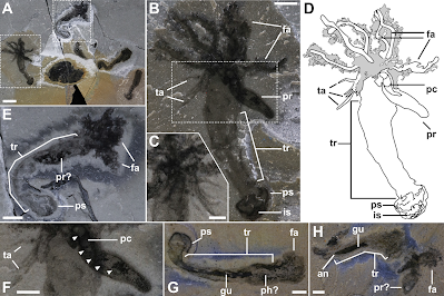
 
Morphology of Gyaltsenglossus senis from the Burgess Shale. (A) ROMIP 65606. Four specimens of Gyaltsenglossus senis preserved on the same surface (an additional three specimens are out of frame on the same surface). The two best-preserved specimens are in the dashed boxes. (B) Holotype (ROMIP 65606.1). Close-up of boxed specimen on the left in (A). Most complete specimen, showing full length of the proboscis, six feeding arms, a cluster of thin appendages located on the dorsal surface of the trunk, and the posterior structure with dark internal structures. (C) Close-up of the thin appendages on the counterpart of ROMIP 65606.1. The thin appendages are unlike the feeding arms; they are much shorter and entirely bare. (D) Line drawing of (B). (E) ROMIP 65606.2. Close-up of boxed specimen on right in (A). The individual arms cannot be clearly differentiated, but the tentacles surrounding them give the area a tufted appearance. A dark, oblong structure may be the proboscis recurved over the trunk, and the posterior structure is bulbous. (F) Close-up of boxed area in (B). The proboscis coelom is indicated by arrowheads. The thin appendages are filamentous and without tentacles. (G) ROMIP 65607.2. The arms appear retracted and cannot be clearly differentiated. The anterior portion of the gut appears more thickly walled and may represent a pharynx, but the internal organization is unclear. (H) ROMIP 65607.1. The terminal point of the gut is most clearly preserved in this specimen, ending before the posterior-most point of the animal. The feeding arms are significantly degraded, as is sub-triangular structure, which may be a decayed proboscis. Abbreviations: an, anus; fa, feeding arms; gu, gut; is, internal structures; ps, posterior structure; pc, proboscis coelom; ph?, possible pharynx; pr, proboscis; tr, trunk; ta, thin appendages. Scale bars (A) 5 mm; (B), (E), (G), and (H) 2 mm; (C) and (F) 1 mm. Nanglu et al. (2020).

Identification of a proboscis is based on three primary factors. First, it is highly similar in morphology to that of the extant Hemichordate class Enteropneusta, particularly to that of the genus Saccoglossus because it is significantly longer than wide (roughly 4:1). Second, it is the anterior-most structure within the tripartite, Hemichordate body plan of Gyaltsenglossus. Finally, it is preserved similarly to the proboscises of the Cambrian Hemichordates Spartobranchus and Oesia. In particular, it is preserved darkly in comparison with the rest of the body, presumably due to the density of muscle tissue that characterizes the Hemichordate proboscis.

 
Feeding Arms and Thin Appendages of Gyaltsenglossus senis. (A) Close-up of four feeding arms on the holotype specimen, ROMIP 65606. (B) Line drawing of (A). Only the base of the tentacles adjoining the feeding arm is usually visible. The arms and tentacles are surrounded by apparently degraded tissue, likely representing decayed tentacles. (C) ROMIP66000. A poorly preserved specimen, showing two thin appendages and degraded feeding arms. (D) Counterpart of (C), showing feeding arms more closely. Abbreviations: fa, feeding arms; ps, posterior structure; ta, thin appendages. Scale bars 2 mm. Naglu et al. (2020).

Although several external characters are well preserved, other features, such as the darker mass within the proboscis and the tissues basal to the proboscis, are less well preserved. These may represent remnants of the heart-kidney-stomochord complex and of the collar, respectively. Although not preserved, Naglu et al. infer the position of the mouth to be on the ventral side of the Animal between the base of the proboscis and the putative collar, as it is in all modern Hemichordates and as has been inferred in Cambrian Hemichordates previously. In addition, Naglu et al. find no evidence of gill bars or gill pores in any of the 33 specimens of Gyaltsenglossus observed. Such features, however, especially the gill pores, were likely affected by taphonomic bias. The gill bars are internal structures that may be obscured by the ectoderm in cases of pristine preservation. However, Naglu et al. do find abundant evidence of gill bars preserved in the similarly sized and approximately coeval Burgess Shale Hemichordates Spartobranchus tenuis and Oesia disjuncta, which share a common preservation pathway with Gyaltsenglossus. Additionally, the collagenous gill bars are the most decay-resistant feature of the Hemichordate body plan, making it unlikely that they would have been preferentially lost before soft tissue characters such as the proboscis. While the relatively small sample size of Gyaltsenglossus in comparison to Spartobranchus and Oesia does not preclude the possibility of finding new specimens with gill bars preserved, Naglu et al. consider this character to be absent in Gyaltsenglossus. With regard to ectodermal gill pores, these are among the Hemichordate features most susceptible to decay, and they have not been found in any Hemichordate fossils described thus far even though the gill bars of Spartobranchus and Oesia are clearly preserved; gill bars would presumably require gill pores to function. Gill pores are therefore regarded as equivocal in all Cambrian enteropneusts, including Gyaltsenglossus.

Another Hemichordate character that warrants additional discussion based on potential taphonomic biases is the collagenous tube, or tubarium. The tubarium is a feature of all Pterobranchs (except for the genus Atubaria), including Graptolites, where it is secreted by the cephalic shield, a homolog of the Enteropneust proboscis. Spartobranchus and Oesia, despite their Enteropneust-like gross morphology, also produced tubes, which Naglu consider to be homologs of the Pterobranch tubarium. Notably, only a subset of the total number of Worm specimens are found inside of a tube, and many tubes have been found lacking a Worm. A variety of factors may contribute to this phenomenon; for instance, the Animals may have abandoned or been dislodged from their tubes prior to fossilisation, or tube dwelling may have been facultative. Though it is possible that Gyaltsenglossus was tubicolous even though its tubes were not found, Naglu et al. consider the character of the tubarium as absent in their study.

Both Bayesian and strict consensus parsimony analyses resolve the Enteropneusta and Pterobranchia classes as reciprocally monophyletic (posterior probabilities of 0.91 and 1.0, respectively). This result is supported by the most recent molecular phylogenies, though it is not without debate. Spartobranchus and Oesia are resolved as stem-group Enteropneusts, while Gyaltsenglossus is resolved as a stem-group Hemichordate in Naglu et al.'s Bayesian analysis. Support for Gyaltsenglossus being a total-group Hemichordate is high (posterior probability of 0.96 in Naglu et al.'s Bayesian analysis), disproving an affinity with other tentaculate taxa included in these analyses, including Annelids, Cnidarians, and Echinoderms. However, support for its exact position within this phylum is lower (posterior probability of 0.56 in Naglu et al.'s Bayesian analysis, and found in a polytomy with Enteropneusts and Pterobranchs in Naglu et al.'s parsimony analysis, owing to its shared morphological characteristics with both Enteropneusta and Pterobranchia.

 
Ambulacrarian Phylogeny Incorporating Cambrian Fossils and a Schematic of Hemichordate Homologies (A) Phylogenetic position of Gyaltsenglossus senis and other Cambrian Ambulacrarians using majority-rules Bayesian analysis of 113 characters and 32 taxa. The tube-building Oesia and Spartobranchus are stem-Enteropneusts, while Gyaltsenglossus senis is stem to Hemichordata. Numbers at nodes represent posterior probabilities. (B) Homologous structures among fossil and extant Hemichordates. (i): 33 h larva of Rhabdopleura normani with homologous structures in the adult Rhabdopleurid body plan shown in (ii). The cells of the adhesive organ attach the larva to the benthos and develop into a stalk. (iii): Cephalodiscus. (iv): While Gyaltsenglossus senis has feeding arms derived from the collar, as in Rhabdopleura and Cephalodiscus, it lacks a U-shaped gut and distinctive cephalic shield, instead having an Enteropneust-like proboscis. (v): Oesia has an Enteropneust body plan, although with a unique and distinctive posterior bilobed attachment structure and gill bars that extend the length of the trunk. (vi): Spartobranchus has a Harrimaniid-like anatomy, with the exception of a pronounced post-anal bulbous structure. (vii): A 4-week-old juvenile of Saccoglossus horsti, with a distinct post-anal tail used for crawling and adhering to the benthos. Naglu et al. (2020).

The unique morphology and phylogenetic placement of Gyaltsenglossus suggest that paired feeding arms derived from the dorsal collar were present in the Hemichordate last common ancestor. As in modern Pterobranchs, the arms and tentacles were likely used to capture and transport suspended particles from the water column to the mouth. Feeding arms provide an advantage to exploit food sources in the water column by epibenthic organisms across a wide range of body sizes, from Ciliates to Crinoids. Notably, the long proboscis also suggests that Gyaltsenglossus was a deposit feeder like the extant Acorn Worm, Saccoglossus. Gyaltsenglossus provides the clearest example of the morphology of the Hemichordate last common ancestor and suggests that a bimodal feeding mechanism may have been an early feature of the phylum. In this context, feeding arms were lost in the lineage leading to Spartobranchus, Oesia, and the crown-group Enteropneusts, concomitant with a gradual specialisation away from tentaculate filter feeding and, eventually, toward infaunal niches.

While gill bars were likely present in the Hemichordate last common ancestor (and are indeed most likely to be a Deuterostome plesiomorphy), Naglu et al. found no evidence of them in the Gyaltsenglossus specimens studied. Computational fluid dynamics analyses of Hemichordate pharynx models showed that Pterobranchs, which also lack gill bars, may have abandoned their gill pores for feeding arms because very small gill pores impose a functional constraint on pharyngeal filter feeding as the energetic cost of pumping fluids becomes prohibitive. Gill pores appear to be non-functional in the Pterobranch Cephalodiscus and were lost in the diminutive Rhabdopleura. Gyaltsenglossus possibly represents a second example of gill loss in the Hemichordates, but the organism is sufficiently large that this energetic constraint would not have applied. Instead, pharyngeal filter feeding may have been simply redundant with the tentaculate suspension feeding and proboscis deposit feeding modes of Gyaltsenglossus, allowing for the loss of gill pores.

The gut of Gyaltsenglossus is straight like that of an Enteropneust, rather than U-shaped like that of a Pterobranch, and it ends just before the muscular post-anal attachment structure. A straight gut has been inferred in some stem-Echinoderms such as Ctenoimbricata, suggesting that it may be a plesiomorphic ambulacrarian trait. The bulbous structure at the posterior end of Gyaltsenglossus is consistent with the posterior attachment structures of the Burgess Shale Enteropneusts Spartobranchus tenuis and Oesia disjuncta. This bulbous structure appears to be a ubiquitous feature of Cambrian Hemichordates and is likely plesiomorphic for the phylum. Morphologically, it is most similar to that of Spartobranchus, being approximately round to ovoid and without the bifurcating attachment form that Oesia used to grasp its secreted tube. Considered in conjunction with its morphology and position, Naglu et al. hypothesise that Gyaltsenglossus was able to use this structure for attachment as an anchor to access the water column to feed on plankton and possibly for locomotion. The most comparable structure in extant Acorn Worms is the postanal tail of juvenile Harrimanids, which are glandular, ciliated, and used for attachment and locomotion. This tail was lost in derived crown-group Enteropneusts (Spengelids, Ptychoderids, and Torquaratorids), and is believed to be a homolog of the Pterobranch stalk. If this is true, then the Pterobranch stalk is an exaptation of the posterior bulbous structure seen in Gyaltsenglossus, Oesia, and Spartobranchus. This exaptation was a major event in Pterobranch evolution because it permitted coloniality, making possible the abundance of Graptolites that characterised Palaeozoic seas.

Artistic Reconstruction of Gyaltsenglossus senis. The image in the foreground shows Gyaltsenglossus as it would appear while motile. The background illustrates the posture of the animal while attached to the seafloor and suspension feeding. Emily Damstra in Naglu et al. (2020).

The arms of Pterobranchs, and presumably of Gyaltsenglossus, develop from paired extensions of the collar coeloms, or mesocoels. These mesocoels are homologous with those of Echinoderms, but only the left coelom (hydrocoel) is elaborated through metamorphosis of the living Echinoderm classes to become the water vascular system (and hyponeural ring sinus). The feeding arms of Crinoids, Asteroids, and Ophiuroids are then not homologous with the arms of Hemichordates, including Gyaltsenglossus. Support for this idea is the absence of feeding arms among the earliest diverging Echinoderm lineages, including the bilateral symmetric Ctenocystoids, which presumably would have had paired mesocoels. The thin appendages in Gyaltsenglossus have no clear homolog with any extant Hemichordate, although dorsal elaborations of the trunk are common in Acorn Worms (i.e. hepatic caeca and gonadal wings).

Further study of a number of putative fossil Ambulacrarians, including the Cambroernids Herpetogaster and Eldonia, may contribute to further disentangling of early Ambulacrarian evolution. However, their body plans are so diverse and bizarre that homologous characters are difficult to code, and their phylogenic positions remain uncertain. The impressive disparity of Cambrian body plans is thought to be due to a combination of open ecological niches to exploit and few developmental constraints, perhaps due to greater flexibility in early gene regulatory networks. While modern Hemichordates are not particularly diverse, Enteropneust and Pterobranch body plans are so different that the traditional literature largely regarded them as distant relatives. Gyaltsenglossus, representing a combination of both Enteropneust and Pterobranch morphologies, demonstrates starkly the unique insights the fossil record provides through direct evidence of long-extinct body plans.

See also...


Online courses in Palaeontology. 

Follow Sciency Thoughts on Facebook.

Follow Sciency Thoughts on Twitter.


Sunday, 12 February 2017

Ovatiovermis cribratus: A Luolishanid Lobopodian from the Middle Cambrian Burgess Shale Lagerstätte of British Columbia.

The Arthropods are grouped together with two other living phyla, the Onychophorans (Velvet Worms) and Tardgrades to form a higher group called the Panarthropods, a group of limbed organisms thought to have evolved from a single Worm-like ancestor. The early origins of these groups are less than clear however; a variety of other Panarthropod groups, such as the Anomalocarids, which may have been early Arthropods, and the Lobopodians, which are thought to have been ancestral to the modern Onychophorans.

In a paper published in the journal BMC Evolutionary Biology on 31 January 2017, Jean-Bernard Caron of the Department of Natural History (Palaeobiology Section) at the Royal Ontario Museum, and the Department of Ecology and Evolutionary Biology and Department of Earth Sciences at the University of Toronto, and Cédric Aria, also of the Department of Ecology and Evolutionary Biology at the University of Toronto, describe a new species of Lobopodian from the Middle Camrian Burgess Shale Lagerstätte of Walcott Quarry in British Columbia.

The new species is named Ovatiovermis cribratus, where 'Ovatiovermis' means 'ovation-Worm' in reference to the presumed life posture of the species, standing on its hindlimbs with its forelimbs waving in the air, and 'cribratus' means 'to sieve'. The species is described from two specimens, one 30 mm in length and the other 12 mm. The species shows diffentiation of the limbs with short hind limbs and longer forelimbs covered in elongate claws, which Caron and Aria interpret as having been used for filter feeding (standing on hindimbs and sieving the water with upraised forelimbs).

Ovatiovermis cribratus from the Burgess Shale, Royal Ontario Museum (ROM) 52707: (a–e, i, j) part, (g, h) counterpart, (f) reconstructed death pose. Close-ups indicated by white rectangles. (a, b) full specimen under direct (a) and polarized (b) lighting conditions. (c–e) superposed elemental maps of carbon (red) and calcium (purple) before preparation of the 8th left lobopod (lL8—see a). The lighter colours represent higher concentrations of elements: parts of the gut, proboscis, pharyngeal area and claws are preserved in carbon whereas the rest of the body is preserved in calcium. (g–j) details of the anterior part of the body showing internal organs in Lobopods (g), pair of visual organs (g, h), spinules and bifid claws (g, i, j). Digital single-lens reflex (DSLR) images taken using direct light (a, i, j) and cross-polarized light (b, g, h), all under dry conditions except (b, g and h). A, annulations; C, claw; Ds, dark stain; E, “eye” (visual organ); G, gut; H, head; I, internal organ; L, lobopod (l, left; r, right; x,y, lobopod position); Lu; foregut lumen; M, mouth; Pr, proboscis; S, spinules. Scale bars: 5 mm (a–c, g, i), 1 mm (e, d, h, j). Caron & Aria (2017).

Previous reconstructions of the phylogeny of Cambrian Lobopodians has shown the presence of two distinct groups, the armoured Hallucigenians and the unamoured Luolishanids, which show greater limb diferentiation and are thought to have been ancstral to the modern Velvet Worms. A phylogenetic reconstruction made by Caron and Aria including Ovatiovermis cribratus placed the new species firmly within the Luolishanid group, but also suggested that both groups in fact split off from the other Panarthropods before the split between Tardigrades, Velvet Worms and Arthropods, suggesting the common ancestor of these groups was in fact Lobopodian-like.

Artistic interpretation of Ovatiovermis cribratus as a living animal. Danielle Dufault in  Caron & Aria (2017).

The closet relatives of the Panarthropods are thought to be the Nematode, Priapulid and Palaeoscorid Worms, with the common ancestor of the Panarthropods thought to be something similar to Paucipodia, a Loboopd from the Early Cambrian Chengjiang Lagerstätte of China which resembles a Nematode with legs. However, while Caron and Aria's phylogeny did recover Paucipodia as close to the common ancestor of the living Arthopods, Velvet Worms and Tardigrades, it also suggests that the Luolishanids split from the other groups before the Hallucigenians, and that the Hallucigenians split off before Paucipodia, making the biology of the earliest Panarthropodian ancestor more uncertain than before.

 Consensus tree of a Bayesian phylogenetic analysis of our morphological matrix composed of 38 taxa and 59 characters (four runs, 10,000,000 generations, burn-in fraction: 0.20). Total-group Onychophorans highlighted in green, Hallucigeniids in blue and Luolishaniids in red. Microdictyon and Facivermis are considered potentially included within Hallucigeniids and Luolishaniids, respectively, which is denoted by the coloured dashed lines. Numbers above branches represent posterior probabilities. Caron & Aria (2017).

See also...

http://sciencythoughts.blogspot.co.uk/2017/01/preserved-trilobte-eggs-from-ordovician.htmlhttp://sciencythoughts.blogspot.co.uk/2016/11/mimetaster-florestaensis-new-species-of.html
http://sciencythoughts.blogspot.co.uk/2016/10/utahcaris-orion-and-origin-of.htmlhttp://sciencythoughts.blogspot.co.uk/2016/10/antennipatus-montceauensis-velvet-worm.html
http://sciencythoughts.blogspot.co.uk/2016/05/dyrnwynia-conollyi-new-species-of.htmlhttp://sciencythoughts.blogspot.co.uk/2016/01/scathascolex-minor-palaeoscolecid-worm.html
Follow Sciency Thoughts on Facebook.

Tuesday, 16 August 2016

Oesia disjuncta: Enigmatic Cambrian fossil re-interpreted as a tub-dwelling vermiform Hemichordate.

The Hemichordates are Deuterostome Animals. members of the group which also includes Chordates (including Vertebrates) and Echinoderms. As such the origin of the Hemichordates is of great interest to palaeontologists trying to understand the origins of the other Deuterostome groups. Unfortunately the nature of the earliest Hemichordates has been hard to discern; all known fossil Hemichordates fall into one of the two groups still alive today, the tube-dwelling, colonial Pterobranchia, the group which includes the extinct Graptolites, in which the individual members of the colony share a common gut, and the free-living vermiform (worm like) Enteropneusta, the group which includes the modern Acorn Worms.

In a paper published in the journal BMC Biology on 7 July 2016 Karma Nanglu of the Department of Ecology and Evolutionary Biology at the University of Toronto, Jean-Bernard Caron of the Department of Ecology and Evolutionary Biology at the University of Toronto and the Department of Natural History Palaeobiology at the Royal Ontario Museum, Simon Conway Morris of the Department of Earth Sciences at the University of Cambridge and Christopher Cameron of the Département de sciences biologiques at the Université de Montréal, report the discovery of a significant number of specimens of the enigmatic Burgess Shale fossil Oesia disjuncta from the Marble Canyon fossil locality in Kootenay National Park in British Columbia, and re-interpret this fossil as a Hemichordate.

Oesia disjuncta was originally described from the Walcott Quarry site by Charles Walcott in 1911, and has been variously described as an Annelid Worm, an Appendicularian Tunicate (free-swimming Tadpole-like animals related to Sea Squirts) or a Chaetognath (Arrow Worm). However none of the Walcott Quarry specimens is well preserved and the identity of the species has remained a mystery.

Nanglu et al. describe the discovery of 45 new specimens of Oesia disjuncta from the Marble Canyon locality, plus another six from Raymond Quarry (close to the original Walcott Quarry site). These enable a far better inspection of the anatomy of Oesia disjuncta than was previously possible, enabling a revision of the taxonomic affinities of this fossil.

General morphology of Oesia disjuncta from the Burgess Shale. Specimens in (d), (e) and (j) come from the Walcott Quarry; all other specimens come from Marble Canyon). (a) Note bilobed posterior structure and extended pharyngeal area (ROM 63737, part and counterpart are superimposed at the dashed line). (b), (c) Tripartite body plan and internal organs in the proboscis (ROM 63711). (d), (e) Large proboscis and possible nuchal skeleton (USNM 509815). (f) Well-developed bilobed posterior structure (ROM 63713). (g)–(i) Details of the pharyngeal area; (h) partial counterpart of (g), highlighted by vertical dashed line; (i) is close-up of framed area in (g), (ROM 63710). (j) Left and right pairs of gill bars preserved in lateral view (USNM 277844). Direct light images: (a), (b), (h); polarised light images: (c)–(g), (j); SEM image: (i). Co: collar, Cr: circum-collar ridge, Dg: digestive groove, Dm: dorsal midline, Gb: gill bars, Hks: heart-kidney-stomochord complex, Ll: lateral side left, Lr: lateral side right, Ns: nuchal skeleton, Pr: proboscis, Ps: posterior structure, Tr: trunk. Scale bars: a = 10mm, b–e = 1 mm, f–h = 5 mm, i = 500 μm, j = 2 mm. Nanglu et al. (2016).

Oesia disjuncta has a fairly typical Hemichordate bodyplan, with a distinct proboscis, collar and trunk, but has a bilobed structure behind the pharynx, rather than an elongate gut. The specimens range from 2.4 mm to 120 mm in length, with an average of 53 mm.

Schematic anatomy of Oesia disjuncta. Co: collar, Cr: circum-collar ridge, Dg: digestive groove, Pr: proboscis, Hks: heart-kidney-stomochord complex, Gb: gill bars, Gp: gill pores, Mo: mouth, Po: pores, Ps: posterior structure, Tr: trunk, Tu: tube. Dashed lines indicate transverse cross sections. Nanglu et al. (2016).

Many of the Oesia disjuncta specimens were found in association with another fossil, Margaretia dorus, which has previously been thought likely to have been a Green Macroalga (Seaweed), and which is known not just from the Burgess Shale deposits in Canada but also from the contemporary Spence and Wheeler Shales of Utah.

Margaretia dorus tubes and associations with Oesia disjuncta from the Burgess Shale. Specimens in (a) and (d) come from the Raymond Quarry; all other specimens come from Marble Canyon. (a–h) Taphonomic gradient of the Worm inside its tube from generally poorly preserved (a) to better preserved (h); the tubes tend to preserve more poorly at Marble Canyon relative to tubes from the Raymond Quarry showing similar amounts of decay of the Worm. (a) Margaretia dorus with Worm preserved as a dark/reflective band along the central axis of the tube (USNM 83922). (b), (c) Small fragments of tubes containing worms showing only few recognisable features (b): (ROM 63955), (c): (ROM 63956). (d) Part of a tube excavated to reveal a poorly preserved Worm inside (ROM 63715). (e) Tripartite body plan recognisable but Worm heavily decayed (ROM 63953). (f) Clear posterior structure but indistinct proboscis and trunk (ROM 63957). (g) Poorly preserved trunk and faded tube (ROM 63952). (h) Close-up of framed area in g on counterpart, showing gill bars readily visible. (i), (j) Specimen showing clear tripartite body plan and evidence of gill bars (ROM 63715). (k) The extant Acorn Worm Saccoglossus pusillus after 48 hours of decay at 25 °C showing dissociated parts, although the tripartite body plan is still recognisable. (l), (m) Oesia disjuncta outside of its tube, showing extreme signs of decay comparable with (k). Direct light (l) is contrasted with polarised light (m) to reveal different aspects of fossil morphology (ROM 63954). The ectoderm is fraying off, the proboscis is indistinct and the trunk has lost turgidity. Most Worms preserved inside their tubes show a similar level of preservation. Direct light images: (a), (b), (d), (l); polarized light images: (c), (e)–(i), (m). Bi: node of bifurcation, Fe: fibrous elements, Wo: Worm, other acronyms see keys to above images. Scale bars: (a)–(c), (e)–(g), (k)–(m) = 10mm, (d), (i) = 5 mm. Nanglu et al. (2016).

Nanglu et al. note that Margaretia dorus does not appear to resemble any other Cambrian (or extant) Green Macroalga, with a tubular structure and fibrous composition, together with an elaborate arrangement of pores, and interpret this as being a tubular structure, built and dwelt in by the Worms.

Life reconstruction with hypothetical closed terminal ends of the tubes — part of one tube partially removed to show a Worm. Marianne Collins in Nanglu et al. (2016).

However Nanglu et al. also note that many of the Oesia disjuncta specimens are not found in tubes, indeed many of those at Marble Canyon are found in groups on a single bedding plain in a way that suggests some sort of mass mortality event. They therefore suggest that the Worm was capable of leaving its tubes, either spending part of its natural life cycle outside the tube, or possibly abandoning its home as some sort of emergency response when threatened.

See also...

http://sciencythoughts.blogspot.co.uk/2016/01/scathascolex-minor-palaeoscolecid-worm.htmlScathascolex minor: A Palaeoscolecid Worm from the Middle Cambrian Burgess Shale of British Colombia.                            Palaeoscolecids are a group of vermiform animals (worms) known from Cambrian to Silurian deposits. They had rings similar to...
http://sciencythoughts.blogspot.co.uk/2015/12/eokinorhynchus-rarus-kinorhynch-from.htmlEokinorhynchus rarus: A Kinorhynch from the Early Cambrian of Sichuan Province, China.                                                 Kinorhynches are tiny (at most 3 mm) worm like animals found in marine sediments, with segmented tube- or barrel-shaped bodies, separate head and neck regions and evertable pharynxes. They are related...
http://sciencythoughts.blogspot.co.uk/2015/05/yuganotheca-elegans-early-cambrian.html Yuganotheca elegans: An Early Cambrian Lophophorate Animal with affinities to Brachiopods and Phoronids.                   Lophophorates are animals which feed using a filte... 
Follow Sciency Thoughts on Facebook. 

Tuesday, 12 January 2016

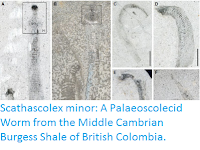
Scathascolex minor: A Palaeoscolecid Worm from the Middle Cambrian Burgess Shale of British Colombia.

Palaeoscolecids are a group of vermiform animals (worms) known from Cambrian to Silurian deposits. They had rings similar to those of Annelid Worms, but were covered in mineralized sclerites (scales), and had evertable probosces similar to those of  Priapulids. They are considered to have been Ecdysozoans, members of the group that includes Arthropods, Tardigrades, Velvet Worms, Nematodes, Nematomorphs, Kinorynches, Loriceferans and Priapulids, though exactly how they are related to other members of the group is unclear.

In a paper published in the journal Palaeontology on 15 October 2015, Martin Smith of the Department of Earth Sciences at University of Cambridge describes a new species of Palaeoscolecid Worm from the Middle Cambiran Burgess Shale of British Colombia, Canada.

The new species is named Scathascolex minor, where 'Scathascolex' refers to the Dragon Scatha from the writings of JRR Tolkien, and 'minor' refers to the small size of the specimens and the fact that some of the specimens were referred to species 'Ottoia minor'. The species is described from three specimens, though seven specimens are referred to the species. Only one of the specimens is complete, this measures 20 mm in length and is 1.3 mm in width, though the largest specimen is 2.4 mm in width and therefore thought to have been around 40 mm in length.

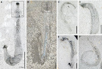
Scathascolex minor from the mid-Cambrian Burgess Shale. (A) NMNH 83939d, anterior section. (B) NMNH 83939b.1, anterior section. (C) NMNH 196193, medial section. (D) NMNH 202689, anterior section, proboscis perpendicular to plane of fossil. (E) NMNH 203005, posterior section showing evidence of extensive decay. (F) NMNH 83939b.2, medial section. Smith (2015).

The trunk section of Scathascolex minor is covered by bands of scales 75–150 μm across separated by scaleless areas 50-100 μm across. The scaled areas each have about 100-200 disk-shaped scales which are about 15-20 μm in diameter, Smith suggests that these may have bee used to gain traction during movement. though it is unclear if the living worms lived within the sediment or on its surface.

See also...

http://sciencythoughts.blogspot.co.uk/2015/12/eokinorhynchus-rarus-kinorhynch-from.htmlEokinorhynchus rarus: A Kinorhynch from the Early Cambrian of Sichuan Province, China.                                                 Kinorhynches are tiny (at most 3 mm) worm like animals found in marine sediments, with segmented tube- or barrel-shaped bodies, separate head and neck regions and evertable pharynxes. They are related...
http://sciencythoughts.blogspot.co.uk/2015/06/quisarctus-yasumurai-new-species-of.htmlQuisarctus yasumurai: A new species of Tardigrade from a submarine cave in the Okinawa Islands.                                                  Tardigrades, or Water Bears, are minute (generally less than 1 mm and always less than 3 mm) aquatic animals related to Arthropods, Nematodes...
http://sciencythoughts.blogspot.co.uk/2015/05/priapulid-worms-from-middle-cambrian-of.htmlPriapulid Worms from the Middle Cambrian of Canada.                                                Priapulid Worms are a form of marine worms with denticle-covered pharynxes which can be everted to form proboscises. They are a minor element of..
Follow Sciency Thoughts on Facebook.

Saturday, 9 May 2015

Priapulid Worms from the Middle Cambrian of Canada.


Priapulid Worms are a form of marine worms with denticle-covered pharynxes which can be everted to form proboscises. They are a minor element of modern marine faunas, largely restricted to deep marine muds and other environments where oxygen levels are low and they face less competition from other forms of Worms. However in the Cambrian Period they appear to have been a major component of marine faunas in many areas. The palaeontologist Charles Doolittle Walcott described the Priapulid Worm genus Ottoia from the Middle Cambrian Burgess Shale in 1911, originally designating three species, Ottoia prolifica, Ottoia minor and Ottoia tenuis, however Ottoia minor has since been moved to a separate genus, Ancalagon, and Ottoiatenuis has been recognized as a (quite unrelated) Enteropneust, leaving only a single species in the genus. A variety of Priapulid fossils from other Cambrian deposits have also been assigned to the genus, though most of these are somewhat dubious.

In a paper published in the journal Palaeontology on 6 May 2015, Martin Smith of the Department of Earth Sciences at the University of Cambridge, Thomas Harvey of the Department of Geology at the University of Leicester and Nicholas Butterfield, also of the Department of Earth Sciences at the University of Cambridge re-examine the genus Ottoia, concluding that there are in fact two species present in the Burgess Shale, and examine the status of Priapulid denticles from other Cambrian localities.

Smith et al. paid particular attention to the denticles of Ottoia, noting that it has several regions on its pharynx, each with denticles with a different morphology. While these were consistent across all the Burgess Shale fossils, one horizon within the deposits contained (exclusively) specimens with a distinctive morphology present in one of these bands, which they refer to as the Type B teeth.

The new species is named Ottoia tricuspida, in reference to its Type B teeth, which have three cusps. In the other described species the Type B teeth have a large central cusp surrounded by four to eight smaller ones, decreasing in size away from the centre. In addition the new species has a double row of Type A teeth, whereas Ottoia prolifica has only a single row.

Ottoia tricuspida. (A) Complete specimen. (B) Enlargement of proboscis. Scale bars represent 10 mm (A); 2 mm (B). Smith et al. (2015).

All of the known specimens of Ottoia tricuspida come from a single horizon within the Burgess Shale, 120–130 cm below the base of the Phyllopod Bed, with a single possible occurrence in the Upper Walcott Quarry. Smith et al. consider the possibilities that it may represent members of the species Ottoia prolifica of a different sex or developmental stage as all the specimens on this horizon appear to belong to Ottoia tricuspida with no specimens of Ottoia prolifica present, whereas on other horizons the reverse is true, and because a wide range of specimen sizes are found in both morphologies.

Schematic diagrams of Ottoia sclerite morphologies. Smith et al. (2015).

Specimens assigned to Ottoiahave also been reported from the Pioche Shale in Nevada, the Marjum Formation and Spence Shale of Utah, the Conasauga Formation of Georgia, the Chancellor Basin of Canada and the Kaili Formation of China. However none of these fossils has been fully described, and most are somewhat dubious, making confident assignment to the genus impossible until such time as these fossils can be properly examined and assessed.

However disarticulated Priapulid denticles are a common feature of Cambrian deposits, and the potential exists to detect Ottoia from such denticles. Smith et al. examined denticles from the Pika Formation of westernmost Jasper National Park, Alberta, the Deadwood Formation of Saskatchewan, and in both cases determined that Ottoia prolifica was present.

Small Carbonaceous Fossils from the Deadwood and Pika formations assigned to Ottoia prolifica. (A–M) Teeth from a single sample of the Deadwood Formation (Riley Lake drillcore at 1300 m), interpreted as tail hooks (A–B), introvert hooks (C–D), a coronal spine (E), a Type A tooth (F), a Type B tooth (G), a Type B tooth with surrounding cuticle (H), morphologies intermediate between tooth types B, C and D (I–L), and a Type D tooth (M). (N–U)Teeth from the Pika Formation, interpreted as an introvert hook (N), an introvert hook without submarginal denticles (O), a Type A-like tooth (P), a Type B tooth (Q), a tooth possibly intermediate between Type B and Type C (R), a Type C tooth (S), an intermediate between Type C and Type D morphologies (T), and a Type D tooth (U). Scale bar represents 50 μm. Smith et al. (2015).

See also…

Vetulicolians are an enigmatic group of Cambrian fossils known from the Chengjiang and Guanshan biotas of South China, Sirius Passet in Greenland and the Burgess Shale in western Canada. They have...
 
In 1997 Stefan Bengtson of the Department of Palaeozoology at the Swedish Museum of Natural History and Yue Zhao of the  Institute of Geology of the Chinese Academy of Geological Sciences announced the discovery of fossilized animal embryos from basal...
 
 
The Lophotrochozoa are a diverse group of Invertebrate animals indicated to have a common ancestry by genetic analysis. The group includes the Annelida, Mollusca, Bryozoa, Cycliophora, Brachiopoda, Entoprocta and Phoronida. Within this group several groups are united by the presence of a crown of tentacles (the Lophophore) surrounding the mouth, which continuously opens and shuts while...
Follow Sciency Thoughts on Facebook.