Showing posts with label Poland. Show all posts

Tuesday, 5 May 2026

Meteorite hunters may have found the largest known chunk of the Bronze Age Kaali Meteorite.

Two Polish meteorite hunters, Filip Nikodem and Andrzej Owczarzak, have recovered what they believe to be a fragment of the Kaali Meteorite, a large body which impacted the Estonian island of Saaremaa about 3500 years ago. The piece is reported to weigh about 40 kg, with a second fragment weighing 15 kg also found. The largest potential fragment of the meteorite discovered to date weighed 5.7 kg, and was found by Filip Nikodem in the spring of 2024.

A 40 kg potential fragment of the Kaali Meteorite found in Estonia by two Polish Meteorite Hunters. Z głową w gwiazdach/Facebook.

The Kaali Impact Structures are a series of nine circular crater lakes on the island of Saaremaa, the largest of which has a diameter of 110 m and a depth of 22 m. These are thought to have been caused by an object with a mass of between 20 and 80 tonnes entering the atmosphere at a velocity of somewhere between 10 and 20 km per second, and exploding in a fireball between 10 and 5 km above the ground. This explosion is thought to have largely vaporised the bolide, as well as removing about 81 000 m³ of rock on the ground, and incinerating vegetation up to 6 km from the impact site.  

The lake in the main Kaali Meteorite Crater on Saaremaa Island. The crater is about 110 m across, but the lake only occupies the central part of it. Wikimedia Commons.

This is thought to have happened about 3500 years ago, when the island was occupied by a Bronze Age population, although attempts to date the site and meteorite fragments from it have produced dates of between 5600 and 400 BC. A Bronze Age arrowhead made from meteoric iron which was found in Switzerland in the 1870s has been linked to the Kaali meteorites on the basis of its metallurgy, making a date of around 1500 BC more plausible.

(a) Overview of the Mörigen arrowhead (A/7396). Note adhering bright sediment material. Remnants of an older label on the left of the sample number. Total length is 39.3 mm. ( b) Side view of the Mörigen arrowhead. Layered texture is well visible. Point is to the right. Thomas Schüpbach in Hofmann et al. (2023).

Prior to 2023, the largest fragment of meteorite found at the Kaali sites weighed 6.21 g. However, in the autumn of that year Filip Nikodem obtained a search permit for the area, and, using a metal detector, found a series of pieces of iron which he believed to be meteoric in origin. These were handed to the Estonian National Heritage Board, who sent them to the University of Tartu, where a rapid X-ray fluorescence analysis found that the iron pieces contained between 0.6% and 4.8% nickel, as well as traces of titanium and vanadium, which is typical of meteoric iron.

The four pieces of meteoric iron handed to the Estonian National Heritage Board  by Filip Nikodem in 2023. Kristo Oks/University of Tartu.

In the spring of 2024, Filip Kikodem returned to the Kaali Lakes, collecting several more fragments with a total mass of 10.5 kg, the largest of which weighed 5.7 kg, making it (at that time) potentially the largest chunk of the Kaali bolide ever discovered. However, despite requests from the Estonian National Heritage Board, these pieces have never been surrendered to them. Instead, the Estonian authorities believe that the pieces were taken to Poland, where they are being analysed at a Polish university. Furthermore, the National Heritage Board received a letter from a lawyer in Poland, asserting Nikodem's ownership rights over the meteorite fragments.

A 5.7 kg possible meteorite fragment found by Filip Nikodem at Kaali Lakes in the spring of 2024. Filip Nikodem.

In the autumn of 2024 Filip Nikodem returned to Kaali Lakes, this time accompanied by meteorite hunter Andrzej Owczarzak, who did not posses a permit to search in Estonia, and was therefore in breach of Estonian heritage laws. At this time concerns were raised by the Estonian newspaper Saarte Hääl, which observed that the original search permit had been given to Filip Nikodem in relation to a two week project run by the University of Tartu in 2017, in which metal detectorists were given a short training course by the university then allowed to collect fragments for a display at a visitor centre, with the collectors being allowed to keep some fragments as a reward.

Saarte Hääl also noted that suspiciously large chunks of 'Kaali Meteorite' had begun to be offered for sale in Poland, and that some Polish enthusiasts had raised concerns that these might be fragments of the more common Morasko Meteorite, which fell near Potsdam about 5000 years ago. The newspaper also noted that Estonian law provides no specific protection for meteorites found in the country, although there are some restrictions upon where a metal detector can be used. 

Following the announcement of these discoveries, Jüri Plado of the University of Tartu applied for funding for a study on the feasibility of changing the law to protect meteorites found in Estonia, reasoning that such large objects would be a significant piece of national heritage and ought to be studied at an institute within the country, but this application was rejected. 

Local farmers talked to by Saarte Hääl stated that the Poles had shown them permits and promised to take any material found to the University of Tartu. However, several expressed doubts that the iron found was in fact meteoric, as it came from shallow depths on farmland, likely to have been disturbed since the Bronze Age, and resembled bog iron (impure iron deposits which precipitate out of solution in boggy soils). The newspaper contacted Kristo Oks of the Estonian National Heritage Board, who confirmed that it would be impossible to tell the difference between bog iron and meteorites without laboratory analysis. 

See also...

Tuesday, 2 July 2024

Using bone histology to understand the lifestyle of the Triasic Diapsid Ozimek volans.

The outcrops of Late Triassic rock in the village of Krasiejow in western Poland have been the subject of palaeontological investigations since the 1990s, yielding a large number of important Tetrapod fossils, including the Temnospondyls Metoposaurus krasiejowensi and Cyclotosaurus intermedius, the Phytosaur Paleorhinus cf. arenaceus, and the Dinosauroform Silesaurus opolensis. One notable Krasiejow Vertebrate is the Diapsid Ozimek volans, which has been interpreted as a potential gliding Animal on the basis of its very long and graceful forelimb and hind-limb bones. 

Ozimek volans was originally interpreted as a Sharovipterygid, grouping it with the Middle-Late Triassic Sharovipteryx mirabilis from Kyrgyzstan, also thought to have been as a glider, but more recently has been considered to be a member of the Tanystropheidae, a group of early Archosauromorphs often exhibiting long necks and tails, at least some of which may have been aquatic.

No close relative of Ozimek volans has a similar limb arrangement; a similar pattern is seen in Sharovipteryx mirabilis, but this can be ruled out as a close relative on the basis of other parts of its anatomy, suggesting the similarity is the result of similar ecological adaptations, rather than common ancestry. Interestingly, Sharovipteryx mirabilis has a preserved membrane reaching the end of its hind limbs, leading weight to the idea that this species was adapted to gliding, and therefore that the similar morphological adaptations seen in Ozimek volans could also be adaptations to aerial behaviour. 

In Birds and Pterosaurs, flight was achieved not just by the elongation of the limbs, but by numerous adaptations to reduce the weight of the skeleton. The most notable of these is the appearance of pneumatic bones, in which the dense marrow filling of the bone is replaced with a hollow, air-filled cavity. This is known in living Birds, and has been demonstrated in Mesozoic Birds, as well as closely related Theropod groups, and Sauropods, although it is only inferred in Pterosaurs, on the basis of the large size these Animals achieved. Both Birds and Pterosaurs have extremely thin bone walls, made of dense fibrolamellar bone tissue, which is very strong and better able to withstand the stresses of flight.

Notably, neither the presence or absence of pneumatic bone nor fibrolamellar bone tissue can be used as absolute determiners of flying behaviour. Fibrolamellar bone tissue is found in a number of Archosaur groups which show no other adaptations to flying, but absent in both small Birds and Bats, while Bats also lack pneumatic bone, something found in the (clearly non-areal) Sauropods. Thus, to establish the flying capacity of an extinct Animal it is necessary to look at its whole morphology, including skeletal and muscular adaptations and the possible presence of flight membranes and/or feathers.

Gliding is a form of flight in which the Animal has active control of aerodynamic forces, but is unable to gain altitude by muscular activity. Such behaviour can be hard to determine in an Animal simply from its morphology, as it is achieved in different ways in a very wide range of organisms, with no single set of adaptations common to all. Indeed, many gliding organisms are morphologically little different to their closest non-gliding relatives, achieving flight purely through behavioural changes. Examination of living gliding Mammals suggests that long humeri and femora improve the aspect ratio of gliding Animals, and that this becomes more important as the Animal becomes larger. Little is currently known about the bone histology of gliding Mammals, although some Flying Squirrels have a light-weight humerus and more circular diaphysis in cross-section compared to non-gliding taxa, which helps to resist torsional loads and provide resistance to multidirectional bending.

Ozimek volans has been noted to have had very thin bone walls, an adaptation associated with weight-reduction and flight, but its histology has not, to date, been examined, limiting our understanding of the living Animal's growth and life history.

In a paper published in the journal Palaontology on 26 June 2024, Dorota Konietzko-Meier of the Institute of Organismic Biology at the University of Bonn, Elżbieta Teschner, also of the Institute of Organismic Biology at the University of Bonn, and of the Institute of Biology at the University of Opole, Agnieszka Tańczuk of the Department of Zoology and Nature Protection at the Maria Curie-Skłodowska University in Lublin, and Martin Sander, again of the Institute of Organismic Biology at the University of Bonn, present the results of a study of the bone histology of Ozimek volans, and the implications of this for the life history and behaviour of the living Animal. 

Most of the material from which Ozimek volans was described is held in the collection of the Institute of Paleobiology of the Polish Academy of Sciences in Warsaw, though two blocks containing bones assigned to the species are held in the collection of the Institute of Biology at Opole University, and it is material from one of these blocks which is used in Konietzko-Meier et al.'s study. The block contains a cluster of bones interpreted as a partial skeleton, including articulated cervical vertebrae, possible coracoids and a pes, as well as a humerus and a femur, although Konietzko-Meier et al. note that the humerus is only 46% of the size of the largest known humerus, and therefore have been presumed to have come from a juvenile, while the femur is the largest known femur assigned to the species, and at least twice as long as the humerus, making it unlikely that they were from the same Animal.

Konietzko-Meier et al. took three thin sections, each about 40μm thick, from the midshaft of the humerus, one transverse, one longitudinal and one oblique tangential, and two from the femur, the bone here being too thin for an oblique section to be taken. These were then examined under both light and scanning electron microscopes at the University of Bonn.

Simplified phylogeny and long bones of Ozimek volans from the Late Triassic of Krasiejów (Poland). (A) Simplified phylogeny showing the position of Ozimek volans among Archosauromorpha, the black silhouette on the right represent the skeletal restoration of Ozimek, with the flight membrane stretched between the elongated forelimbs and hindlimbs. (B) Left humerus UOPB 1148a in medial view. (C) Right femur UOPB 1148b in anterior view. The white lines mark the cutting planes: 1, transverse section; 2, longitudinal section; 3, oblique tangential section. Scale bars represent 10 cm (A) and 10 mm (B) and (C). Konietzko-Meier et al. (2024).

The humerus has a thickness of 3.4 mm at the site where it was sampled, with a cortex (outer layer of bone) thickness of 0.5 mm. This cortex is made up of two layers, the periosteal portion, which is laid down on the exterior as the bone grows, and a compact endosteal layer, formed from the inner surface outwards as a secondary structure, by reworking of the original bone. The outer periosteal is more heavily mineralized, and made up of fibrous lamellae with simple vascular canals, with several lines of arrested growth (marks left in growing bone by slowed growth at one time of year, typically winter in a temperate climate). The fibres which make up this layer are not arranged at random, but have a longitudinal orientation. No sign of pneumosteum can be seen anywhere on the bone.

Microstructure and histology of the humerus UOPB 1148a midshaft of Ozimek volans from the Late Triassic of Krasiejów (Poland). (A)–(C) Overview of the histological framework visible in the transverse section in: (A) normal light; (B) polarized light; (C) with lambda filter. (D) Schema of the distribution of the growth marks visible in the transverse section of the humerus midshaft. (E)–(G) closeup of the posterior part of cortex with clearly visible lamellae and growth marks in: (E) normal light; (F) polarized light; (G) with lambda filter. (H)–(J) Close-up of the anterior fragment of cortex with visible island of coarse compact cancellous bone and growth marks in: (H) normal light; (I) polarized light; (J) with lambda filter. (K) Scanning electron microscope photograph showing the osteocyte lacunae surrounded by a network of canaliculi. (L)–(M) Overview of the histological framework visible in the longitudinal section in: (L) normal light; (M) polarized light; note the vascular canal. (N) Close-up of the fragment of cortex visible in the longitudinal section in polarized light. (O)–(Q) Overview of the histological framework visible in the oblique tangential section in: (O) normal light; (P) polarized light; (Q) with lambda filter. Continuous lines indicate reversal lines; dashed lines mark lines of arrested growth. Abbreviations: A, annulus; CCCB, coarse compact cancellous bone; eA, endosteal annulus; eZ, endosteal zone; PO, primary osteon; SV, simple vascular canal. Scale bars represent: 500 μm (A(–(D), (L), (M), (O)–(Q); 200 μm (N); 100 μm (E)–(J); 20 μm (K). Konietzko-Meier et al. (2024).

The femur is 2.7 mm in width at the point where it was sampled, with a cortex thickness of 0.2 mm. The structure of this bone is less complex, with signs of bone resorption on the inner surface, but no endosteal cortex layer. The periosteal layer is again lamellar and highly mineralized, with several lines of arrested growth. Vascular canals are more tightly backed than in the humerus. 

Microstructure and histology of the femur UOPB 1148b midshaft of Ozimek volans from the Late Triassic of Krasiejów (Poland). (A)–(C) Overview of the histological framework visible in the transverse section in: (A) Normal light; (B) polarized light; (C) with lambda-filter. (D) Close-up of the cortex with visible well-organized lamellae system, image in polarized light. (E)–(F) Close-up of the cortex with preserved fragments of coarse compact cancellous bone in: (E) normal; (F) polarized light. (G)–(I) Close-up of the anterior fragment of cortex with visible growth marks in: (G) normal light; (H) polarized light; (I) with lambda filter; the arrow indicates the second zone. (J)–(L) Overview of the histological framework visible in the longitudinal section in: (J) normal light; (K) polarized light; (L) with lambda filter. Continuous lines indicate reversal lines; dashed lines mark lines of arrested growth. Abbreviations: A, annulus; CCCB, coarse compact cancellous bone; PO, primary osteon; SV, simple vascular canal; Z, zone. Scale bars represent: 500 μm (A)–(C), (J)–(L); 100 μm (E)–(I); 50 μm (D). Konietzko-Meier et al. (2024).

Konietzko-Meier et al. recognise that their sample size is small, but propose thar some simple observations about bone growth in Ozimek volans can be made. Bone growth is typically driven from the periosteal surface, where lamellae of new cortex tissue are laid down. A line of bone reworking called the Haversian substitution front progresses outwards from the medullary (inner) edge of the bone, producing a layer of secondary tissue including Haversian bone. At the same time, a resorption line also works outwards from the inner surface, removing bone tissue, and sometimes overtaking the Haversian substitution front.

This three-front model of bone growth was first observed in Sauropods, but has now been found in a wide range of Tetrapod groups. However, the compact endosteal layer is a tissue not seen in Sauropods (or most other Tetrapods) and may represent a fourth growth front (i.e. lamellar bone/Haversian bone/compact endostreal bone/resorbtion line). Notably, the Hacersian growth front in Ozimek volans appears to be very slow, generally being obliterated by the endostreal growth front and resorption line.

Lamellar bone is common in Tetrapods, particularly non-endothermic ones, although it is also found in many small Mammals. However, the lamellar structure of Ozimek volans is exceptionally well-developed, unlike anything seen in any other fossil group. However, something similar has been observed in living Bats, and is thought to be an adaptation enabling the formation of lone, lightweight, and strong bones needed for flight in a group which has not evolved pneumatic bone (is has also been suggested that the adoption of strong lamellar bone rather than pneumatic bone has placed a restriction on the size to which bones can grow). It is therefore quite possible that this adaptation in Ozimek volans was also an adaptation to flight (although in this case unpowered).

See also...

Thursday, 1 February 2024

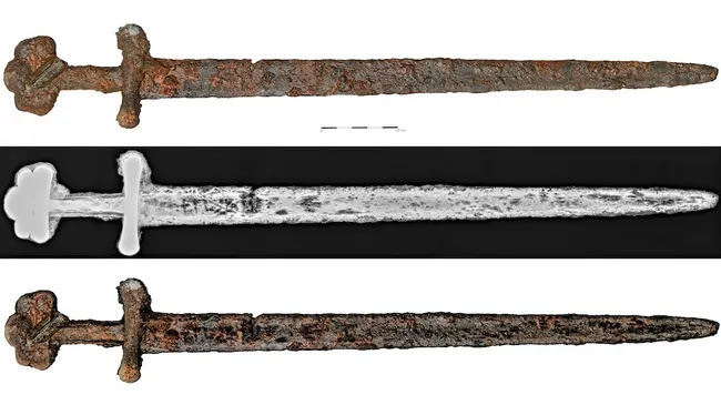
Medieval Ulfberht sword recovered from Polish river.

Workers carrying out a dredging operation in the River Wisła in Włocławek in central Poland in the second week of January 2024 recovered an example of a type of sword thought to be over a thousand years old along with a load of river silt. The sword is double-edged with a three-lobed pommel, which are distinctive features of these swords, and when viewed in x-ray could be seen to bear the inscription +VLFBERTH+, which have been found on about 170 similar swords from across Northern Europe and Scandinavia.

This is the eighth Ulfberht sword found in Poland; the country which has produced the largest number of these swords is Norway, with about 40 known to date. This has led to these swords sometimes being referred to as 'Viking' swords, although historians are fairly confident that they are of Frankish origin, and probably from the southern part of the Frankish realm, in southern Germany or possibly even France. These swords date from the ninth and tenth centuries, and are considered to be an intermediate stage between the earlier swords used across northern Europe and the knightly swords of Late Medieval Europe. They are typically made of superior steel to that used in earlier swords, which would have made them stronger and sharper.

The sword shortly after its discovery. Provincial Office for the Protection of Monuments in Toruń.

A ninth or tenth century date for the sword would suggest it was in use around the time of the founding of the modern state of Poland, by the Piast Dynasty, who, according to legend, unified the Slavic tribes of the region, fought for independence from the area's German rulers, defended against raiders from Scandinavia, and introduced Christianity to Poland.

See also...


Wednesday, 31 January 2024

Alienacanthus malkowskii: A highly specialised Placoderm Fish from the Late Devonian Rheic Ocean.

Placoderms are thought to have been the earliest jawed Vertebrates, first appearing in the Silurian and rising to become the most diverse group of Fish in the Devonian, before their extinction at the end of that period. During the Devonian the Placoderms, and in particular the Arthrodires (the most abundant and diverse Placoderm group) produced a wide range of forms, implying an equally diverse range of ecological and feeding strategies. However, Placoderms are known almost entirely from their hard parts, with only a single specimen with a body outline known, and no known stomach contents or soft parts, limiting our ability to interpret the ecology of these diverse early Fish. The jaws of early Placoderms show tend to be similar, apparently adapted to rapid snatching of prey, but later members of the group are much more varied, and have been interpreted to reflect a range of feeding styles from filter feeding to durophagy (the crushing of hard foodstuffs, such as shellfish).

In a paper published in the journal Royal Society Open Science on 31 January 2024, Melina Jobbins of the Department of Palaeontology at the University of ZurichMartin Rücklin of the Naturalis Biodiversity Cente and the University of LeidenMarcelo Sánchez Villagra, also of the Department of Palaeontology at the University of Zurich, Hervé Lelièvre of the Muséum National d’Histoire NaturelleEileen Grogan of the Department of Biology at Saint Joseph’s University, Piotr Szrek of the Polish Geological Institute, and Christian Klug, again of the Department of Palaeontology at the University of Zurich, redescribe a species of Late Devonian Placoderm previously only known from fragmentary material from the Holy Cross Mountains of Poland, on the basis of new material from the eastern Anti-Atlas of Morocco.

Alienacanthus malkowskii was originally described from fragmentary material from two quarries in Poland, as composing large, possibly paired, spines of uncertain origin. Jobbins et al.'s redescription of the species is based upon a nearly complete skull, the left side of a second skull, and a number of more fragmentary remains from sites in Morocco, which reveals the 'spines' to be part of the lower jaw of a large Eubrachythoracid Placoderm.

Alienacanthus malkowskii, skull, PIMUZ A/I 5239. In right (a), (b), left (c), (d) and dorsal (e), (f) view; inferognathals, PIMUZ A/I5238, in lingual (g), lateral (h) and dorsal (i) view. Each bone is differentiated by a separate colour. Black arrow points to lingualdepression. Scale bars correspond to 100 mm. Jobbins et al. (2024).

The inferognathal bones, which form the lower jaws in Placoderms protrude significantly beyond the upper jaw, reaching about twice the length of the rest of the skull, reaching a pointed tip. These jaw elements run closely parallel to one-another over about 60% of their length, although they are not fused at any point. The teeth of both jaws are posteriorly recurved, with the 'teeth' (actually bony protrusions, as in all Placoderms) of the lower jaw continuing forward of the upper jaw, but a significant distance short of the tip of the bone.

Extremely elongated lower jaws are known in a variety of other extant and fossil Fish and marine Tetrapods, including the Carboniferous Chondrichthyan Ornithoprion, the extant ray-finned Halfbeaks, which have a fossil record dating back to the Palaeogene, and the Pliocene Porpoise Semirostrum. Although in none of these are the lower jaws as elongated as they are in Alienacanthus malkowskii, with the longest examples being found in some species of Halfbeak, which can reach about 1.6 times the length of the skull.

Live reconstructions of Alienacanthus. Based on the body morphology of extinct and modern Fish with elongated jaws (elongated, fusiform, bodies). Beat Scheffold & Christian Klug in Jobbins et al. (2024).

The recurved teeth of Alienacanthus malkowskii are strongly suggestinve of a diet of live Fish, mirroring the shape of teeth seen in many other Fish-eating groups, including Ichthyosaurs, Snakes, Choristoderans, and other living and extinct Fish species. However, the lower teeth of Alienacanthus malkowskii continue beyond the upper jaw, with up to twelve teeth forward of the mouth in observed specimens. 

Teeth forward of the mouth are known in a number of Condrichthyan groups, including Sawfish, Sawsharks, and Rajiform Rays. All of these have teeth on the upper jaw rather than the lower, and are equipped with electroreceptive sensory organs which enable them to detect prey-Fish and strike them with a rapid side-motion of the rostrum. However, thin sections of the jaw of Alienacanthus malkowskii show no signs of the additional neural canals which would be associated with such a system, and the teeth of Alienacanthus malkowskii are directed upwards, rather than sideways, making it unlikely that the elongated jaw was used in the same way as seen in Sawfish. Instead, Jobbins et al. suggest that the presence of teeth forward of the mouth in Alienacanthus malkowskii is a product of the way the living Animal grew, with formerly useable oral teeth being carried forward as the jawbone elongated, probably in a short burst of growth as the Fish approached maturity, although it is still possible that the long lower jaw was used to strike at prey, and that the forward teeth could have inflicted damage on soft-bodied Animals.

See also...


Thursday, 7 December 2023

Collection of Bronze Age axes discovered in Pomerania, Poland.

Archaeologists in the Starogard Forest District of Pomerania, northern Poland, have recovered a collection of five Bronze Age Axes, which were found by an amateur metal detectorist. Denis Konkol, a history enthusiast, was detecting with official permission (required by law in Poland) when he made the discovery, and informed the local authorities. 

Five Bronze Age axes discovered in the Starogard Forest, Poland. Nadleśnictwo Starogard.

The axes are of a type known as tautušiai, which are large axe-heads with a slender handle with elevated edges and a broad blade, which were made in eastern Poland and Lithuania between about 1700 and about 1300 BC. Small groups of these axes are thought to have been buried together for religious reasons, although this is the first such collection uncovered in Poland for about 20 years. More common from the same period, are small amounts of jewelery, also ritually buried.

One of the five tautušiai axes uncovered in Pomerania. Nadleśnictwo Starogard.

The axes will be taken to the Archaeological Museum in Gdańsk for study and preservation. 

See also...

Follow Sciency Thoughts on Facebook.

Follow Sciency Thoughts on Twitter.