
The epidermis is a fundamental cellular tissue of Metazoans (Animals) which acts as an interface between the external medium and the body interior. It includes epithelial cells and extracellular material. Epithelial cells form a tissue with complex junction systems and secrete two types of extracellular substrates: basally, the extracellular matrix, and apically, the glycocalyx (layers of proteins and sugar) and the cuticle. Although absent in Placozoans and Ctenophores, the glycocalyx plays a crucial role as a matrix on which other extracellular molecules (e.g. chitin, collagen) can anchor and eventually form a cuticular structure. A cuticle occurs in all present-day Ecdysozoans (Nematoida, Scalidophora, Panarthropoda), with a consistent three-layered structure. Ecdysozoans display a great variety of cuticular structures often associated with sensory organs (e.g. setae). Cuticular moulting is common to all extant representatives of Ecdysozoa and is a key diagnostic feature of the group. Exuviae found in early Cambrian Scalidophoran worms from China (roughly 535 million years old) suggest that ecdysis has a very ancient origin and evolves through a suite of genetic innovations in the three Ecdysozoans clades (Scalidophora, Nematoida, Panarthropoda).
In a paper published in the Proceedings of the Royal Society Series B: Biological Sciences on 6 May 2020, Deng Wang of the State Key Laboratory of Continental Dynamics and Shaanxi Key Laboratory of Early Life and Environments at Northwest University, and the University Claude Bernard Lyon 1, Jean Vannier, also of the Université de Lyon, and Xiao-guang Yang, Jie Sun, Yi-fei Sun, Wen-jing Hao, Qing-qin Tang, Ping Liu, and Jian Han, also of the State Key Laboratory of Continental Dynamics and Shaanxi Key Laboratory of Early Life and Environments at Northwest University, describe two types of micro-reticulate cuticular patterns in lowermost Cambrian Scalidophoran Worms from the Kuanchuanpu Formation, which have direct counterparts in extant Priapulid Worms such as Priapulus caudatus and Halicryptus spinulosus, suggesting that the reticulation observed on the external surface of the Cambrian Worms is the direct expression of the epidermis cellular pavement.
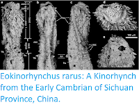
Cambrian Scalidophorans have very diverse cuticular features that are best preserved in Burgess Shale-type Lagerstätten and small carbonaceous fossil and small shelly fossil assemblages in which (small shelly fossil) scalids, platelets and pharyngeal teeth are preserved in three dimensions as exemplified by stem-group Scalidophorans such as Eopriapulites, Eokinorhynchus and Palaeoscolecids. These microscopic cuticular elements have been studied in detail in various groups of early Cambrian Scalidophorans and provide diagnostic characters useful to their classification. A recent study found a new type of micro-ornament in Scalidophoran worms from the Early Cambrian Kuanchuanpu Formation (roughly 535-million-years-old). It consists of very fine reticulation on both the external and internal surface of exuviae. The relationships between the external cuticular features and the underlying epidermal tissues of ancient Animals have been largely overlooked. In extant Arthropods such as Centipedes, the polygonal surface pattern of the cuticle faithfully reproduces the geometry of the underlying epidermal cells. A comparable epidermis-to-cuticular correspondence has
been established in Crustaceans such as Ostracods, Isopods, and Decapods, but has never been observed in other Ecdysozoans groups such as the Scalidophora and the Nematoida.
been established in Crustaceans such as Ostracods, Isopods, and Decapods, but has never been observed in other Ecdysozoans groups such as the Scalidophora and the Nematoida.
The studied specimens were collected from Bed 2 of the early Cambrian Kuanchuanpu Formation (Fortunian Stage, Terreneuvian Series) at the Zhangjiagou Section near Dahe town, Xixiang County, Shaanxi Province. The Xixiang areawas palaeogeographically located on the northwestern margin of the Yangtze Platform during the Ediacaran and Cambrian periods. The fossil-bearing horizon correlates to the Anabarites trisulcatus–Protohertzina anabarica assemblage biozone, which indicates the Meishucunian Stage (equivalent to the Fortunian Stage). Uranium-lead dating of zircons from tuff layers (Meishucun Section, Yunnan Province) gives an estimated age of 536.5 million years. Several horizons of the Kuanchuanpu Formation have yielded abundant secondarily phosphatised small shelly fossils that are extracted fromcalcareous rocks through digestion in 8–10% acetic acid and picked out from residues under a binocular-microscope. These small shelly fossil assemblages contain abundant fossils with Cnidarian affinities, possible Deuterostomes, diverse Scalidophoran Worms, Cyanobacteria, and problematic forms. Wang et al.'s study is based on 22 specimens in which cuticular ornament is particularly well preserved. All specimens are deposited in the collections of the Early Life Institute of Northwest University, Xi’an, China.These specimens were mounted on stubs, coated with gold and observed under a FEI Quanta 400 FEG Scanning Electron Microscope at the State Key Laboratory of Continental Dynamics at Northwest University, China. X-ray microtomography was also performed at the State Key Laboratory of Continental Dynamics in order to reconstruct the threedimensional morphology of the reticulate microstructures of two specimens (ELIXX93-335 and ELIXX95-85).
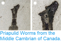
Priapulus caudatus was collected from the Gullmarsfjord near the Kristineberg Marine Research Station of Göteborg University, Sweden, and Halicryptus spinulosus from the brackish waters of the Baltic Sea, near the Askö Laboratory of the Baltic Sea Centre of Stockholm University. Priapulus caudatus was dredged from muddy, poorly oxygenated sediments and kept in tanks at low temperature before being fixed (glutaraldehyde) and desiccated in a Leica Critical Point Dryer for microscopic observations. Halicryptus spinulosus was prepared via comparable standard methods. Priapulus caudatus and Halicryptus spinulosus secrete mucus which sticks sedimentary particles and detritus to the external surface of the trunk thus preventing clear observation of the cuticular microstructures. Fine cuticular structures were best observed in freshly moulted specimens and exuviae. Images of dissected and whole specimens were taken with a Zeiss Merlin compact scanning electron microscope at the Centre Technologique des Microstructures of University Claude Bernard Lyon 1. Some specimens were embedded in resin, sliced and stained with Toluidine blue for observation of cellular and cuticular features using optical microscopy.
Scalidophoran Worms are frequent elements of small shelly fossil assemblages from the Early Cambrian Kuanchuanpu Formation, and are represented by at least five different species and more than nine unnamed forms. They consist of relatively rare complete specimens and more frequent fragmentary tubular remains, all secondarily preserved in calcium phosphate and showing very fine external details of cuticular features (e.g. sclerites, scalids and pharyngeal teeth). Some of these tubular elements are exuviae which display either a positive or a negative relief, indicating that these worms were able to turn their flexible cuticle inside out during ecdysis in a manner similar to that of extant Priapulids. Scanning electron microscope observations have revealed very fine reticulate ornament over the entire external surface of the cuticle, characterised by a polygonal network with interconnected narrow ridges or furrows. This micro-ornament may be locally less well defined due to factors of preservation. A second type of reticulate pattern, characterised by wavy ridges, occurs on the inner surface of exuviae.

Reticulation in Scalidophoran Worms from the early Cambrian Kuanchuanpu Formation, China. (a)–(c) Reticulation on the external surface of sclerites; ELIXX106-520, general view and close-ups showing polygons with straight interconnected ridges. (d)–(f) Reticulation on the internal surface of sclerites; ELIXX78-205, general view and close-ups showing polygons with interconnected furrows. (g), (h) ELIXX93-335, general view and X-ray microtomograph image of a virtual section through the specimen (dotted line); each ridge on the outer surface corresponds to a furrow on the inner surface. (i)–(l) ELIXX73-212, reticulation on the external surface of flexible cuticle; (i) general view and close-ups showing reticulation with microfolded interconnected ridges (9–26 V-shaped microfolds); (j) a broken part of phosphatised cuticle with no visible microstructures. (m)–(q) ELIXX95-85, general view and close-ups showing reticulation with punctuated interconnected furrows (10–22 pits) and elongated punctuated interconnected furrows between sclerites (external surface of an inverted exuvia; (p), (q) X-ray microtomography images of a virtual section through the specimen (dotted line in (e)). Abbreviations: cu, cuticle; fo, fold; fu, furrow; PIF, punctuated interconnected furrows; pt, pit; ri, ridge. Scale bars represent: 500 μm (a), (e), (g), (i), (m), 200 μm (p), 100 μm (e), (h), 50 μm (b), (k), 20 μm (c), (f), (l), (n), (o), (q), 4 μm (j). Wang et al. (2020).
Reticulation is well defined in the two types of exuviae, especially around the basal part of sclerites. In exuviae with a positive relief, it consists of five- to seven-sided polygons delimited by straight interconnected ridges, which form a rather regular network. In some cases, elongate polygons or polygons with a more irregular shape are evident. Straight interconnected ridge relief decreases towards the apex of the sclerites. Measurements of more than 200 polygons were made in 10 different specimens. The diameter of the polygons ranges from 4 to 12 μm, their perimeter length is from 14 to 31 μm and straight interconnected ridge width varies from 1.3 to 2.0 μm. The exact counterpart of this polygonal network was observed in exuviae with a negative relief (i.e. sclerites pointing inwards instead of outwards) in which straight interconnected ridges faithfully correspond to interconnected furrows. Interconnected furrows encircle slightly convex polygonal areas. Measurements of about 200 of these polygons in 5 specimens give values similar to those obtained from sclerites with a positive relief (polygon diameter and perimeter, 4–15 and 20–43 μm, respectively). Virtual tomographic sections show that straight interconnected ridges are the exact counterparts of interconnected furrows.
A different type of reticulate pattern is evident in areas where the cuticle is devoid of sclerites and supposedly more flexible. It is characterized by strongly microfolded interconnected ridges which contrast with the straight outline of straight interconnected ridges. These ridges bear tiny V-shape, evenly distributed microfolds. The diameter of these polygons (based upon 130 in 7 different specimens) ranges from 5 to 19 μm with a wall thickness of 0.4–2.0 μm. Their perimeter varies from 19 to 47 μm. Individual polygons have 9 to 26 microfolds (microfold length between 3.0 and 4.6 μm).
Exuviae with a negative relief show a reticulate pattern characterized by punctuated interconnected furrows around polygonal areas. As for straight interconnected ridges and interconnected furrows (observed on sclerites), punctuated interconnected furrows are the inverted counterparts of microfolded interconnected ridges. The smooth microfolded walls of microfolded interconnected ridges match exactly with the punctuated outline of punctuated interconnected furrows as confirmed by measurements (100; polygon diameter: 5–15 μm; number of pits: 10–22) and tomographic cross-sections. Some polygonal areas seem to be stretched in one direction.
In Priapulus caudatus, reticulation occurs along both the trunk and the proboscis, with polygonal diameters of 9–14 and 10–21 μm, respectively. The three to five-sided polygons observed on the trunk are delimited by straight weakly elevated interconnected ridges and form a rather regular network. Microfolded polygons (resembling the microfolded interconnected ridges-type in fossils) preferentially occur in the strongly contracted areas of the trunk. Their diameter and thickness are 7–11 μm and 1.2–1.6 μm, respectively. Microfolds are evident both on the walls and inner areas of the polygons.

Reticulation and epidermal cells in Priapulus caudatus and Halicryptus spinulosus from Sweden. (a)–(g) Priapulus caudatus. (a) General view of a live specimen on sediment. (b) Transverse section through the cuticle (details of epicuticle surface obscured by sediment particles and debris). (c), (d) Oblique section showing the layered structure of the cuticle (each layer with reticulate network); general view and close-up. (e) Reticulate surface affected by microfolds (on both ridges and the floor of the polygons). (f) Scaly reticulation on the proboscis. (g) Close-up of (f). (h)–(k) Halicryptus spinulosus. (h) General view of a moulting specimen with old cuticle partly detached from the body. (i) Transverse section through trunk showing cuticle epidermal cells and circular muscles. (j) Newly moulted cuticle with reticulation and unsclerotised tubuli. (k) Reticulation along the internal surface of an exuvia. (l)–(t) Priapulus caudatus from Sweden; microtome sections of specimens embedded in resin (toluidine blue staining). (l) Trunk annuli of Priapulus caudatus. (m) Simplified diagram of trunk to show location of transverse and longitudinal sections. (n) Transverse section through trunk cuticle (cuticle, epidermal cells, circular muscles, longitudinal muscles in light grey, red green and white, respectively). (o) Transverse section showing epidermis (single layer) between cuticle and circular muscle. (p), (q) Sections showing correspondence between the boundaries of epidermal cells and the walls of cuticular reticulation. (r)–(t) Intermediate section showing boundaries between annuli (dark blue lines) and reticulation within annuli; general view and details showing subhexagonal network near the boundaries and more elongated one in central part of annulus. Abbreviations: an, annulus; ba, boundaries of annuli; ca, caudal appendage; cm, circular muscle; cu, cuticle; ec, epidermal cells; la, layer; lm, longitudinal muscles; ls, longitudinal section; oc, old cuticle; pr, proboscis; tr, trunk; ts, transverse section; ut, unsclerotised tubuli. Light grey, purple and light green colours represent the cuticle, epidermal cells and circular muscles, respectively. Scale bars represent: 2 cm (a), 500 μm (n), (r), 100 μm (o), 20 μm (c), (g), (i), (j), (k), (p), (q), (s), (t), 10 μm (b), (d), (e) and 5 μm (h). Wang et al. (2020).
Transverse sections observed under the scanning electron microscope show that the cuticle of Priapulus caudatus sits above the epidermal cells and is made of numerous, densely packed laminated layers. The cuticle thickness varies from 4.7 to 10 μm. However, clear subdivisions (e.g. exocuticle, endocuticle) could not be observed in scanning electron microscope images. Reticulation seems to affect the most superficial part of the cuticle, presumably the epicuticle. Sections through embedded specimens reveal the configuration of epidermal cells and also the relation between the epidermis and the cuticle. Transverse sections show that the epidermis has a single layer of subrectangular cells. However, the height (10–20 μm) and width (approx. 10 μm) of these cells depend on the location and orientation of the microtome sections and therefore show some variations. The boundaries between epidermal cells correspond to the external polygonal ridges observed on the external surface of the cuticle. Longitudinal sections through the epidermal layer show that the layer of epidermal cells forms a well-defined pentagonal and hexagonal network (cell size between 8 and 25 μm in diameter).
A comparable reticulation was observed in freshly moulted specimens of Halicryptus spinulosus, the diameter of polygons (7–16 μm) being virtually the same as in Priapulus caudatus. The strongly wrinkled appearance of the cuticle is probably due to its extreme thinness and disappears after a few days following ecdysis. In Halicryptus spinulosus reticulation also occurs along the inner surface of fresh exuviae, with five- to seven-sided polygons of 5–16 μm and 0.8–1.3 μm in size and wall thickness, respectively.
Straight interconnected ridges/interconnected furrows and microfolded interconnected ridges/punctuated interconnected furrows clearly represent the positive/negative expressions of two types of cuticular micro-ornament. The consistent size range of polygons (4–19 μm) overlaps with that of epidermal cells in Priapulus (8–25 μm). Moreover, as seen in Priapulus, epidermal cells form a regular polygonal network in which cell boundaries match with the walls of the reticulation. These comparative studies with modern Priapulids suggest that the reticulation evident on the cuticle of Cambrian Scalidophorans similarly replicates the pavement of underlying epidermal cells from which the cuticle was secreted.
In present-day Ecdysozoans, epidermal cells secrete a cuticle characterised by three distinct layers: a basal layer with chitin, a middle layer of homogeneous structure and an apical layer having three very thin lamellae, establishing a faithful correspondence in size and shape between the network of epidermal cells and that of the cuticular reticulation. A typical example is in the case of Centipedes which display a regular hexagonal network over the external surface of their cuticle. In these Myriapods, reticulation appears on the epicuticle and clearly replicates the epidermal cell pavement evident at the beginning of moulting. A comparable geometrical replication occurs in Crustacean Ostracods and other extant Arthropod groups. Prior to moulting, Ecdysozoans secrete specific enzymes that loosen contact between the basal part of the old cuticle and the epidermal cells. The growth of epidermis via mitosis results in new epidermal cells relatively smaller than in their pre-moult state. Their apical surface characterised by a wavy appearance bears microvilli and starts to secrete individual cuticular patches which fuse together to eventually forma newcuticular layer as the epidermis stretches horizontally.

Idealised diagram to explain cuticular reticulation in Cambrian Scalidophoran worms, based on analogues in extant Priapulids (Priapulus and Halicryptus). (a)–(e) Successive steps in cuticle formation: (a) old cuticle splitting from the epidermis layer (ecdysis); (b) mitosis within epidermis (dividing cells) and formation of small cuticle patches at the boundary between cells; (c) epicuticle layer is formed and bears external reticulation; underlying cuticular layer starts growing; (d) intermediate step; (e) whole cuticle is formed. (f)–(i) Block diagrams successively showing ecdysis, cell division and initial step of cuticle secretion and expansion (red arrows) of epidermal layer and cuticle, leading to final reticulate pattern; polygons have the same size in (f) and i). Epicuticle is depicted in red/pink; undifferentiated exo- and endocuticle in yellow; epidermal cells in grey; and circular muscles in light green. Wang et al. (2020).
The accumulation of material secreted by microvilli at the boundary between adjacent epidermal cells most probably induces the formation of cuticular ridges in this particular boundary area. This process leads to an exact correspondence between the epidermal and the cuticular reticulate networks. Although Wang et al.'s fossil material does not show details of the cuticle ultrastructure and underlying epidermal cells, they hypothesise that the cuticle of early Cambrian Scalidophoran Worms was formed through a comparable process. Thus, fundamental aspects of ecdysis and cuticle formation seem to have been established early in the evolution of Ecdysozoans (at least not later than 535 million years ago), possibly before the rise and major diversification of Arthropods.
Wang et al. suggest that the microfolds associated with the reticulation of the cuticle of Cambrian Worms may be original features. The cuticle of extant Scalidophorans such as Priapulus caudatus and Halicryptus spinulosus is very thin (approximately 4.7–10 μm) and responds in a flexible manner to the contraction and relaxation of circular and longitudinal muscles by creating transverse microfolds, parallel to annuli. The reaction of Worms to fixing chemicals (e.g. glutaraldehyde, formaldehyde) often results in extreme body contraction and cuticular folding which, when exerted on reticulate structures, generate microfolds along the walls of the polygons. The microfolded features observed on the cuticle of Cambrian Worms, exemplified by microfolded interconnected ridges/punctuated interconnected furrows, may be similarly explained by muscular contractions, possibly induced by environmental stress that caused the animal’s death. However, these microfolds are not seen in the inner areas (floors) of the polygons. In addition, the V-shaped microfolds observed along the polygonal ridges have a consistent amplitude and a virtually constant number. By contrast, the microfolds seen in extant Priapulids are distributed more irregularly and seem to affect the whole cuticular structure. Wang et al. suggest that the microfolds associated with the cuticle reticulation evident in Cambrian Worms may be original features. They may characterise freshly moulted Worms in which the cuticle is extremely thin and wrinkled as seen in extant Halicryptus spinulosus, or result from unknown external or internal factors. Interestingly, microfolded interconnected ridges/punctuated interconnected furrows are mostly found in areas where the cuticle is supposed to be thinner and flexible, whereas straight interconnected ridges/interconnected furrows preferentially occur around thick sclerites. Microfolds may be seen as a physical solution to absorb plastic deformation due to muscular contractions, especially where the cuticle is thin. That would explain why non-deformable sclerites lack such wavy appearance. Similarly, Eopriapulites sphinx, another Scalidophoran worm from the Kuanchuanpu Formation, shows a complex wrinkled cuticular pattern with closely spaced annuli and short longitudinal folds running across each annulus. This pattern may represent another solution to absorb contraction exerted by both circular and longitudinal muscles. An alternative third hypothesis is that the microfolded walls of polygons might be the external replica of interdigitated junctions between epidermal cells. Such junctions might have strengthened contacts between cells in body areas subjected to frequent contractions.
In summary, both reticulation types support the hypothesis that early Cambrian Scalidophorans secreted their cuticle in a similar way to that of modern Ecdysozoans, that is: (i) that individual epidermis cells served as a matrix for the deposition of molecules such as chitin, which resulted in the formation of the cuticular layer; and (ii) that the reticulate pattern of the cuticle replicate the boundaries between epidermal cells. Straight interconnected ridges/interconnected furrows seem to be mainly associated with relatively thick cuticular structures, typically trunk sclerites, whereas microfolded interconnected ridges/punctuated interconnected furrows preferentially occur in areas where the cuticle is thinner and more flexible. microfolded interconnected ridges/punctuated interconnected furrows may result from the absorption of body contraction exerted by underlying muscles, cuticular shrinkage during the early step of cuticle formation or (more improbably) from a special type epidermal cell junctions. Reticulation also appears in other Ecdysozoan groups (e.g. Palaeoscolecids, Panarthropods)

The oldest known body fossil record of Ccdysozoans is at about 535 million years ago, as exemplified by the diverse stem-group Scalidophoran Worms from the Kuanchuanpu Formation. No other Ecdysozoan group (e.g. Panarthropods) is known from this early Cambrian (Terreneuvian) biota. This suggests that Scalidophorans diverged early in the evolutionary history of Ecdysozoans and that the last common ancestor of Ecdysozoans has yet to be found in the Precambrian. Various phylogenies based on genomic and morphological data have been proposed over the recent years and reveal important uncertainties and controversies in the divergence pattern and relationships of major Ecdysozoan lineages. In the simplified phylogenetic tree used by Wang et al. to plot the occurrence of reticulate patterns of the cuticle through geological times, the Panarthropoda (Arthropoda, Onychophora, Tardigrada) and Cycloneuralia (Scalidophora, Nematoda, Nematomorpha) are sister groups. The stem-group Scalidophora which accommodates various Worm categories (e.g. Palaeoscolideans and taxa from the Kuanchunapu biota) and the stem-group Onychophora (Lobopodian Panarthropods) were added to the tree. It is evident that various types of cuticular patterns developed during the Cambrian Period within both the Scalidophora and the Panarthopoda (e.g. Arthropods and Lobopodian stem-group Onychophorans). The oldest known types of reticulation occur in Scalidophorans from the Kuanchuanpu Formation and consist of simple hexagons and other polygons with either straight or microfolded walls. Although the nature of the last common ancestor of Ecdysozoans remains to be defined, Wang et al. suggest that this Animal had a cuticle, grew through successive moulting stages (ecdysis) and replicated its epidermal cellular pavement to form a regular cuticular network. Cuticular reticulation in Ecdysozoa appears to be nothing than an initial by-product of cell division during cuticle growth. However, this basic process evolved to produce many different variants through time. An ancient differentiation may have occurred when cuticles became more complex and thicker (e.g. strengthening features) as exemplified by the straight interconnected ridges/interconnected furrows and microfolded interconnected ridges/punctuated interconnected furrows types in Kuanchuanpu Scalidophorans are found in the rigid and flexible cuticular areas, respectively. During the course of evolution, the increased complexity of cuticular features (e.g. sclerotisation, mineralisation) associated with possible epidermal differentiation gave rise independently in almost all groups (except Nematodes) to a great variety of patterns of reticulation. In contrast with Arthropods, the cuticular ornament of Scalidophoran Worms has remained virtually unchanged and poorly diversified since the early Cambrian.
See also...
In summary, both reticulation types support the hypothesis that early Cambrian Scalidophorans secreted their cuticle in a similar way to that of modern Ecdysozoans, that is: (i) that individual epidermis cells served as a matrix for the deposition of molecules such as chitin, which resulted in the formation of the cuticular layer; and (ii) that the reticulate pattern of the cuticle replicate the boundaries between epidermal cells. Straight interconnected ridges/interconnected furrows seem to be mainly associated with relatively thick cuticular structures, typically trunk sclerites, whereas microfolded interconnected ridges/punctuated interconnected furrows preferentially occur in areas where the cuticle is thinner and more flexible. microfolded interconnected ridges/punctuated interconnected furrows may result from the absorption of body contraction exerted by underlying muscles, cuticular shrinkage during the early step of cuticle formation or (more improbably) from a special type epidermal cell junctions. Reticulation also appears in other Ecdysozoan groups (e.g. Palaeoscolecids, Panarthropods)

Occurrences of cuticular patterns of reticulation plotted on a phylogenetic tree of Ecdysozoa showing two sister groups (Cycloneuralia and Panarthropoda). The stem-group Scalidophora to which the present fossil material belongs and the stem-group Onychophora (Lobopodian Panarthropods) are added to the tree. The Scalidophoran fossil record goes back to the lowermost Cambrian (e.g. body fossils from the Kuanchuanpu Formation, Terreneuvian) but may extend to the Precambrian–Cambrian boundary as exemplified by trace fossils probably made by stem-group Scalidophoran worms (e.g. Treptichnus). Black question mark and dotted lines represent the first fossil record crown Onychophora. Some Lobopodian Panarthropods (e.g. Hallucigenia) are likely to belong to the stem-group Onychophora. Whether Onychophorans from the Carboniferous Mazon Creek and Montceau-les-Mines Lagerstätten belong to the crown-group Onychophora or not is currently debated. The chronology of divergence between groups below the basal Cambrian is hypothetical. Reticulation of the cuticle is found in all Ecdysozoan clades except Nematoda (red question mark) and may be present in the last common ancestor of Ecdysozoans. Green bars represent the appearance of reticulation. Microreticulation corresponds to polygon size of 10–60 μm that replicates epidermal cell boundaries; macroreticulation to polygons of larger size that may result from more complex cellular processes. Wang et al. (2020).
The oldest known body fossil record of Ccdysozoans is at about 535 million years ago, as exemplified by the diverse stem-group Scalidophoran Worms from the Kuanchuanpu Formation. No other Ecdysozoan group (e.g. Panarthropods) is known from this early Cambrian (Terreneuvian) biota. This suggests that Scalidophorans diverged early in the evolutionary history of Ecdysozoans and that the last common ancestor of Ecdysozoans has yet to be found in the Precambrian. Various phylogenies based on genomic and morphological data have been proposed over the recent years and reveal important uncertainties and controversies in the divergence pattern and relationships of major Ecdysozoan lineages. In the simplified phylogenetic tree used by Wang et al. to plot the occurrence of reticulate patterns of the cuticle through geological times, the Panarthropoda (Arthropoda, Onychophora, Tardigrada) and Cycloneuralia (Scalidophora, Nematoda, Nematomorpha) are sister groups. The stem-group Scalidophora which accommodates various Worm categories (e.g. Palaeoscolideans and taxa from the Kuanchunapu biota) and the stem-group Onychophora (Lobopodian Panarthropods) were added to the tree. It is evident that various types of cuticular patterns developed during the Cambrian Period within both the Scalidophora and the Panarthopoda (e.g. Arthropods and Lobopodian stem-group Onychophorans). The oldest known types of reticulation occur in Scalidophorans from the Kuanchuanpu Formation and consist of simple hexagons and other polygons with either straight or microfolded walls. Although the nature of the last common ancestor of Ecdysozoans remains to be defined, Wang et al. suggest that this Animal had a cuticle, grew through successive moulting stages (ecdysis) and replicated its epidermal cellular pavement to form a regular cuticular network. Cuticular reticulation in Ecdysozoa appears to be nothing than an initial by-product of cell division during cuticle growth. However, this basic process evolved to produce many different variants through time. An ancient differentiation may have occurred when cuticles became more complex and thicker (e.g. strengthening features) as exemplified by the straight interconnected ridges/interconnected furrows and microfolded interconnected ridges/punctuated interconnected furrows types in Kuanchuanpu Scalidophorans are found in the rigid and flexible cuticular areas, respectively. During the course of evolution, the increased complexity of cuticular features (e.g. sclerotisation, mineralisation) associated with possible epidermal differentiation gave rise independently in almost all groups (except Nematodes) to a great variety of patterns of reticulation. In contrast with Arthropods, the cuticular ornament of Scalidophoran Worms has remained virtually unchanged and poorly diversified since the early Cambrian.
See also...


Follow Sciency Thoughts on Facebook.