
The Fuxianhuiids occupy a crucial position in the upper stem-lineage of Euarthropods, and have gained considerable notoriety in recent years, due in large part to the discovery of exceptionally preserved specimens possessing putative nervous tissues, and vascular remains. Most members of this group have been described to possess a head with a wide carapace, covering a hypostome, a pair of antennae, and a pair of specialised post-antennal appendages; a trunk differentiated into a thorax bearing multipodomerous appendages, and a limbless abdomen ending in a telson flanked by a pair of lateral processes. The thorax is divided into the prothorax consisting of a series of anteriorly reduced tergites and the wider opisthothorax bearing pleura. The prothorax is usually covered by the posterior margin of the carapace. Although temporally and geographically restricted to the Early Cambrian (Series 2, Stage 3 to Stage 4), of Yunnan, southwest China, at least seven species of Fuxianhuiid have been unequivocally recognised. The youngest of these is Guangweicaris spinatus, from the Cambrian (Series 2, Stage 4) Guanshan Biota. This species occurs within the Wulongqing Member of the Canglangpu Formation, which outcrops in the vicinity of Kunming. Although known from a large number of specimens (at least 150), this taxon is probably the poorest known of all the Fuxianhuiids.
In a paper published in the journal Acta Palaeontologica Polonica on 28 February 2020, Hong Chen of the Yunnan Key Laboratory for Palaeobiology and MEC International Laboratory for Palaeobiology and Palaeoenvironment at Yunnan University, and the School of Biological Sciences and Technology at Liupanshui Normal University, David Legg of the School of Earth and Environmental Sciences at the University of Manchester, and Da-You Zhai, Yu Liu, and Xian-Guang Hou, also of the unnan Key Laboratory for Palaeobiology and MEC International Laboratory
for Palaeobiology and Palaeoenvironment at Yunnan University, present a redescription of Guangweicaris spinatus based upon the type material, and a large collection of new specimens.
All currently known specimens of Guangweicaris spinatus, except for those deposited at the Early Life Institute at Northwest University, were examined and photographed using a Canon EOS 5DSR with an MP-E 100 mm objective lens. These include a total of 51 new specimens, all deposited in the collection of the Yunnan Key Laboratory for Palaeobiology, five previously described specimens, also held in the collection of the Yunnan Key Laboratory for Palaeobiology, and three specimens from the collection of the Nanjing Institute of Geo logical and Palaeontological Sciences. The original type material of the species (i.e. the material from which the species was first described, stored in the collection of the Yunnan Institute of Geological Sciences, was also examined.

Type specimens of Guangweicaris spinatus from the Gaoloufang Section of the Wulongqing Member (Canglangpu Formation), in the vicinity of Guangweicun (Guangwei village), Guandu District of Kunming (Yunnan, China). (A) Holotype, dorsal view, Kgs-1-26. (B) Paratype, lateral view, Kgs-1-42a. (C) Paratype, lateral view, Kgs-1-41. (D) Paratype, lateral view, Kgs-1-40. (E) Paratype, dorsal view, Kgs-1-29. Chen et al. (2020).
All newly studied specimens were derived from the Gaoloufang Section of the Wulongqing Member (Canglangpu Formation), situated near Guangwei village in the vicinity of Kunming, Yunnan, China. Associated Trilobites, specifically Palaeolenus douvillei and Palaeolenus mansuyi, indicate placement within the Palaeolenus Trilobite Zone, which is between 513 and 512 million years old. Thisr occurs within the upper Canglangpuan regional stage, which correlated to lower Stage 4 of Cambrian Series 2.
The Family Fuxianhuiidae is placed within the Order Fuxianhuiida. Based upon their new material Chen et al. emend the diagnosis of the Family Fuxianhuiidae to include Fuxianhuiids with a subtrapezoidal cephalic carapace at least 2–2.8 wider than long, covering a prothorax composed of three tergites. The remaining trunk is divided into an anterior, limb-bearing, opisthothorax with well developed, subtriangular pleural margins, and a caudal, limbless abdomen, the posteriormost segment of which is elongate and subtriangular. Endopods with a rounded termination.
A sister-taxon relationship between the genera Guangweicaris and Fuxianhuia is well established, as is a sister-taxon relationship between the Fuxianhuiidae and the Chengjiangocarididae, which Chen et al. believe does not need discussing further. However, diagnostic features of the Fuxianhuiidae, as originally described, need to be emended, based on new observations. The original description stated the length to width ratio of the head shield to be 1:4. Whilst this is true of some specimens attributed to species of Fuxianhuia, this is not the case for Guangweicaris, which has a ratio closer to 1:2.5. This is still greater than that observed in Chengjiangocaridids, however, and so is emended rather than deleted from the diagnosis of this group. Likewise, the original description stated that the endopods (the endopod is the inner branch of the a two-branched limb in an Arthropod) of Fuxianhuiids did not extend beyond the pleural margins of the parent tergite and/or carapace, however, this feature could not be actually and accurately determined for Guangweicaris as appendages are rare, and when present, appear to extend beyond the tergite lateral margins, although it is unclear if this is a genuine feature or due to post-mortem detachment.
The diagnosis of Guangweicaris spinatus is also emended, to include Fuxianhuiids possessing a wide carapace with a medial cephalic bulge; a pair of specialized post-antennal appendages and a tripartite hypostome (mouthpart); an opisthothorax (hind part of the thorax) composed of five segments, the posterior four of which possess an extensive posteromedial axial spine and posterolateral spinose extensions of the pleurae; opisthothoracic appendages composed of, at least, 11 podomeres (limb segments), each bearing a subpentagonal spine; an abdomen consisting of seven tergites (segments covered by sclerotized plates) all bearing a posteromedial axial spine and a terminal abdominal segment bearing a subtriangular telson, two pairs of posteroventral spines, and a pair of lateral, phylliform processes.

Tergal morphology of Fuxianhuiid Arthropod Guangweicaris spinatus from the Lower Cambrian Guanshan Biota, China. (A) YKLP 11564a, a nearly complete specimen in dorsal view, showing three prothoracic tergites, five opisthothoracic tergites and seven abdominal tergites. (B) YIGS Kgs-1-36 (paratype), prothoracic tergites in dorsal view. (C) YIGS Kgs-1-37 (paratype), isolated opisthothoracic tergite in dorsal view. (D) YKLP 11140, last two prothoracic and five opisthothoracic tergites in lateral view, appendages are detached from the body. (E) YKLP 11162b, last prothoracic, five opisthothoracic and first two abdominal tergites in lateral view. Chen et al. (2020).
The carapace of Guangweicaris spinatus is subreniform (somewhat kidney-shaped) in outline with a broad anterior margin, and widely curved lateral margins, which abruptly change angle at their postero-lateral edge, resulting in an acute margin that then curves gently towards the posteromedial axis. The carapace is between 2.0 and 2.8 times wider than long, with most variation in aspect ratio caused by differences in burial orientation, which also causes the anterior margin to appear more strongly curved in some specimens. Lateral wrinkling is no doubt the result of compaction, indicating the anterior-medial area of the carapace was somewhat bulbous. A medial hinge or suture is lacking. It is unclear, due to a lack of ventrally preserved specimens, if a marginal doublure is present. The posterior margin of the carapace entirely covers the prothoracic segments and overlaps the anterior margin of the first opisthothoracic tergite.

An almost complete and exquisite specimen of Fuxianhuiid Arthropod Guangweicaris spinatus from the Lower Cambrian Guanshan Biota, China. Specimen in dorsal view showing the antennae, eyes, carapace and trunk (A₁), explanatory drawing (A₂), close-up of the left margin of the carapace, showing the flap of an exopod (A₃). Chen et al. (2020).
The trunk is divided into three distinct sections, or pseudotagmata: a prothorax composed of three segments, an opisthothorax bearing five segments, and finally an abdomen of seven segments. The anterior-most prothoracic tergite is the least commonly preserved; it is very small, measuring about half the length and a third the width of the second tergite, and almost semi-circular in outline. The second tergite is subtrapezoidal with a slightly bowed anterior, and expansive and rounded lateral margins. The third and final prothoracic tergite curves around the second tergite resulting in an almost trapezoidal outline with anteriorly deflected posterolateral margins. The second tergite is roughly two thirds the length, and three quarters the width of the third prothoracic tergite. The third prothoracic tergite is nestled within the anteriomedial margin of the first opisthothoracic tergite, and is the widest prothothoracic tergite.
The first opisthothoracic segment is over twice as wide as the preceding tergite, and between two and three times as long. It is almost semi-circular in outline. Its rounded lateral margins slope away from anterior tergal boundary at roughly 50°, before terminating at an acute posterolateral edge. Although the posterior margin of the first opisthothoracic tergite is often poorly preserved, presumable occurring during excavation and preparation, well preserved specimens indicate that it was relatively straight and featureless. The lateral margins of the second opisthothoracic tergite are contiguous with those of the preceding segment.
Isolated opisthothoracic tergites demonstrate that the anterior margins were relatively straight, with an observed medial deflection attributable to deformation caused by post-burial compression. With only a few exceptions, which can be attributed to taphonomic variance, the second opisthothoracic tergite is always the widest. The second opisthothoracic tergite is only a fraction larger than the adjacent tergites, with a more pronounced decrease in width occurring in tergites 4 and 5. The latter measures just two-thirds the width of the second tergite. Like the first opisthothoracic tergite, the lateral margins of the remaining opisthothoracic tergite are rounded, however, unlike the former their posterolateral margins are extended into a small subtriangular spine. The second opisthothoracic tergite, and all more posterior tergites, possess an extensive medial axial spine. These spines are often broken in dorsally preserved specimens, and are best observed in laterally preserved specimens, e.g., YKLP 11140, and YKLP 11162. These specimens reveal that the longest axial spines typically belong to the second opisthothoracic tergite, with successive tergal spines progressively decreasing in length. The fifth axial tergal spine measures roughly 60% the length of the first. Both the observed length of the axial spines and their angle of deflection, measured along the posterior spine margin, are controlled by their angle of burial, appearing shorter and more posteriorly arched in obliquely preserved specimens. The angle of posterior deflection typically measuring between 130–150° (compared to the dorsum of parent tergite), with the more posterior spines showing a greater degree of displacement.
The abdomen is defined by an abrupt change in width compared to the preceding tergite, typically measuring 60–80% of the fifth opisthothoracic tergite. As with all post-prothoracic tergites, the first abdominal tergite possesses a posteromedial axial spine. The posterior margin of this tergite, and the following abdominal tergites, curve in a posteromedial orientation towards this spine. This spine is typically longer than that of the preceding tergite and with an increased posterior deflection. The terminal abdominal spine measured nearly three times the length of the first abdominal spine. Posterior spines also show a greater degree of posterior deflection, ranging 120–140° from the anterior tergites to the posterior. This is taken to the extreme in the terminal abdominal tergite, in which the main body of the tergite is more than twice as long as the preceding tergite, and the accompanying spine is orientated parallel to the dorsum of its parent tergite. The terminal abdominal segment preserved in lateral aspect and demonstrates that at least one spinose process occurs on the posteroventral margin. Such processes also appear to be present in YKLP 11566, where two pairs of triangular spines consisting of an enlarged outer spine, and a subordinate inner spine, are present on the posteroventral margin.

Terminal abdominal tergite and associated structures of Fuxianhuiid Arthropod Guangweicaris spinatus from the Lower Cambrian Guanshan Biota, China. (A) YIGS Kgs-1-62 (paratype), incomplete abdomen in lateral view. (B) NIGPAS Kgs-1-137, nearly complete specimen in lateral view (B₁), close-up (B₂). Arrows indicate the position of the posteroventral spine. (C) YKLP 11566, complete trunk and telson in lateral view (C₁), close-up (C₂), explanatory drawing (C₃), white arrows, outer spines; black arrows, inner spines. Image (B) courtesy of Shixue Hu of the Chengdu Centre of the Geological Survey of China. Chen et al. (2020).
YKLP 11566 subtriangular extension, presumably a telson under the posterodorsal spine. This structure, and the associated spine, were previously identified as the furcae of a 'tail' (lateral processes), however, the medial extension is in fact flanked by two, albeit poorly preserved, phylliform projections, herein interpreted as lateral processes, comparable to those in other specimens. These processes are each associated with the aforementioned pair of triangular spines.
The eyes, and associated structures, are preserved in a single specimen, YKLP 11141. These eyes are small and ovoid, measuring just 1.25 mm, along their widest axis, compared to the carapace width, which is 21 mm. The left eye is the better preserved, although it is doubtful if an associated structure represents the right eye that appears to be attached to a small, subrectangular eyestalk. although the base of this structure is poorly preserved. Anterior sclerite could be recognised and partially covered by a carapace. Unlike, other Fuxianhuiids, it appears that the eyes did not project far beyond the anterior of the carapace.
Like other Fuxianhuiids, Guangweicaris spinatus possessed two pairs of post-ocular head appendages. Antennae are present in four specimens, and consist of at least 21 podomeres. The proximal podomeres are wider than long, with successive podomeres becoming smaller and gradually more elongate, resulting in a more rectangular shape in the distal segments, accompanied by a reversal in the length to width ratio when compared to the proximal elements. Accessory antennal spines, or setae are not present in the described material. A tripartite hypostome and a pair of specialised post-antennal appendages forming the mouth part are well preserved in only one specimen, where the carapace is completely separated from the head appendages. Posterior to the anterior sclerite is a large boomerang-shaped region. The middle area of the hypostome is subrhombic, and the lobes on both sides are suboval. The mouth opening is expected to situate at the posterior margin of the hypostome, where indications of tiny teeth can be observed. A pair of robust appendages, the specialised post-antennal appendages, are partly covered the hypostome. The medial margin of the basal podomere appears spinose. The proximal and middle part is transverse, and the distal part is curved backwards.

Carapace and appendages of Fuxianhuiid Arthropod Guangweicaris spinatus from the Lower Cambrian Guanshan Biota, China. (A) YKLP 11201, detached carapace in ventral view and the organisation of head appendages (A₁); close-up of the head appendages (A₂, photograph; A₃, explanatory drawing); white arrows, the lateral lobes of the hypostome; black arrow, the possible section in specialized post-antennal appendage; close-up of the spinose medial margin of the specialized post-antennal appendage (A₄), white arrows indicate the spinose medial margin. (B) NIGPAS Kgs-1-137, nearly complete body with well-preserved appendages in lateral view; white arrows, the anterior appendages; black arrows, the appendages belonging to the first and second opisthothoracic segments; white arrowheads, lateral subpentagonal spines of an endopod; black arrowheads, the attachments of the proximal podomeres of appendages. (C) YKLP 11202, opisthothoracic appendages in dorsal view; black arrow, the terminal subtriangular podomere; white arrowheads, lateral subpentagonal spines of an endopod; black arrowheads, the attachments of the proximal podomeres of appendages. (D) NIGPAS Kgs-6-108 the carapace, antennae and trunk appendages in ventral view (D₁), close-up of trunk appendages showing 11 podomeres in the endopod of one biramous appendage (D₂), arrowheads point to hollow nodes that indicate the insertions of the spines along the inner margin of the endopod. Images (B) and (C) courtesy of Shixue Hu of the Chengdu Centre of the Geological Survey of China. Chen et al. (2020).
Biramous trunk appendages are preserved in seven specimens, of which five are figured by Chen et al. The endopod of every biramous trunk appendage is composed of 11 podomeres. Most podomeres are subrhombic to subtrapezoidal, each possessing a medial, subpentagonal spine. The distal podomere, by contrast, is in subtriangular shape and without a spine. Due to different angles of compression, nodes instead of spines are observed in other specimens. The most proximal part of the appendage is only partly revealed, being hidden by the proceeding appendage. The anterior appendages are presumably derived from the prothoracic segments, with the lager, succeeding appendages presumably derived from the first and second opisthothoracic segments, akin to the arrangement seen in other Fuxianhuiids. More posterior appendages become shorter, progressively. The number of attachments at the proximal podomeres of appendages indicates that each opisthothoracic segment possesses two pairs of appendages. Flaps indicating exopods can be observed in two specimens. Apart from these, no more evidence for the exopod is observed in Chen et al.'s material.
The new information presented herein was used to produce a new reconstruction of Guangweicaris spinatus.
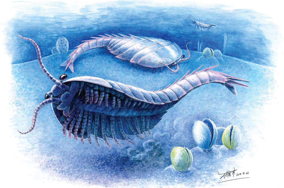
Artistic reconstruction of Guangweicaris spinatus from the Lower Cambrian Guanshan Biota, China. Illustration by Xiaodong Wang of the Yunnan Zhishui Corporation, Kunming, China. Chen et al. (2020).
The discovery and restudy of material attributed to Guangweicaris spinatus has allowed Chen et al. to produce a more accurate depiction of its external morphology and provide a more detailed diagnosis of this taxon. Among their new findings, the tripartite hypostome and the specialised post-antennal appendages in Guangweicaris spinatus indicate that these structures may be present in all Fuxianhuiids (even in Liangwangshania), probably being a shared character of this group. Moreover, the specialised post-antennal appendages can serve as a reliable diagnostic feature of Fuxianhuiids. The basal podomeres of specialised post-antennal appendages are covered by hypostomal flaps, and this would limit the movement of specialised post-antennal appendages, thus reducing the possibility that they were used for active prey capture. The basal podomere of the specialised post-antennal appendages carries gnathobasic edges with several spinose endites, are very like that in Chengjiangocaris kunmingensis, and this was very helpful for food processing. It has been reported that each prothorax segment of Fuxianhuiids corresponds to one pair of appendages, such as Fuxianhuia protensa, Chengjiangocaris kunmingensis, and Alacaris mirabilis. Due to the preservation of our specimens of Guangweicaris spinatus, we are not able to clearly define the correspondence between the prothoracic segments and appendages. Nonetheless, according to the number of anterior appendages, we speculate that each prothoracic segment corresponds to one pair of appendages. Due to the burial direction, widths of the exposed appendage are different, which indicates that the appendages of Guangweicaris spinatus are not cylindrical like those of Fuxianhuia protensa, but similar to the broad and thick oars, which are very suitable for swimming. The abdominal segments show a considerable degree of deflection, implying that the abdomen could have adjusted the direction of swimming. The dorsal axial spines on the opisthothoracic and abdominal segments, on the other hand, might have served as a defensive function.
Chen et al.'s new study of Guangweicaris spinatus is based on both new and previously published specimens, and provides important new information in the head organisation, the body segmentation and trunk appendages of this species, and improves our understanding of the autecology of Guangweicaris spinatus and the phylogenetic relationships among Fuxianhuiids. Chen et al. suggest that further work can aim at synthesising the morphological data of Fuxianhuiids and generating more complete pictures of the phylogenetic connections among Fuxianhuiids and other important evolutionary lineages of Euarthropods, as well as the ecological positions of different Fuxianhuiids in early Cambrian oceanic ecosystem, based on functional morphological and biostratigraphical analyses.
See also...


Follow Sciency Thoughts on Facebook.