Showing posts with label Sweden. Show all posts

Thursday, 11 April 2024

Hoard of medieval silver coins found on Visingsö Island, Sweden.

Archaeologists from the Jönköping County Museum carrying out surveying work ahead of the installation of geothermal heating system in a church on Visingsö, an island on Lake Vättern in south-central Sweden, have uncovered a pair of medieval graves, one of which contained a hoard of silver coins. The coins were found close to the foot of the skeleton of a man thought to have been between 20 and 25 when he died. Similar finds are common in earlier, pre-Chistian burials in Scandinavia, but their inclusion in what is thought to have been a Christian burial is very unusual.

A hoard of silver coins discovered on Visingsö Island, Sweden. Jönköping County Museum.

The hoard comprises 170 coins of a type known as silver bracteates, which had a stamped motif on one side only, which were common in Germany and Scandinavia after between the twelfth and fifteenth centuries. These coins retained their value for only a limited time, then had to be taken back to the issuing authority and restamped, which was intended as a way of ensuring that money continued to circulate and couldn't be hoarded. The coins in the Visingsö hoard are thought to date to between 1150 and 1180, and contain many stamps not previously seen by modern archaeologists.

See also...

Sunday, 7 August 2022

Understanding the threat to social cohesion presented by the emergence of a warrior caste in the Nordic Bronze Age.

The emergence of a warrior caste is seen as one of the defining features of the European Bronze Age, accompanied by concepts of a warrior ideal. The precise role of such warriors within society is debatable, and likely to have varied from place to place, but it is generally accepted that a ruling warrior elite appeared in Southern Scandinavia, northern Germany, and parts of Poland and the Netherlands. However, the exact nature of this ruling class is uncertain, with centralised kingdoms, local chiefdoms, extended ruling families, and flatter societies in which one's status was entirely dependent on personal prowess have all been suggested. 

Whatever the truth of this, the widespread occurrence of Warrior Burials (individual warriors buried with weapons and other grave goods indicative of social status), buried weapons hoards, and rock art depicting the actions of warriors, all clearly send a message that these were an important group of people.

Warriors are considered to have been important figures in the trade in metals. They are known to have taken part in (sometimes lengthy) journeys by both land and sea. Moreover, the presence of items associated with personal grooming in Warrior Burials suggests that they were image conscious, and possibly felt the need to distinguish themselves from other members of the community. Such a desire to distinguish themselves may have related to the membership of an elite caste which controlled access to some forms of knowledge, possibly concerning navigation, the symbolic interpretation of rock art, or the use of weapons.

Studies of the Bronze Age societies which produced these warriors have not generally considered the impact of their activities upon those societies. Warriors are assumed to have gone to war, fought, and then returned and continued with their lives, something which seems unlikely given our knowledge of later warrior societies, and the difficulties that modern soldiers often have re-integrating into their societies after taking part in conflicts.

In a paper published in the European Journal of Archaeology on 3 August 2022, Christian Horn of the Department of Historical Studies at the University of Gothenburg, considers the threat that such warriors may have presented to their own societies, and how those communities may have dealt with that threat.

Horn considers the evidence for warfare during the Nordic Bronze Age, and considers against whom such conflicts are likely to have been fought. He then considers the impact that the presence of the warriors who fought in these wars may have had on the societies which produced them, and looks for evidence for those societies having taken steps to cope with this threat.

Warfare appears to have been a common practice during the Nordic Bronze Age. The Early Bronze Age saw the appearance of the first swords, weapons which were to become the preferred status symbol of warrior classes for the next three millennia, as well as the refinement of spears, which had been around since at least the Neolithic, as weapons of war.

Bronze weapons are less prone to corrosion than iron weapons, which has allowed the examination of many Bronze Age weapons for traces of use. This has uncovered significant evidence of these weapons being used regularly (rather than just held as status symbols), including V-shaped notches, likely to have been caused by blows from other blades, U-shaped notches, variously interpreted as being caused by impacts against axes, shields, or bone, and signs of the weapons having been regularly repaired and maintained to keep them battle-ready.

(a)–(b) Early Bronze Age spears from Torsted (Denmark, NM K B15118.19); (c) Late Bronze Age spear from Bad Oldesloh (Germany, LMSH KS923); (d) Early Bronze Age full-hilted sword from Bragby (Sweden, SHM 14759); (e) Early Bronze Age sword from Vreta Kloster (Sweden, SHM 10419:211). Horn (2022).

Evidence of violence is also fairly common in burials from the Early Bronze Age of southern Scandinavia. Examples of this include burial from Over-Vindinge in Denmark, where the skeleton of a man aged between 50 and 60 buried between 1600 and 1500 BC was found to have a broken spear tip embedded in his pelvis. Another burial site, at Kråkerøy in Norway, produced the body of a man who was suffering from malnutrition, but had apparently died as a result of several sword blows to the head and neck. Another male body showing signs of having died from sword blows to the head and arm was found on a former seafloor at Granhammar in Sweden, possibly indicating that he died in a conflict at sea in about 800 BC.

Larger scale sites are also known from the region. At Sund in Norway the bodies of 22 men, women, and children, apparently all killed in a single incident, were found. These remains all showed signs of long-term malnutrition and hard labour, making it likely that they were slaves or other low status individuals. Finally in the Tollense Valley in Germany, a battlefield site has produced 140 sets of remains to date, with roughly 5-7% showing signs of violent injuries (this does not mean that the other remains are those of people who died peacefully, as fatal injuries to soft tissue will not be preserved in skeletonised remains). Many of these injuries appeared to be old wounds, which had healed at the time of the battle, implying warriors who had taken part in more than one conflict.

Weapons, and Human remains, give a good idea about the physical effects of violence in Early Bronze Age Scandinavia, but art has the capacity to reflect how this violence was perceived and imagined by the people involved. Rock art dating to the period is widespread across Southern Scandinavia, and includes over 6000 known depictions of Human figures, as well as more than 20 000 depictions of boats. Not all aspects of life are shown in this art. Pottery and houses are never shown, and agricultural activities are known form only a few sites. 

In order to analyse the representation of violence in these images, Horn looked at a dataset of 4000 images held by the Swedish Rock Art Research Archive. Images which were damaged or unclear were excluded from the study, leaving 3742 Human figures.  Of these, 977 could be identified as male, while 32 can be identified as female. Offensive weapons were carried by 74% of the male figures, 34% of the unidentified figures, and 6% of the female figures, while defensive weapons were carried by 575 figures, 226 of which also had an offensive weapon; of these 417 could not have their sex identified, while 158 were male (i.e 16% of unidentified figures and 15% of males had defensive weapons).

A network analysis carried out by Horn found that male figures were strongly associated with exaggerated calves, swords and boats; axes, spears, and shields were also common. Female figures were also strongly associated with exaggerated with exaggerated calves and boats, but their association with swords was much lower, and other weapons absent. Long hair was also important for female figures.

Weapons are far more common in male burials than female ones in the Scandinavian Bronze Age, and violence-related injuries more frequent on male bodies, both of which suggest a male-dominated society in which social status was linked to the ability to wage war. Many pieces of rock art also depict figures in which weapons are fused with male sexual organs, apparently indicating the concept of sexual violence, and not just against women; these scenes depict both heterosexual and homosexual couplings, as well as couplings between Humans and Animals.

Scenes potentially showing sexualized violence. (a) A warrior with erect penis stabs a sexless figure with a sword or spear (Tanum 158:1); (b) Scene depicting various forms of sexual intercourse involving armed figures, and a figure with a raised spear in a fighting position (Kville 182:1). Horn (2022).

The depiction of acts of sexual violence, combined with evidence of violence against low-status individuals, makes it unlikely that these Bronze Age Scandinavian fighters were 'clean' warriors bound by some sort of noble code. Rather, these individuals appear to have been capable of using severe violence and brutality to subdue opponents. The depictions of weapons being carried and presence of weapons in burials probably indicate that the open carrying of weapons was normal, something well documented in more recent warrior-dominated societies. The widespread depictions of violence also make it likely that violence was celebrated rather than sanctioned. In such an environment it would be very easy for warriors to turn violence against weaker groups within their own society, in order to protect or bolster their own social standing.

The Battle of Tollense took place in the thirteenth century BC, a time also noted for numerous other conflicts. This has been linked to the appearance of the Urnfield Culture, which brought with it new belief systems and practices which clashed with local religions and customs. Some of the fighters from Tollense have been shown to have come from far from a long way away from the battle, suggesting they were invading outsiders. However, the much of the conflict occurring during this period still seams to have been local in nature. 

All of the fighters appearing in Bronze Age Scandinavian Rock Art are depicted in the same way, armed with similar weapons. The Vitlycke Panel at Tanum in Sweden depicts two identical warriors fighting across a boundary, shown by a row of spots. This may imply a standardised way of drawing such fighters, regardless of their actual origin or appearance, but Horn believes that is part of a body of evidence suggesting most conflict was in fact local in nature. The spear tip which killed the man at Over-Vindage was of local manufacture, and the man buried at Granhammar appears to have been killed by a local axe. The victims of the massacre at Sund were also probably killed in some local conflict, possibly linked to slaving.

Scene showing three or four warriors with similar bodies and equipment in a fight across a border depicted by a row of cupmarks at Vitlycke (Tanum 1:1). Horn (2022).

Warriors in Bronze Age Scandinavian rock art are often closely associated with boats, which again appear standardised in form, with very little variation. A scene depicting a Horse battle at Litsleby in Sweden, thought to date to the end of the Bronze Age, shows two groups of mounted warriors, again with no difference between the figures or their equipment. All of this appears to represent Scandinavian warriors fighting other Scandinavians, rather than invaders from elsewhere in Europe with different weapons and cultural items.

Fighting scenes. (a) Warriors with spears and two boats, Massleberg (Skee 614:1); (b) Warriors on horseback armed with shields and spears, Litsleby (Tanum 72:1). Horn (2022).

Warriors crossed boundaries into the territories of other groups to commit acts of violence, and used violence to defend the boundaries of their own group. Thus they were often absent from the group, and frequently tainted by death, which many societies have regarded as spiritually polluting. This can make warriors somewhat outsiders in their own communities, figures to be venerated, but also figures viewed with a degree of fear and suspicion.

The threat presented by warriors returning to their communities may also have been more tangible. A group of warriors returning from battle in a state of arousal and being treated with high kudos can present a threat to rulers even today, with plenty of modern warlords having ceased power under these circumstances. Even where local rulers were already from a warrior class, they could easily be overthrown by returning, battle-hardened young warriors with greater fighting skills or ruthlessness. If this happens repeatedly it can seriously threaten the stability of a society, something which is likely to have been a serious risk in the Nordic Bronze Age.

Furthermore, violence can be an addictive activity, particularly in societies where it is generally applauded, and when the use of violence becomes a successful political tool it can be very hard to stop. This may have been the cause of events like the massacre at Sund and the violent killing at Grenhammar. If the majority of conflicts being fought in Scandinavia at this time were local in nature, then the communities to which warriors were returning would have closely resembled the societies which they had been fighting, further undermining any social taboos against violence at home.

If returning warriors were, as Horn theorises, a threat to their own societies both spiritually and politically, then those societies can reasonably have been expected to take measures to minimise that threat, something for which he believes he has evidence.

Horn suggests that the burial of weapons, common in Scandinavia and other areas of Europe during the Bronze Age, could represent a ritual sacrifice associated with a right of passage which enabled warriors to give up their warrior status and move on to other roles in society. Such sacrifices may have involved the ritual destruction of the weapons. In Scandinavia and Northern Europe weapons burials almost always occur within 20 km of navigable waters; i.e. less than a day's walking distance. Thus potentially these burials could represent the giving up of the weapons of an entire group of warriors involved in maritime raiding.

Interpreting ritual activities from ancient sites is difficult, and requires careful excavation of the site. Many older weapons hoards in Scandinavia (and other areas) were discovered before archaeologists had an appreciation of this, resulting in a loss of information from these sites. However, some archaeologists, even in the nineteenth century, did provide careful context of their discoveries, enabling some interpretation to be made.

The Smørumøvre Hoard was excavated by Danish Archaeologist Jens Jacob Asmussen Worsaae in the 1850s. Worsaae recorded that the hoard comprised a group of bronze spearheads tightly packed together, and embedded in the floor of a former lake, possibly indicating that the had been bound into a package without their hafts before ritual deposition. The Torstead Hoard, again from Denmark, was uncovered in the 1960s, and provides better context. This hoard comprises forty spearheads and seven axeheads, deposited between 1800 and 1600 BC, within a small stone structure, which could not have held the hafts of any of these weapons. The removal of the hafts of these weapons could be a way of ritually 'killing' them by rendering them into a useless state. Some of the spears buried at Torstead also had their tips broken off, as did several swords from a hoard at Dystrup, with other swords being damaged in other ways. The Torstead Hoard was even buried in a small stone cist, similar to the ones in which Human remains were deposited at the time, re-enforcing the idea that these weapons may have been considered dead.

This ritual killing of weapons may have represented a way for warriors to shed their identities and re-integrate into their communities, something which has been recorded in some more recent societies, and proposed as an explanation for similar hoards from the Chalcolithic of Europe and Late Bronze Age of Britain.

Rock art may also have played some role in the ritual reinstatement of warriors into society. Curiously, areas where numerous hoards of Bronze Age weapons have been found tend to have very few rock paintings, while areas with large numbers of rock paintings often lack depositions of weapons, although both tend to be local to water. 

In Sweden, the majority of Bronze Age rock art is found along the stretch of coast between Bohuslän to Mälardalen, and long the large rivers of this area, and around lakes Vänern and Vättern. At Gerum a large panel depicts 95 boats, 43 figures, 28 animals, 16 footprints (some of them shod), and 187 cupmarks. The land here has been uplifting since the end of the last Ice Age, and the panel was likely completely covered by water until shortly before carving began, with carvings being made close to the water, possibly from a boat, constantly working their way down the rock face as it became exposed.

The carvings at the Berget III site in the Tyrifjorden-Randsfjorden area of Norway are also thought to have been made from a boat. This carvings are 1.7-1.8 m above sealevel today, but when they were carved would have been only slightly above the water when they were carved. This is also thought to have been true of the rock art at Flögen in Sweden, where a series of boats (and a single Bull), were depicted over the course of the Bronze Age. These are now between 11.66 and 12.51 m above sealevel, but would have been slightly above the water when made, making it likely that the artists were either in a boat or standing on frozen ice.

(a) Density of Nordic Bronze Age metalwork based on published large catalogues. (b) Density of Nordic Bronze Age rock art. Horn (2022).

All of the rock art is placed close to water, but never in sight of the open sea. Instead it is placed within straits, fjords, and bays in places it can be seen from land, but hidden from the sea, presumably indicating that the scenes were directed inwards, for the benefit of the local community, but hidden from outsiders.

In the Early Bronze Age, warriors (and others) were buried in stone barrows and cairns, where weapons and other grave goods could be easily placed. By the Late Bronze Age, however, cremation had taken over as the predominant way of disposing of the dead, severely limiting the potential for sending metal goods with the dead. In areas where rock art was common, the amount of grave goods placed in tombs was lower, and the tombs tended to be placed close to the panels, and often directly above them, possibly indicating that these could serve as a substitute for grave goods. The proximity of these tombs and art to the sea, while at the same time being hidden for it and visible to the community may have in some way been symbolic of a warrior's final journey.

Grave goods included in Warrior Burials were often diverse and numerous, including weapons, equipment for personal grooming, and decorative items. In contrast, the weapons depicted in rock art are generally very simple in their execution. Curiously, many weapons were drawn in the earliest Nordic Bronze Age were subsequently reworked and modified extensively through the remainder of the period, sometimes having Human figures added.

Once the practice of burning the dead became predominant in the Late Bronze Age, the amount of grave goods buried with the cremated dead shrank, and became less varied. At the same time the burials themselves became less conspicuous, typically on hillslopes, or as secondary burials inside existing tombs. The process began between about 1300 and 1100 BC, when detailed images of warriors began to appear in the rock art, with these detailed warriors becoming most common and elaborate between about 950 and 720 BC.

Horn interprets this as an attempt to depict a warrior ideal in art, as this medium took over from elaborate burials as the main way to remember the dead. This art may well have been used in conjunction with other rituals, and helped to convey older narratives about warriors and heroes, but this does not mean that such rituals were not also intended to help warriors find their correct place in society, or step into and out of the role of warrior when this was required. 

Nordic Bronze Age rock art appears to have been used to celebrate an ideal of maritime warriorhood. The placement of this art, at the entrance/exit to enclosed waterways, combined with a ritual link to metalworking, may suggest that this art played a role intended to help men change their roles, from fierce maritime warriors on the outside, to responsible members of the community when at home. 

The long-distance raiding abilities of early Scandinavian cultures are often considered in historical studies, but shorter raids against nearby communities would have been much more common, and no less dangerous. Furthermore, those involved in such nearby conflicts would have had far less time for any associated bloodlust to subside before returning to their home communities, making them more dangerous at home. This could have created a need for a ritual means to re-integrate these warriors into their home communities. Horn theorises that the location of the rock art, combined with the fact that this was periodically reworked, strongly implies that this art was involved in the journey-related rituals, the most likely of which to need a ritual intervention were war raids.

The ritual sacrifice of weapons, combined with the retelling and embellishment of local warrior legends, performed at the boundaries of communities and the outside world, could have been a way for men to keep their identities as warriors in the outside world separate from their roles within their home community, thereby reducing the danger of internal conflicts within small communities.

See also...

Follow Sciency Thoughts on Facebook.

Follow Sciency Thoughts on Twitter.


Saturday, 30 October 2021

Estimating the benefits of agroforestry to European wildlife.

The term agroforestry is used to denote practices in which the cultivation of trees is integrated either with the rearing of livestock (in which case it is called silvopasturalism) or other plant crops (silvoarablism). This is a traditional practice across much of Europe, where methods such as grazing livestock in orchards are very widespread, with newer methods being developed more recently, such as short-rotation coppicing being carried out alongside rows of other crops. Systems in which productive trees are grown around the edges of fields are also sometimes considered to be agroforestry, although in these cases the trees are managed separately to the other produce, and may be under separate ownership.

 
Pigs grazing in an open Oak forest system in Spain, a system known as a 'dehesa'. Álvarez (2016).

Europe has suffered particularly severe losses of biodiversity compared to other parts of the world, and this is particularly severe in areas where intensive agriculture is prevalent. Agroforestry promotes a more diverse landscape than arable monoculture, potentially resulting in higher biodiversity. Quantifying the benefits of this could potentially lead to the system being more heavily prioritised under the European Common Agricultural Policy or any successor system.

Agroforestry systems have been well studied in tropical environments, where the evidence suggests that the system offers significant advantages in biodiversity preservation over intensive monocultural systems, but nevertheless tends to lead to reduced biodiversity compared to both primary and secondary forests. The system is less well studied in temperate regions, with most studies tending to concentrate on single groups of Animals, such as Birds or Insects. This leaves the benefits of such systems in Europe somewhat unclear, particularly as the definitions of agroforestry can vary, leading to differences in what systems are included in studies, making comparisons between studies difficult. 

 
Hazel short rotation coppice system alongside crops in Suffolk, UK. Smith et al. (2014).

In a paper published in the journal BMC Ecology and Evolution on 23 October 2021, Anne‑Christine Mupepele of Nature Conservation and Landscape Ecology and Biometry and Environmental System Analysis at the University of Freiburg, and Matteo Keller and Carsten Dormann, also of Environmental System Analysis at the University of Freiburg, present the results of a meta-analysis which combined results from a number of studies of agroforestry systems across Europe.

Mupepele et al. sought to answer three questions, 'What is the effect of agroforestry on biodiversity relative to forests, pastures, cropland or abandoned, shrub-encroached agroforestry?', 'Is the effect of agroforestry on biodiversity influenced by environmental variables, specifically the kind of agroforestry system (silvopasture or silvoarable), sampling method, the specific measure of biodiversity, sampling year, country, climate and the reference used?' and 'How strong and robust is the underlying evidence of these results?'

To which end they located 1411 previous studies of agroforestry systems in Europe, 50 of which were eventually included in the study, representing 69 individual agroforestry sites. Each of these had a direct comparison of a type of agroforestry (silvoarable or silvopastoral) to forests, cropland, pasture, and/or abandoned agroforestry systems.

 
Map of Europe with the number of effect sites per country. Mupepele et al. (2021).

The studies included in the analysis covered sites across Europe where agroforestry systems have been studied between 1984 and 2019. The majority of these sites were caried out in Iberia and the Mediterranean region, with twelve studies from Spain, eight from Portugal, five from Italy, one from France and one from Turkey. Temperate central Europe was represented by six studies from the UK, four from Romania, two each from France, Germany, and Switzerland, and one each from Belgium and northern Italy. The northern boreal region was represented by four studies from Sweden and two from Finland.

Thirty six of the included studies looked at silvopastoral systems, with thirty six studies looking at 52 sites, while silvoarable systems were the subject of thirteen studies looking at seventeen sites. The biodiversity of agroforestry was most commonly compared to that of pasture (23 sites), or forests (21 sites), then abandoned agroforestry systems (thirteen sites) and cropland (12 sites).

 
Sheep grazing in a plantation of Pine and Eucalyptus in Spain. Monica Pelliccia/Mongabay.

The different studies measured biodiversity in different ways, and concentrated on different groups. In order to make a comparison between these diverse studies, Mupepele et al. divided the measured wildlife into five groups, Arthropods, Birds, Bats, Plants, and 'Fungi plus Lichens and Bryophytes', Most of the included studies measured biodiversity at the 'species richness level', although other measures were used.

Mupepelele et al.'s results showed no overall benefit for biodiversity compared to the average derived from all systems. However, silvoarable systems were found to host considerably more biodiversity than other croplands, although they generally hosted less biodiversity than forests. Silvopastoral systems produced less clear results, with measures often producing conflicting results in different studies (i.e. one study might show higher Avian biodiversity in a silvopastoral system than a forest, while another showed the reverse.

Birds and Artropods were typically found at higher levels of diversity in agroforestry envoronments than other systems, Where the original group sorted Arthropods into different groups (e.g. Bees, Beetles and Spiders', then this biodiversity increassed, although this was across all environments, with no change in the beneficial effect of agroforestry.

 
Cereal crops grown alongside trees in Bedfordshire, UK. Agroforestry Research Trust.

Mupepele et al. note that the quality of the studies they were referencing varied somewhat, with some using replicated experimentation with clear controls, whilst others were more observational in nature. To compensate for this, they tried applying a statistical weighting method that gave more value to the more statistically strong studies, but found this made no difference to the overall result. They also carried out funnel plot and Egger’s regression tests for undetected biases in their data, but did not find bias was a problem.

A previous  meta-analysis led by Mario Torralba of the Department of Geosciences and Natural Resource Management at the University of Copenhagen found that agroforestry had a much stronger impact on biodiversity, which caused Mupepele et al. to consider the differences between their findings and that of the earlier study. They note that Torralba et al.'s study was published in 2016, and contained the results from two studies published in 2015 on the benefits of agroforestry in Mediterranean ecosystems, both of which produced very strong positive results, and that if these were excluded from Torrialba et al.'s data then the result was closer to that of Mupepele et al. who included several post 2015 studies with less clear results.

Properly done, meta-analyses can provide a powerful tool for understanding ecological systems in ways not possible from individual studies or unsystematic literature searches. However, the robustness of these results is dependent on the methods used to analyse the data, and in particular the use of weighting to take into account the quality of the studies being referenced. This needs to be done carefully, as failure to apply the right weighting can often lead to very different results. This said, applying weighting to Mupepele et al.'s results resulted in no significant change in the outcome of the study, which strongly supports the robustness of their findings. 

The application of repeated meta-analyses to the same data set can reveal changes over time, as new studies add to the overall picture, dampening the results from atypical studies that might have a profound impact on a smaller data-set. By building a cumulative model in which data were added in chronological order, Mupepele et al. were able to demonstrate that the impact of agroforestry upon biodiversity remained essentially unchanged over time, despite the presence of some anomalous data. They do, however, note that silvoarable systems make up a relatively small proportion of the whole, and that the addition of a higher proportion of studies of these systems in future might change the results of the meta-analysis.

 
Merino Sheep under a Cork Oak in a montado silvopastoral system in Portugal. European Agroforestry Foundation.

The ability to reproduce results is an important principle in science, but can be difficult in fields like ecology, which look at complex natural systems, no two of which are ever completely the same. Mupepele et al.'s results differed strongly from the earlier results of Torralba et al., resulting in their drawing different conclusions; Torralba et al. concluded that agroforestry has a general positive impact upon biodiversity, while Mupepele et al. concluded that this benefit was only clear when agroforestry was compared to croplands, despite both studies having used much of the same data. Mupepele et al. note that Torralba et al. included hedgerows and woody riparian buffers to agricultural land as agroforestry, while Mupepele et al. excluded them on the basis that they are not emplaced for silvicultural purposes (i.e. the trees used in these settings are grown for their value as boundaries, not as a crop in themselves). Neither did Torralba et al. include data from studies which suggested agroforestry had a negative impact on biodiversity. Mupepele et al. believe that scientists should be very clear about what data they are including in meta-analyses, the criteria for choosing this data, and the reasons to do so, in order to help policy-makers judge the significance of different studies. 

Mupepele et al. conclude that silvoarable systems produce an increase in biodiversity compared to conventional croplands, particularly with regard to Birds and Arthropods, but that this increase is not large, and there was no overall positive benefit of agroforestry to all other settings. Notably, silvopasturalism showed no clear benefit over either forestry or conventional pasturelands. Where previous studies have produced enthusiastic support for agroforestry, and strongly suggested these systems are linked to a significant increase in biodiversity, Mupepele take a more cautious approach, noting that relatively few studies find an unqualified link between agroforestry and increased biodiversity, and that literature reviews and meta-analyses need to be careful to include both the positive and negative impacts of systems when drawing on data from multiple studies. Nevertheless, they do conclude that agroforestry can have a positive impact on biodiversity under some circumstances, as well as providing carbon sequestration and other ecosystem services, and that a better understanding of how these systems work could lead to more informed future decisions by policy makers.

See also...


Follow Sciency Thoughts on Facebook.

Follow Sciency Thoughts on Twitter


Monday, 29 June 2020

Using high-resolution computed-tomography to study pathologies in Belemnites.

The term pathology refers to malformations of specimens of a population or species that are caused by exogenic or endogenic processes. Exogenic processes include injuries due to a predator attack, parasite infections, and the colonisation of the shell during lifetime. Endogenic processes include infections, mutations, or other illnesses. The term palaeopathology is used pathological features of fossils. To ensure accurate palaeoecological interpretations it is required to distinguish between pathologies and pseudopathologies, e.g. tectonically deformed Belemnite rostra. Fractures of tectonically deformed rostra are filled with blocky calcite cement instead of biogenic radial fibrous calcite. Such rostra, therefore, do not contribute to our understanding of Belemnite palaeoecology. The visible reactions of an affected specimen are called symptoms while several abnormalities that occur in a single specimen are described as complex syndromes. As a rule, only reactions to the disturbing factor should be considered to characterise pathological symptoms or syndromes. The disturbing factor (pathogen) underlying the palaeopathological features of fossil hardparts is only rarely identified. In exceptional cases, specific bite traces with a distinct shape or morphology of shell breakage are preserved and allow the identification of specific producers such as Crustaceans, Coleoids, Fishes, or Reptiles.

Pathologies of modern and fossil Cephalopods have received proportionally large attention. The first reports on malformed Cephalopods were made by Johann Samuel Schröter in 1774, although he did not recognise them as such. Later, pathological specimens were regarded as monstrous natural curiosities. Later, pathological specimens were regarded as monstrous natural curiosities. The potential factors causing abnormal morphologies were first discussed in the 1890s. Consequently, exploration of the disturbing factors was increasingly used to reconstruct the palaeobiology and palaeoecology of the affected specimens and helped in reconstructing phylogenetic processes. 

Due to their internal shell, Coleoid Cephalopods such as Belemnites are not affected by epizoans that settle on the external shells of Nautiloids or Ammonoids. In externally shelled Cephalopods, damage to the phragmocone can result in uncontrolled flooding of the chambers, which in turn leads to lethal drowning by shell implosion. In contrast, the internal shell of Coleoids often remains in the mantle sac after being fractured. In case the animal survived the attack, damaged skeletal parts are often successfully repaired.

Data gained from palaeopathological studies of Cephalopods helped in estimating the efficiency of the buoyancy apparatus. Synecological factors can be reconstructed by the recognition of parasite infections or the presence of epizoa which in turn helped to reconstruct potential habitats and life habits. Lethal or sublethal injuries also add information on predator-prey interactions. Recent studies have provided a detailed overview on palaeopathological Belemnite rostra and introduced forma aegra-types for recurring kinds of abnormalities following the scheme developed for Ammonoids. It should be noted that forma aegra-types do not refer to taxonomic entities but are a type of open nomenclature for the classification of pathologies. For fossil coleoid cephalopods, including belemnites, three categories of palaeopathologies can be distinguished based on their causes: (i) regeneration of injuries including inflammatory infection often caused by an unsuccessful predatory attack, (ii) prod traces due to collisions, (iii) parasite infection.

Most malformations of Belemnite rostra resulted from mechanical injuries, often caused by failed predation attempts, and less commonly from parasite infestations. It has been argued that rostra broke due to mechanical stress induced during active digging in the sediment with the rostrum. Due to the likely nektic lifestyle of belemnites and the counterweight function of the rostrum that allows a horizontal swimming position, injuries due to digging are highly unlikely.

In a paper published in the journal Acta Palaeontologica Polonica on 24 March 2020, René Hoffmann and Kevin Stevens of the Institut für Geologie, Mineralogie und Geophysik at the Ruhr-Universität Bochum, Marie-Claire Picollier of St. Pantaly d’Excideuil, Jörg Mutterlose, also of the Institut für Geologie, Mineralogie und Geophysik at the Ruhr-Universität Bochum, and Christian Klug of the Paläontologisches Institut und Museum at Universität Zürich, demonstrate that nano-computed-tomography scanning of pathological Belemnite rostra provide data with an excellent resolution, allowing a detailed description of internal features including malformations.

High resolution and good signal to noise ratios in the resulting computed-tomography-images are important for the successful study of pathological belemnite rostra because of the continuous deposition of concentric growth increments. That mode of growth resulted in a successive attenuation of the primary morphology of the injury and the resulting symptom due to the deposition of post-traumatic growth increments. Based on computed-tomography-data, Hoffmann et al. provide short descriptions of the different types of palaeopathologies and discuss their potential causes. Furthermore, they demonstrate additional applications such as the recognition of a heterogeneous or homogenous composition of Belemnite rostra due to diagenesis, sedimentary fill, structures of the rostrum surface, and internal structures of the phragmocone such as septal spacing and the position of the siphuncle.

A total of 18 pathological belemnite rostra from Lower Jurassic (Toarcian) to Upper Cretaceous (Campanian) deposits, were computed-tomography-scanned. The material can be attributed to the following genera (in alphabetical order, number of studied specimens in parenthesis): ?Acrocoelites sp. (1), Belemnellocamax spp. (4), Belemnitella sp. (2), Duvalia emerici(1), Goniocamax sp. (1), Gonioteuthis spp. (5), Hibolithes jaculoides (2), Neoclavibelus subclavatus (1), and Pseudobelus sp. (1).

Hoffmann et al. describe various pathologies encountered according to their morphological expressions including two-tipped specimens, blisters and pearls, broken rostra, which healed straight, rostra with a kink or bent rostra. Differences in grey scale values in the images suggest different materials such as calcite, silica, pyrite, or sediment. The growth increments of the rostrum, for example, are visible in computed-tomography-images due to the varying amount of organic matter, which has significant lower absorption properties compared to pure calcite. This implies that organic-rich growth increments are darker compared to those composed of pure calcite.

Rostrum of Belemnite ?Acrocoelites sp., PIMUZ 37346, Toarcian, Altdorf (southwest-Germany) with apex malformation. (A) Overview (A₁), (A₂). (B) Close-up of the two apices, showing the radial furrows covering the apex that represent the 'normal' tip of the rostrum (B₁)–(B₅). Hoffmann et al. (2020).

One ?Acrocoelites sp. specimen (PIMUZ 37346) and one Gonioteuthis sp. specimen (RUB-Pal 11264) rostrum are characterised by two divergent apices. In both specimens one apex represents the continuation of the original rostrum axis and bears radial striation on its outer surface. The second apex of ?Acrocoelites sp. bears one strong dorsal furrow. Superficially, the arrangement of the ?Acrocoelites sp. apices resembles a crustacean claw. Unfortunately, the computed-tomography-data did not reveal the course of growth increments or other internal structures, which suggests an irregular formation of the skeletal calcite.

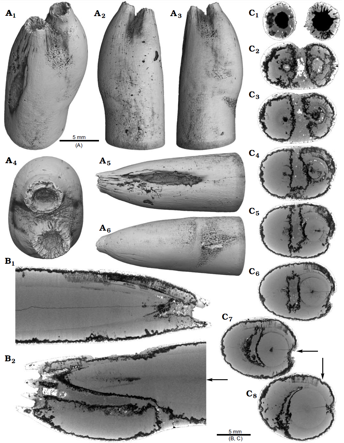
Rostrum of Belemnite Gonioteuthis sp., RUB-Pal 11264, Campanian, Höver (northwest-Germany) with apex malformation. (A) Surface images showing the hollow opening of the two apices, and the partially dissolved or poorly mineralized rostrum (A₁)–(A₆); arrow in (A₅) indicates meandering trace fossil. (B) Longitudinal section showing growth increments, apical line, and irregular mineralized layers (B₁), (B₂). (C) Cross sections with increasing distance to the apices (C₁)–(C₈), showing the process of apex separation, only the straight continuous apex (arrows) develops distinct growth increments and the apical line (see also the arrow in B₂), note: the area between the apices is filled with sediment (diffuse grey area) and pyrite (white area), black area silicified. Hoffmann et al. (2020).

The continuation of the normal rostrum growth in the Gonioteuthis sp. is supported by the presence of the apical line and growth increments. The rostrum surface of this Gonioteuthis sp. partially shows dissolution features or poorly mineralized layers exposing triangular structures. Some of these features suggest the presence of meandering trace fossils. Both apices were partially hollow and subsequently filled with sediment. Development of the two apices is reflected in the image series based on virtual cross sections. Areas darker than the calcitic parts of the rostrum and mostly restricted to the rostrum surface indicate the presence of silica.

One Neoclavibelus subclavatus (SNSB-BSPG-83264) and one Belemnitella sp. (RE 551.763.333. A 5238) rostrum each show a bump-like structure. Virtual cross sections of the Neoclavibelus subclavatus rostrum reveal the presence of sediment inside the rostrum. This debris was subsequently overgrown by calcite precipitated by the Belemnite, causing the malformation. The longitudinal section shows that the contaminant covers parts of the juvenile rostrum, thereby causing a deviation in growth increment orientation. White areas suggest the presence of pyrite, which completely absorbs the X-rays. The bump-like structure in the Belemnitella sp. rostrum was probably caused by a similar process. In this case, the foreign material caused the bending of dorsolateral depressions covering the rostrum surface. Darker areas along the margin of the rostrum suggest the presence of silica. For both species, Hoffmann et al. also provide high-resolution computed-tomography-images. A large part of the Goniocamax sp. rostrum shows signs of exfoliation. Hoffmann et al.'s computed-tomography-based examination of the area where outer rostral layers thin out reveals, that sediment was deposited between distinct growth increments of the belemnite rostrum. Both specimens show hollow areas, partially filled with carbonate crystals, within the rostrum forming blisters. The Goniocamax sp. blister is elongated with a spiral morphology. The sediment filled blister contrasts with the massive bump-like structures of another two specimens, undetermined specimen CASP K9068 and Duvalia emerici. The undetermined rostrum shows a distinctive layer from which the growth perturbation starts in longitudinal- and cross section. This structure resembles the phenomenon described for Neoclavibelus subclavatus and Belemnitella sp. For Duvalia emerici, no such perturbation has been identified.

(A) Rostrum of Belemnite Neoclavibelus subclavatus, SNSB-BSPG-83264, Toarcian, Mistelgau (southwest-Germany) with blister malformation. Surface images showing the bump-shaped irregular rostrum growth (A₁)–(A₄); longitudinal sections showing presence of sediment (diffuse grey) and pyrite (white) within the rostrum, and increasing irregular growth increments (A₅), (A₆). (B) Rostrum of Belemnite Belemnitella sp., RE 551.763.333 A 5238, Late Cretaceous, northwest-Germany with forma aegra bullata (coll. Baschin). Surface images showing the bump-shaped irregular rostrum growth and imprints of an organic network on the rostrum surface in dorsolateral (B₁), (B₂) and ventrolateral (B₃), (B₄) views; longitudinal sections (B₅), (B₆), and cross section (B₇). (B₅)–(B₇) showing silification (black) along the rostrum surface but also along the malformed area. Hoffmann et al. (2020).

One Belemnite assigned to Gonioteuthis sp. (RUB-Pal 11301) and one assigned to Hibolithes sp. (RUB-Pal 11303) have fractured rostra. Due to strong silification, the results from the computed-tomography-scans remain inconclusive for the Gonioteuthis sp. rostrum. The apical line appears to form a continuous line without the formation of a blunt rostrum. Parts of the rostrum increments seem to be poorly mineralised, causing parts of the juvenile rostrum to stick out where normally the alveolus is situated. This example demonstrates the potential of the computed-tomography technique to better understand the mineralogical composition of Belemnite rostra. In Hoffmann et al.'s specimen silification rings correspond to darker areas in virtual section images. Virtual sections of the Hibolithes jaculoides rostrum reveal a broken juvenile rostrum with the apex missing. The fracture was overgrown by irregularly formed rostrum material. While the outer layers show regularly formed growth increments, the internal part appears to be chaotic without forming an apical line. This irregular growth results in a rostrum morphology comparable to the genus Produvalia.

(A) Rostrum of Belemnite cf. Hibolithes jaculoides, SNSB-BSPG-83251, Hauterivian, Heligoland (north-Germany) with blister malformation. Surface images showing the bump-shaped irregular rostrum growth, in lateral (A₁), dorsal (A₂), lateral (A₃), and ventral (A₄) views. Longitudinal section showing open pore space partially filled with carbonate cements (A₅). (B) Rostrum of Belemnite Goniocamax sp., MGUH 32024, Santonian, Bavnodde (Denmark) with blister malformation. Surface images of the rostrum showing the bump-shaped irregular rostrum growth (B₁)–(B₄). Longitudinal section with growth increments, apical line, and the bump-like structure with pore space (black) (B₅). Cross section through the malformed area showing the pore space partially filled with carbonate cement (B₆). Marginal section through the malformed area (B₇). (C) Undetermined Belemnite, CASP K9068, Greenland, Early Cretaceous with blister malformation. Surface images of the rostrum showing the irregular rostrum area (C₁)–(C₃). Longitudinal section showing growth increments, apical line and a distinct growth increment at which the malformation starts (C₄). Cross section that shows the distinct growth increment, and a horizontal fracture (white), as well as grains of pyrite (white) (C₅). (D) Rostrum of Belemnite Duvalia emerici, RUB-Pal 22170, Late Valanginian, Laborel (France) with blister malformation. Surface images of the rostrum showing the irregular rostrum area (half-pearl shaped), in ventral (D₁), lateral (D₂), (D₃), and dorsal (D₄) views. Longitudinal section through the malformed area (D₅). Cross section, both section reveal that the structure is massive, i.e., not a blister pearl (D₆). Hoffmann et al. (2020).

Two specimens of Gonioteuthis spp. display a marked distortion of the rostrum. Both, the rostrum and the phragmocone were affected in one specimen, post mortem overgrown by an Oyster In this case parts of the juvenile rostrum are not preserved (RUB-Pal 11302). The rostrum surface shows faint radial striae. The second specimen (SNSB-BSPG-83246) yields an intact phragmocone but parts of the juvenile rostrum broke off. The phragmocone is partially filled with pyrite (white area) and sediment (irregular greyish). Due to partial silification, the growth increments are well visible. Areas with strong bending are silicified, and the rostrum surface is rough due to dissolution, poor mineralisation, or both. Although strongly bent, the rostrum shows growth increments and an apical line.

A. Rostrum of Belemnite Gonioteuthis sp., RUB-Pal 11301, Campanian, Höver (northwest-Germany) with blunt rostra. Surface images showing no irregularities except for silification rings (A₁)–(A₃). Longitudinal section showing a homogenous internal rostrum with a dark layer along its outer margin (silica) (A₄). Volume rendering image shows a darker centre due to the maximum thickness of the structure but no additional features (A₅). Detail of the rostrum surface showing silification rings (A₆). Cross section with a homogeneous centre and a dark margin (A₇). (B) Rostrum of Belemnite Hibolithes jaculoides, RUB-Pal 11303, Hauterivian, Resse (northwest-Germany) with blunt rostra. Surface images showing the overall irregular rostrum morphology and the blunt and hollow apex in lateral (B₁), (B₂), ventral (B₃), and dorsal (B₄), apical (B₇) views. Longitudinal sections (central, subcentral) showing the broken juvenile rostrum, parts of the preserved phragmocone, and notable the lack of the apical line after the injury took place, note the irregular outline of the hollow central canal (B₅), (B₆). Cross section with the juvenile rostrum, and subsequently deposited homogeneous material, white areas indicate the presence of pyrite (B₈). Hoffmann et al. (2020).

One rostrum each of Gonioteuthis sp. and Pseudobelus sp. are strongly distorted resulting in a change of growth direction. The specimen of Gonioteuthis sp. (SNSBBSPG-83370) deviates from the normal (straight) growth direction by about 90° with its apex pointing slightly towards the alveolus. A small part of the phragmocone, now filled with sediment, is preserved and the rostrum is heavily silicified. A few growth increments and a faint trace of the apical line are visible. The rostrum of Pseudobelus sp. (RUB-Pal 3196) shows the strongest deformation reported here. Computed-tomography-data suggest three traumatic events resulting in fractures and finally a reversion of the growth direction towards the head of the animal. Growth increments and apical line are, however, well visible in the longitudinal section. The cross-section image shows four growth zones representing a temporal sequence implying that the rostrum broke during four successive and independent events.

Rostra of Belemnite Gonioteuthis spp. (A) RUB-Pal 11302, Campanian, Höver (northwest-Germany) with bent rostra. Surface images showing the knee-like morphology of the rostrum, and the attachment-base of an Oyster, in lateral (A₁), (A₄), dorsal (A₂), and ventral (A₄) views; note the weak furrows in (A₃). Median sections perpendicular to each other showing silicified areas (darker) and the broken phragmocone now filled with sediment, no additional internal feature visible (A₅), (A₆). (B) SNSB-BSPG-83246, Campanian, Höver (northwest-Germany) with bent rostra. Surface images showing the knee-like morphology of the rostrum, in ventral (B₁), lateral (B₂), (B₄), and dorsal (B₅) views. Cross section (B₃). Median sections showing silicified areas specifically at places heavily bent (darker) (B₆), (B₇); see also (A₅), (A₆) for the same phenomenon. Black box indicates close up in (B₇), showing the broken juvenile rostrum with growth increment (bent rostra), and the phragmocone partially filled with pyrite (white). Hoffmann et al. (2020).

Four malformed specimens of Belemnellocamax sp. with fractured rostra show weak to strong reactions that occurred during the healing process. One specimen (GM 04.1918) is a knee-like bent rostrum with a spongy, highly porous internal structure. A second specimen (GM 02.1918) has a short rostrum with a blunt apex due to a spherical depression. The virtual section reveals a broken juvenile rostrum associated with a porous area surrounding it. The third specimen (GM 01.1918) also has a broken juvenile septum, which extended beyond growth increments of older parts of the rostrum. Subsequently formed growth increments never covered the juvenile rostrum entirely, forming a callus-like structure. Parts of the alveolar region are preserved allowing for a correct orientation of the rostrum. Specimen four (GM 03.1918) shows a slightly irregular rostrum morphology with curved depressions and a small hole on the rostrum surface. Virtual longitudinal sections reveal that the hole at the surface is connected to a canal that appears to start in the central alveolar region. Growth increments, otherwise well visible, disappear around the canal.

(A) Rostrum of Belemnite Gonioteuthis sp., SNSB-BSPG-83370, Campanian, Höver (northwest-Germany) with hook-shaped rostra. Surface images showing the knee-like strongly bent and irregular morphology of the rostrum (A₁)–(A₄). Median section showing the irregular internal silification of the rostrum indicating poorly mineralized areas, growth and apical line partially visible (A₅). Volume rendering image with the darkest areas represented by the thickest or densest areas (A₆). (B) Rostrum of Belemnite Pseudobelus sp., RUB-Pal 3196, Valanginian, Barret-Meouge (France) with hook-shaped rostra. Surface images showing the strongly bent and irregular morphology of the rostrum with the apex growth in anterior direction (B₃)–(B₆). Median section overview and close up showing growth increments and the presence of pyrite along the apical line (white) (B₁), (B₇), (B₈). Cross section showing four growth center representing a temporal sequence (B₂). Hoffmann et al. (2020).

Like all Coleoid Cephalopods, Belemnites formed their internal shell within the shell sac. Belemnite rostra were secreted by the muscular mantle that is fused above the shell mantle in Endocochleate Cephalopods. Growth increments are rhythmically added to the rostrum by simultaneous accretion on its surface. This mode of biomineralisation is comparable to the formation of the cuttlebone sheath of Sepiids including the spine, which contains high amounts of organic material. The rostrum is composed of radiaxial calcite fibres that cross the concentric growth increments.

Rostra of Belemnite Belemnellocamax spp., Campanian, Ivo Klack (Sweden). (A) GM 04.1918, collar formation. Surface images showing the knee-like morphology of the rostrum (A₁)–(A₄). Longitudinal section showing the porous apical area (black) (A₅). Alveolar region (A₆). Cross section showing pore space within the rostrum (black) (A₇). (B) GM 02.1918, collar formation. Alveolar region (B₁). Cross section with the juvenile rostrum in its centre (B₂). Rostrum surface of the short, stout rostrum with a blunt apex and a concave impression (?dissolution feature), in lateral (B₃)–(B₆) and apical (B₇) views. Central and subcentral longitudinal section with the juvenile rostrum and the surrounding porous area (B₈), (B₉). Hoffmann et al. (2020).

The recent application of non-invasive imaging methods other than magnetic resonance imaging to Belemnite rostra, such as synchrotron radiation based micro-computed-tomography, revealed the presence of a new rostral ultrastructure, suggesting a composition of two distinct calcite phases. Micro-computed-tomography has previously been used to describe bioerosion features (trace fossils), which are often found on Belemnite rostra, and to determine rostrum volumes to reconstruct temporal changes in Belemnite size. The description of a variety of pathological phenomena highlights the advantage of the higher spatial resolution and suitable contrast properties accessible by computed-tomography-derived volume data to explore the three-dimensional nature of pathological specimens.

Rostra of belemnite Belemnellocamax spp. (A) GM 01.1918, Campanian, Ivo Klack (Sweden) with collar formation. Surface images showing the collar-like structure and parts of the juvenile rostrum (A₁)–(A₄). Longitudinal section (A₅) showing the sediment filled phragmocone with the protoconch, the Klähn’sche plane, the juvenile rostrum, and the material added after the traumatic event forming a collar-like structure but not covering the juvenile rostrum completely. Alveolar region (A₆). Opposite side with the juvenile rostrum sticking out (A₇). (B) GM 03.1918, Campanian, Ugnsmunnarna (Sweden) with fcollar formation. Irregular outline morphology of the rostrum (B₁)–(B₄). Longitudinal section to show the canal, growth increments (B₅), (B₆). Cross section through the apical line and the canal (B₇). Hoffmann et al. (2020).

The following malformation categories were used to group the various types of malformation:

Apex malformation describes a duplication or multiplication (up to five apices known so far) of the rostrum apex. It has been argued that due to traumatic events (mechani cal injuries) or the activity of parasites parts of the apex forming mantle epithelium were separated into locally independent secretion centres that formed several apices. Hoffmann et al. report apex malformations in one specimen of ?Acrocoelites sp. and Gonioteuthis sp. each.

Blister malformation includes blister thickenings of the rostrum often enclosing a hollow space, which may indicate the earlier presence of endoparasites. The locally proliferating mineralization of the rostrum can in rare cases lead to the formation of a second rostrum paralleling the primary rostrum. Hoffmann et al. report blister malformations in one specimen of Neoclavibelus subclavatus, Belemnitella sp., Hibolithes jaculoides, Goniocamax sp., an undetermined Belemnite, and Duvalia emerici each.

Blunt rostra malformation contains more or less club-shaped rostra that are thicker and shorter compared to normal specimens. They are the result of broken juvenile rostra. Depending on the timing and severity of the fracture, slightly to heavily deformed rostra result. Often, the earliest parts of the phragmocone including the initial chamber are affected, Extreme shortenings of the rostrum results in a droplet-shaped morphology. Hoffmann et al. report blunt rostra malformation in one specimen of Gonioteuthis sp., one specimen of Hibolithes jaculoides, and two Belemnellocamax sp. specimens.

Bent rostra malformation is characterised by multiple rostrum fractures. The dislocated rostrum fragments were covered by post-traumatic growth increments. Depending on the size and position of the fragments within the mantle sac, distortions of variable angles (within a plane or a screw-like arrangement of the fragments in different planes) resulted. This phenomenon includes specimens with fragments grown together sub-parallel to each other after the attack. Hoffmann et al. report bent rostra malformation in two specimens of Gonioteuthis spp. and one specimen of Belemnellocamax sp.

Hook-shaped rostra are often the result of rostrum fractures that occurred during early ontogeny. Parts of the broken rostrum become dislocated and post-traumatic growth is preferentially oriented towards one side of the rostrum resulting in a change of growth direction. In cases where the rostrum becomes significantly shorter, a form transitional between the hook-shaped rostra and collar formation exists. A 180° reversal of the growth direction of the rostrum towards the head of the animal due to several fractures is documented by Hoffmann et al. for the first time. Hoffmann et al. report Hoffmann et al. in one specimen of Gonioteuthis sp. and Pseudobelus sp. each.

Sceptre-shaped rostra occur where a mechanical injury of the mantle sac epithelium  leads to a partial lack of growth increments. This is related to the disabled function of the injured epithelium to secrete shell material. This symptom is often observed in the apical area of the rostrum causing an incomplete apex. In this case juvenile, pre-traumatic parts of the rostrum form the apex. A damage of the lateral epithelium is related to a thinning of the rostrum in the affected area and result in a sceptre-shaped rostrum. Hoffmann et al. report sceptre-shaped rostra in one specimen of Gonioteuthis sp.

Collar formation deformation describes flap-like outgrowths at the sub-apical rostrum area without visible injury. This symptom has been reported from a variety of Jurassic and Cretaceous Belemnites and possibly results from a parasitic infestation This infestation caused the formation of a collar that is regarded as an effluence channel and potentially indicates an inflammation of the mantle sac epithelium. Hoffmann et al. report collar formation in one specimen of Belemnellocamax sp.

Most studies about modern Molluscan parasites deal with parasites of Bivalves. Bivalves host a diverse group of parasites and disease-causing agents such as, e.g., Viruses, Prokaryotes, Fungi, Protists, Parazoans, and Metazoans (Platyhelminths, Annelids, Molluscs, Bryozoans, and Arthropods). The most abundant parasites are endoparasitic Flukes (Trematoda, Digenea), which have also been observed in Gastropods. Larvae of Digenean Trematodes (Gymnophallidae) sometimes lead to the formation of blisters and have been found in fossil Bivalves dating back to the Triassic. Blister formation, however, can have a variety of causes, some of which are not due to parasites. Identification of a specific parasite taxon, however, is hampered by the fact that parasites are typically small-bodied (submillimetre) and usually lack biomineralised hardparts.

In modern Cephalopods, parasites are known from many species from all major oceans ranging from coastal, shelf, to oceanic and deep-sea environments. This is no surprise given that Cephalopods are a key trophic element in marine ecosystems. One study documented 200 species of endoparasites in Cephalopods with macroparasites such as Nematodes, Copepods, and Isopods. These predominantly affect non-mineralized tissues such as the digestive tract and the gills. The infestation of mainly non-mineralised tissues limits the fossil preservation potential of parasite activity and the resulting host reaction.

For Belemnites, most reported pathologies are due to survived predator attacks (e.g. bent rostra), a few malformations are assigned to the activity of parasites like, blister malformation, collar formation, and apex malformation. Although Hoffmann et al. identify a parasite infection as the most likely trigger of these pathologies, attributing the deformations to a specific parasite taxon is difficult to impossible. While some parasites leave characteristic patterns on their host, this is not necessarily definitive evidence of their presence. Different parasites can leave similar traces and taxonomically distant parasites can inflict similar symptoms on their hosts because of convergence in the evolution of host-exploitation strategies. Differential diagnosis for palaeopathological phenomena may lead to the conclusion that two or more alternative conditions remain as potential triggers.

Hoffmann et al.'s high-resolution computed-tomography-data represent an innovative and non-destructive way to analyse the internal structure of Belemnite rostra. computed-tomography-images revealed internal structures that allow the examination of a variety of pathological phenomena and to differentiate between different types of pathology. Most importantly, the computed-tomography-data allow to infer potential causes of these deformations (predator attack or parasite activity), therefore going beyond a classification of pathologies from the surface of a specimen alone. Hoffmann et al. thereby demonstrated the usefulness and feasibility of the application of non-invasive imaging methods to the field of palaeopathology. The data gained can be further used for the identification of various diagenetic alteration features in Belemnite rostra. computed-tomography-data turned out to perform better than magnetic resonance imaging-data in terms of spatial resolution and contrast.

See also...


https://sciencythoughts.blogspot.com/2020/06/ptychites-embreei-new-species-of.htmlhttps://sciencythoughts.blogspot.com/2020/06/remarkably-intact-giant-squid-found-on.html
https://sciencythoughts.blogspot.com/2020/06/coldwater-lamellorthoceratid.htmlhttps://sciencythoughts.blogspot.com/2020/06/early-ontogenetic-growth-stages-of.html
https://sciencythoughts.blogspot.com/2020/02/sepiella-japonica-paternity-testing.htmlhttps://sciencythoughts.blogspot.com/2020/02/evidence-for-predation-of-soft-bodied.html
Follow Sciency Thoughts on Facebook.