Showing posts with label Hominin. Show all posts

Monday, 14 October 2019

Using morphometric analysis to identify the Lincoln Cave Hominin teeth.

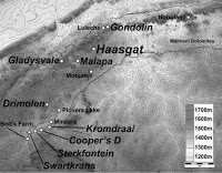
Hominin remains from the Middle Pleistocene of Africa are rare, and those that do exist are often highly fragmentary, frustrating for scientists studying the origin of our own species. One notable, and dramatic, exception to this is the fossils of the Dinaledi Chamber of the Rising Star Cave system, part of the Maropeng Cradle of Humankind complex of caves in Gauteng State, to the northwest of Johannesburg, South Africa, where over 1550 pieces of bone belonging to at least fifteen individuals, were found within a single chamber, dated to between 335 000 and 236 000 years ago. These remains were used to describe a new species of Hominin, Homo naledi. This discovery has led to a renewed interest in the fragmentary remains of similar age found in other caves of similar age in the same area, including the Hominin teeth found in Lincoln Cave between 1997 and 2003, and Sterkfontein Cave L/63, which is undated, but contains similar artifacts and animal remains to Lincoln Cave.


Morphometric analysis is a tool used by palaeontologists, archaeologists, anthropologists and forensic pathologists to analyse and compare specimens. It relies on taking numerous measurements of an object such as a bone or shell, and comparing both these measurements and ratios between measurements to those obtained from other specimens in order to establish relationships between them. Traditionally these measurements have been obtained using tape measures and callipers, but modern scientists often use more sophisticated tools such as structured light scanners, which are capable of building highly detailed three dimensional models of specimens. 

The Lincoln Cave is located in the Lincoln-Fault Cave system adjacent to the Sterkfontein Cave system. It is divided in two by an old ramp made by limestone miners, named Lincoln Cave North and Lincoln Cave South. Lincoln Cave North, consists of calcified deposits while Lincoln Cave South is uncalcified. The cave dates to between 252 600 and 115 300 years ago based on uranium series dating of flowstones, dates comparable to the 335 000 to 236 000 years old estimated for the Dinaledi Chamber.

Flowstone is formed by the deposition of calcium carbonate onto surfaces by evaporating water; typically water that has flowed through limestone deposits then run out onto a surface such as a cave wall or cliff face before evaporating. The most obvious examples of this are stalagmites and stalactites, though many caves have an interior surfaces covered by flowstone.  Uranium-thorium dating works because uranium decays to thorium at a known rate, so that the ratio  of the two elements in minerals that naturally incorporate uranium but not thorium can be used to establish a date for the minerals. Neither uranium nor thorium are typically found in carbonate deposits, but uranium can be absorbed into these minerals as they form, whereas thorium cannot. Thus all the uranium and thorium in a sample of calcium carbonate will have been uranium when the deposit was laid down, and the ratio of the two elements can be used to date the rock (the more thorium there is, the older the rock).

Three Hominin teeth were recovered during the Lincoln Cave excavations, as well as a number of stone tools and a variety of animal remains. These are StW 591 is an unerupted permanent left upper first incisor, StW 592 an unerupted left maxillary first molar, and StW 593 a lower right first incisor. A single tooth was found during the excavation of Sterkfontein L/63, a a right maxillary canine identified as StW 585.

Brophy et al. were able to use two of these teeth for the study, StW 585, which was was directly compared with the Homo naledi maxillary permanent canines from the Dinaledi Chamber at the University of the Witwatersrand, and StW 592, which was compared with Homo naledi maxillary first molars based on the description, image and measurements given in their original description of the Lincoln Cave material by Reynolds et al. (2007).

Lingual view of StW 585 from L/63 area of Sterkfontein Cave (centre) and Homo naledi maxillary permanent canines from the Dinaledi Chamber. Left to right: U.W. 101-337 RC, U.W. 101-908 RC, StW 585 RC, U.W. 101-501 LC, U.W. 101-412 LC. Arrow shows large tuberculum dentale of StW 585. Brophy et al. (2019).

StW 585 and the Homo naledi maxillary permanent canines from the Dinaledi Chamber differ in significant ways. Lingually (on the inner side of the tooth), StW 585 has a large tuberculum dentale (a small elevation of variable size on the crown of a tooth representing a thickened area of enamel or an accessory cusp) while Homo naledi does not. The median lingual ridge of StW 585 divides the crown into small (mesial (front) and large distal (back) fossae (shallow depressions or hollows); while in the Homo naledi canines the mesial fossae is large and the distal fossae small. The distal crest of StW 585 is less convex than that of Homo naledi. StW 585 is more mesiodistally curved (the mesial and distal crown edge curve inward toward the midline of the tooth) than Homo naledi specimens such as U.W. 101-337. The crown of StW 585 is short and robust relative to its overall size while Homo naledi canines appear tall.

Labial view of StW 585 from L/63 area of Sterkfontein Cave (centre) and Homo naledi maxillary permanent canines from the Dinaledi Chamber. Left to right: U.W. 101-337 RC, U.W. 101-908 RC, StW 585 RC, U.W. 101-501 LC, U.W. 101-412 LC. Brophy et al. (2019).

The StW 585 and Homo naledi canines do share several traits, including, lingually, a mesial crest that is shorter than the distal crest, and a mesial shoulder that is more apically placed than the distal shoulder. The labial face is minimally curved incisocervically in both StW 585 and Homo naledi. All have a mesial crest that is more concave than the distal counterpart. Also, the mesial and distal labial grooves are weakly expressed in all canines. A deep groove runs along the mesial length of the root, with a shallow groove along the distal length. StW 585 falls within the absolute size range of variation for Homo naledi. While root length is not a conclusive feature for determining species, StW 585 overlaps in size with the Homo naledi sample.

Mesial view of StW 585 from L/63 area of Lincoln Cave (centre) and Homo naledi maxillary permanent canines from the Dinaledi Chamber. Left to right: U.W. 101-337 RC, U.W. 101-908 RC, StW 585 RC, U.W. 101-501 LC, U.W. 101-412 LC. Brophy et al. (2019).

StW 592 has a prominent C5 (fifth cusp), while Homo naledi maxillary first molars lack a C5 or other accessory cusps. The crista obliqua is continuous between the protocone and metacone (ridge connecting the front outer cusp and the back inner cusp) in Homo naledi, unlike StW 592. The StW 592 crown is larger than all Homo naledi upper first molars. Finally, StW 592 exhibits a more ‘bulbous’ morphology relative to Homo naledi U.W. 101-1305 or U.W. 101-1688.

Occlusal view of Homo naledi U.W. 101-1305 (left) and StW 592. Brophy et al. (2019).

Despite these differences, StW 592 and Homo naledi first molars share a similar size gradient of the principal cusps: the protocone (outer front cusp) is larger than the hypocone (outer back cusp), which in turn is larger than the metacone and the paracone (inner back and front cusps), which are roughly the same size. In addition, occlusal outlines of the StW 592 and Homo naledi molars are rhomboidal with a distolingual projection of the hypocone (projection from the back inner side of the outer back cusp).

Based upon this analysis, Brophy et al. conclude that the Lincoln Cave and L/63 teeth, despite some parallels in depositional and post-depositional contexts, are inconsistent with known samples of Homo naledi. If there is overlap in time, the results would suggest that more than one species of Homo was present in the Late Middle Pleistocene of South Africa. If not, the Lincoln Cave and L/63 teeth may represent an earlier species of Homo. Unfortunately, given the uncertainty of the dates, at present the Lincoln Cave and L/63 teeth offer little support for either scenario.

See also...

https://sciencythoughts.blogspot.com/2018/12/tyto-alba-barn-owl-from-dinaledi.htmlhttps://sciencythoughts.blogspot.com/2018/08/using-dentition-to-determine-number-and.html
https://sciencythoughts.blogspot.com/2017/07/australopithecus-africanus-deciphering.htmlhttps://sciencythoughts.blogspot.com/2016/10/evidence-for-lichen-on-bones-of-homo.html
https://sciencythoughts.blogspot.com/2016/09/an-osteogenic-tumour-in-198-million.htmlhttps://sciencythoughts.blogspot.com/2016/09/evidence-of-lichen-growth-on-bones-of.html
Follow Sciency Thoughts on Facebook.

Sunday, 12 August 2018

Using dentition to determine the number and ages of Homo nedali individuals in the Dinaledi Cave System.

In 2013 scientists in South Africa described the discovery of a remarkable new Hominin species in the Dinaledi Cave System in Gauteng State, South Africa (part of the Maropeng Cradle of Humankind World Heritage Site). Homo naledi was similar to small Modern Humans in size, but had a cranial capacity more typical of an Australopithicene and a post-cranial skeleton similar to that of early members of the genus Homo.The remains have now been dated to between 236 000 and 335 000 years ago, and appear to have been deposited over a very short time period, though it is unlikely they were deposited in a single event. All of the remains were placed in close proximity, and largely disarticulated, making it difficult to assess the total number of individuals present in the chamber.

In a paper published in the South African Journal of Science on 30 January 2018, Debra Bolter of the Department of Anthropology at Modesto Junior College, and the Evolutionary Studies Institute at the University of the Witwatersrand, John Hawks and Barry Bogin, also of the Evolutionary Studies Institute at the University of the Witwatersrand, and of the Department of Anthropology at the University of Wisconsin-Madison, and Noel Cameron, again of the Evolutionary Studies Institute at the University of the Witwatersrand, and of the School of Sport, Exercise & Health Sciences at Loughborough University, use dental information to determine the number indiviuals on the Dinaledi Cave System, and the ages of these individuals.

A total of 190 teeth were recovered from the Dinali Chamber, of which 60 were in situ in sections of jaw, six partial mandibles, one complete mandible, and one partial maxila, with all of the other teeth being loose. Of these loose teeth, sixteen were considered too fragmentary for use in this study, leaving eight jaw fragments with teeth and 114 loose teeth for analysis.

Bolter et al. sorted the molar teeth and jaw fragments into six age classes; infants, in which no permanent molars were erupted, early juveniles, in which the first molar was erupted, late juveniles in which the second molar was erupted, sub-adults in which the third molars were erupted, but where there was little or no tooth wear, young adults, in which all molars are erupted and there is moderate tooth wear, and older adults in which all the molars are erupted, and there is heavy tooth wear.

There were a total of sixty four molar teeth in the assemblage, including those in jaw fragments, which, taking into account tooth type and side, represents a minimum of nine individuals. The jaw fragments comprise a complete mandible which articulates with the single fragment of maxila, and therefore represents a single individual, plus six mandible fragments. In the complete mandible and maxila all the molars are erupted and show moderate wear, leading Bolter et al. to conclude these came from a young adult. Of the seven mandible fragments, one contained an unerupted first molar, and was therefore assessed to be an infant, one had an erupted first and second molars and is therefore considered to be a late juvenile, one has all teeth erupted and heavily worn, and is considered an older adult, and three have all molars erupted, but no appreciable wear, and are therefore considered sub-adults.

(a) Infant left partial mandible with deciduous teeth and M1 crown in the crypt (unerupted). (b) Late juvenile right partial mandible with permanent erupted teeth labelled. (c) Adult full mandible with all permanent teeth labelled. (d) Old adult left partial mandible with permanent teeth very worn. Not to scale. Bolter et al. (2018).

This gave a minimum of seven individuals from the jaw fragments alone. Analysis of the isolated molars, and comparison of these to the jaw fragments revealed the presence of another five individuals, by assuming that each individual could have no more than three upper and three lower molars on each side, and that in each individual all of the molars would have roughly the same state of wear. These additional individuals comprise two infants, two early juveniles and an subadult, bringing the number of individuals to twelve.

Next Bolter et al. looked at the anterior teeth (incisors and canines). These are less useful for assessing age than molars, but the same principles of a maximum number of teeth per individual apply, and some age assessments can be made. Based upon this Bolter et al. deduce the presence of three more individuals, an early juvenile, an adult, and a juvenile or subadult, bringing the number of individuals to fifteen.

Fifteen individuals is too small a sample to undertake a full demographic analysis of Homo naledi population structures, but it is enough for some discussion of the issue. The sample represents three individuals that died as infants (20%), five that died as juveniles or subadults (33%) and seven that died as adults (47%). This is different from the pattern seen in Modern Humans living in hunter-gatherer societies, such as the Hadza people of northern Tanzania, who have an infant mortality rate of 52.7% and a juvenile mortality rate of 12.3%, or the Dobe !Kung of Namibia and Angola, who have infant mortality rate of 33.1% and a juvenile mortality rate of 7.4%. In wild Chimpanzees infant mortality rates vary between 30% and 60%, while the juvenile mortality rate is between 12% and 27%, closer to Modern Human hunter gatherers than Homo naledi. In Australopithecus afarensis specimens from Hadar in Ethiopia a similar pattern is seen, with a higher number of infants than juveniles, which leads Bolter et al to suggest that the Dinali remains may not reflect the true mortality pattern in Homo naledi, but rather an assemblage caused by some unusual event, such as a flash flood.

See also...

https://sciencythoughts.blogspot.com/2018/07/australopithecus-afarensis-early.htmlhttps://sciencythoughts.blogspot.com/2017/07/australopithecus-africanus-deciphering.html
https://sciencythoughts.blogspot.com/2016/10/evidence-for-lichen-on-bones-of-homo.htmlhttps://sciencythoughts.blogspot.com/2016/09/an-osteogenic-tumour-in-198-million.html
https://sciencythoughts.blogspot.com/2016/09/evidence-of-lichen-growth-on-bones-of.htmlhttps://sciencythoughts.blogspot.com/2016/08/malignant-osteosarcoma-in-17-million.html
Follow Sciency Thoughts on Facebook.

Saturday, 28 July 2018

Australopithecus afarensis: An early Hominin foot fossil from Ethiopia.

Whilst extensive searches of likely deposits in Southern and East Africa have provided us with a large number of specimens of early Hominins, most of these are known only from their skulls, and the larger bones of the limbs and torso, with smaller skeletal elements such as the hands and feet being extremely rare. This is unfortunate, as one of the things which most clearly differentiates Humans from their nearest Ape relatives is their upright stance and locomotion, making the transition between the two conditions of great interest to archaeoanthropologists, and while some deductions about this can be made by examining the long bones of the legs, access to the bones of the feet is potentially much more useful.

In a paper published in the journal Science Advances on 4 July 2018, Jeremy DeSilva of the Department of Anthropology at Dartmouth College, Corey Gill of the Department of Anthropology at Boston University, the Department of Radiology at Massachusetts General Hospital and Harvard Medical School, and the Department of Medicine at Icahn School of Medicine at Mount Sinai, Thomas Prang of the Center for the Study of Human Origins at New York University, and the New York Consortium in Evolutionary Anthropology, Miriam Bredella, also of the Department of Radiology at Massachusetts General Hospital and Harvard Medical School, and Zeresenay Alemseged, of the Department of Organismal Biology and Anatomy at the University of Chicago, desctibe a fossil foot from a juvenile specimen of the Hominin Australopithecus afarensis from Dikika in the Afar Region of Ethiopia.

The specimen described DIK-1-1f, was found in association with the previously described DIK-1-1 skeleton in January 2002, however, while its discovery was recorded, the specimen was not properly described as it was embedded in the matrix and it took considerable time and effort to expose it. The specimen is 54.6 mm in length, and comprises the hind part of the foot, to the bases of the metatarsals, though the forward parts of the metatarsals and the phalanges are missing.

Australopithecus afarensis juvenile foot DIK-1-1f shown in (clockwise from top left) medial, dorsal, and lateral views. DaSilva et al. (2018).

This is the first described juvenile foot of Australopithecus afarensis, and with an estimated age of 3.3 million years old is the oldest described foot of any juvenile Hominin. However, a foot of an adult of the same species has previously been described, enabling a comparison of the ontology (growth progress) of Australopithecus afarensis, compared to Apes and Modern Humans. 

The feet of young Apes are typically gracile (slender) and adapted for gripping as well as locomotion, a trait which often becomes more extreme as they age. The feet of young Humans, in contrast, tend to be robust and adapted to load bearing, which again becomes more extreme as we age. The feet of adult Australopithecus afarensis are Human-like, robust and suited to load-bearing during bipedal locomotion, but not for gripping. The foot of DIK-1-1f, however, is more slender and apparently capable of gripping, indicating that the change in lifestyle of adult Hominins occurred before that of juveniles. DaSilva et al. suggest that, like many other Primate species, juvenile Australopithecus afarensis may have been carried by their mothers for an extended period before becoming independent, and that an ability to grip onto their mothers with their feet would have facilitated this.

See also...


https://sciencythoughts.blogspot.com/2018/04/australopithecus-africanus-was-mrs-ples.htmlhttps://sciencythoughts.blogspot.com/2018/01/australopithecus-africanus-does.html
https://sciencythoughts.blogspot.com/2017/07/australopithecus-africanus-deciphering.htmlhttps://sciencythoughts.blogspot.com/2016/10/fossil-bees-nests-from-taung-child.html
https://sciencythoughts.blogspot.com/2016/09/an-osteogenic-tumour-in-198-million.htmlhttps://sciencythoughts.blogspot.com/2016/08/malignant-osteosarcoma-in-17-million.html


Follow Sciency Thoughts on Facebook.

Saturday, 29 July 2017

Australopithecus africanus: Deciphering the osteopathology of a Plio-Pleistocene Hominin.

The Sterkfontein Caves site, part of the wider ‘Cradle of Humankind’ complex in Gauteng State, South Africa, has yielded the largest known collection of specimens assigned to the Plio-Pleistocene Hominin species Australopithecus africanus. The site comprises a series of karstic caves (i.e. caves created by the action of water percolating through soft limestone) that would have been encountered by the Hominins both as cave openings at the surface that could provide potential shelter and as potholes into which they could fall. Specimen StW 431 is a partial Australopithecus africanus skeleton discovered at Sterkfontein in 1987. This specimen's lumbar vertebrae showa number of deformaties, with both bone overgrowth and erosion. This has been diagnosed variously as brucellosis, spondylosis deformans, osteophytic formation,and  osteoarthritis of the facet joints.

In a paper published in the South African Journal of Science on 30 January 2017, Edward Odes of the School of Anatomical Sciences at the University of the Witwatersrand, Alexander Parkinson of the Evolutionary Studies Institute, also at the University of the Witwatersrand, Patrick Randolph-Quinney, also of the School of Anatomical Sciences and Evolutionary Studies Institute at the University of the Witwatersrand, and of the School of Forensic and Applied Sciences at the University of Central Lancashire, Bernhard Zipfel, agian of the Evolutionary Studies Institute, and of the School of Geosciences at the University of the Witwatersrand, Kudakwashe Jakata, again of the Evolutionary Studies Institute at the University of the Witwatersrand, Heather Bonney of the Department of Earth Sciences at the Natural History Museum and Lee Berger, once again of the Evolutionary Studies Institute at the University of the Witwatersrand, re-examine skeleton StW 431, and draw new conclusions about its taphonamy and osteopathology. 

StW 431 – a partial skeleton of Australopithecus africanus discovered at Sterkfontein Caves in 1987. Stw 431 represented only the third partial skeleton attributed at the time to Australopithecus africanus, and represents the only probable male skeleton attributed to this taxon to date. Odes et al. (2017).

Odes et al. examined the fourth and fifth vertebrae of StW 431, using micro computed tomography to examine the internal structures of the bone. They were able to identify areas of both erosion and deposition of bone, though unlike previous studies, which had examined only the exterior of the bones, they conclude that, while the excess bone deposition cleary happened while the individual was alive, the bone erosion was almost certainly post-mortem, and is consistent with damage caused by  Insects, such as Southern African Termite, Trinervitermes trinervoides, or Dermestid Beetle, Dermestes maculatus.

Micro-CT orthoslice views of the L4 vertebra of StW 431: (a) transverse superior, (b) sagittal midline, (c) transverse inferior and (d) anterior. Note the bilateral osteophytic formation and two zones of erosion evident on the inferior endplate surface of the L4 in (c). There is no evidence of sclerosis or new bone formation around the margins of the cavities (c). Also note areas of new bone formation ranging from open woven (c) to sclerotic (d). Odes et al. (2017).

Based upon this Odes et al. conclude that the individual was suffering from osseous proliferation (osteophytosis), cosistant with a degenerative spinal joint disease. Such osseous proliferation is a fairly common condition, typically caused by erosion of the cartilage, which can lead to bone rubbing against bone, leading to bone damage which the body tries to heal through new growth,

Micro-CT orthoslice views of the L5 vertebra of StW 431: (a,b) transverse to the midline (close to surface), (c) transverse to inferior, (d) coronal midline, (e) sagittal superior to bottom. Note new bone formation on the anterior wall (e) and major osteophytic formation at the antero-superior margin indicating remodelling of the cortex, revealing a sclerotic margin, and as crenulated buttresses of porous bone devoid of internal trabeculae (a,b). There is no evidence of sclerosis or reactive bone formation around the cavity margins (a). The inferior endplate (c) exhibits an osteophytic formation as a thin ordered sclerotic rim around inferior circumference with no presence of buttressing. Notice the channel interpreted as invertebrate damage in (e). Odes et al. (2017).

See also...

http://sciencythoughts.blogspot.co.uk/2016/10/evidence-for-lichen-on-bones-of-homo.htmlhttp://sciencythoughts.blogspot.co.uk/2016/09/an-osteogenic-tumour-in-198-million.html
http://sciencythoughts.blogspot.co.uk/2016/09/evidence-of-lichen-growth-on-bones-of.htmlhttp://sciencythoughts.blogspot.co.uk/2016/08/malignant-osteosarcoma-in-17-million.html
http://sciencythoughts.blogspot.co.uk/2016/06/hominin-rib-from-sterkfontein-caves.html
Follow Sciency Thoughts on Facebook.

Monday, 17 October 2016

Evidence for Lichen on the bones of Homo naledi contested.

Homo naledi, a new species of Hominin was described in 2013 from a series of complete skeletons found in the Dinaledi (Rising Star) Chamber at the Maropeng Cradle of Humankind World Heritage Site. The presence of a large number of articulated skeletons in a deep subsurface chamber was considered indicative of organized burial practices by the species, which was surprising as anatomically Homo naledi was considered close to the earliest members of the genus Homo or even later members of the genus Australopithicus, species thought unlikely to have had advanced burial customs. Earlier this year (2016) Francis Thackeray of the Evolutionary Studies Institute at the University of the Witwatersrand published a paper in the South African Journal of Science in which he contended that a mottled pattern of manganese dioxide seen on the surface of the bones could be the product of the action of photosynthetic Lichens, and therefore possibly indicate that the cave had been exposed at to the surface at the time when the bones were laid down.

In a second paper published in the South African Journal of Science on 28 September 2016, Patrick Randolph-Quinney of the School of Forensicand Applied Sciences at the University of Central Lancashire, and the Evolutionary Studies Institute and School of Anatomical Sciences at the University of the Witwatersrand, Lucinda Backwell and Lee Berger, also of the Evolutionary Studies Institute at the University of the Witwatersrand, John Hawks again of the Evolutionary Studies Institute at the University of the Witwatersrand and of the Department of Anthropology at the University of Wisconsin-Madison, Paul Dirks and Eric Roberts, also of the Evolutionary Studies Institute at the University of the Witwatersrand and of the Department of Geoscience at James Cook University, Godwin Nhauro once again of the Evolutionary Studies Institute at the University of the Witwatersrand and Jan Kramers of the Department of Geology at the University of Johannesburg, refute Thakeray's Lichen theory, and instead put forward an alternative explanation for the mottling on the bones of Homo naledi.

 Patterns of mineral staining affecting tibia U.W. 101-996. Note the distribution of manganese (black) and iron (yellow-red) oxides around the circumference of the shaft. Randolph-Quinney et al. (2016).

Randolph-Quinney et al. do not dispute that Lichens grow in the vicinity of the Maropeng location, nor that they absorb manganese from the rocks, nor that they redeposit excess manganese on the rock surface, forming a mottled black pattern. However they do observe that they are not the only organisms to act in this way, noting that chemotrophic Bacteria are well known to secrete both manganese and iron oxides in this way, often resulting in a mottled pattern in old bones, and indeed in the case of older specimens, turning the fossils completely black. Notably, such Bactria do not require light in order to carry out this process (unlike photosynthetic Lichens). This has two main implications; firstly the chamber does not have to have been exposed to light, as Bacteria will quite happily grow in the dark, and secondly the mottling should have a random distribution upon the bones, as to a pattern induced by a photosynthetic Lichen, which would by concentrated on the upper, exposed surface, a distribution which is indeed seen on the bones of Homo naledi.

Randolph-Quinney et al. also make a further observation, that such Bacteria induced manganese oxide mottling will be concentrated along and soil surface the bones were embedded in creating a 'tifde mark', as Bacteria are better able to absorb minerals from the environment along such boundaries. Such 'tide marks' can be seen on the bones of Homo naledi.

 Specimen U.W. 101–419 Cranium A(1) displaying tide lines of mineral staining which extends across different vault fragments. Tide lines mark a contact boundary between the bone surface and surrounding sediment, and indicate the resting orientation of the bone during precipitation of the stains. Randolph-Quinney et al. (2016).

See also...

http://sciencythoughts.blogspot.co.uk/2016/09/an-osteogenic-tumour-in-198-million.htmlAn osteogenic tumour in a 1.98-million-year-old Australopithicine from Malapa Cave, South Africa.                                      Neoplasmic tumors are areas of localized tissue growth where cellular proliferation occurs without the oversight of the bodies growth control...
http://sciencythoughts.blogspot.co.uk/2016/09/evidence-of-lichen-growth-on-bones-of.htmlEvidence of Lichen growth on the bones of Homo naledi.                                                  In 2013 scientists in South Africa described the discovery of a remarkable new Hominin species in the Dinaledi Cave System in Gauteng State, South Africa (part of the Maropeng Cradle of Humankind...
http://sciencythoughts.blogspot.co.uk/2016/08/malignant-osteosarcoma-in-17-million.htmlMalignant Osteosarcoma in a 1.7 million-year-old Hominin Metatarsal from Swartkrans Cave, South Africa.                                  Malignant Cancers are the biggest singe killer of...
Follow Sciency Thoughts on Facebook.

Monday, 19 September 2016

An osteogenic tumour in a 1.98-million-year-old Australopithicine from Malapa Cave, South Africa.

Neoplasmic tumors are areas of localized tissue growth where cellular proliferation occurs without the oversight of the bodies growth control mechanisms. These are split into two categories, malign growths which are often fatal, and which are the second highest cause of death among modern human populations (after coronary disease), and benign growths, which are not typically lethal but which often cause chronic long-term illnesses. Cancers are often considered to be modern diseases, caused by poor lifestyles choices which make us more prone to the genetic damage that causes them, but they are known in both the fossil and archaeological record, albeit at extremely low levels, and probably occurred at higher levels than we are aware of, as only cancers affecting hard tissues such as bone are likely to be preserved.

In a paper published in the South African Journal of Science on 28 July 2016, Patrick Randolph-Quinney of the School of Anatomical Sciences and Evolutionary Studies Institute at the University of theWitwatersrand, describe a benign tumour from a 1.98-million-year-old Australopithicine from Malapa Cave at the Cradle of Humankind WorldHeritage Site in Guateng Province, South Africa.

The tumour is located in the sixth thoracic vertebra of an individual known as MH1 (Malapa Hominin 1), a juvenile male with a development roughly equivalent of a 12-13-year-old Modern Human, which is the specimen from which the species Australopithecus sediba (thought to bebe a likely candidate for the species ancestral to the genus Homo) was described. This specimen is one of two specimens of Australopithecus sediba from Malapa Cave, which, along with a number of other Hominins, are thought to have fallen into the vertical cave and died, rather than used it as a dwelling, as with the other caves at the Cradle of Humankind site.

The tumour measures approximately 6.7 by 5.9 mm and penetrates the bone from the right surface of the bone. The right portion of the vertebrae appears thickened relative to the left, apparently indicating remodelling of the bone in reaction to the tumour. The lesson formed by the tumour widens beneath the entrance hole, then narrows deeper into the bone, deviating slightly to the right. It does not penetrate the vertebral canal, but the position of and bone modification caused by the tumour may have affected the articulation of both the spine and the right shoulder, and could quite possibly have been a source of acute or chronic pain and even muscle spasming.

Sixth thoracic vertebra of juvenile Australopithecus sediba (Malapa Hominin 1). Partially transparent image volume with the segmented boundaries of the lesion rendered solid pink. Volume data derived from phase-contrast X-ray synchrotron microtomography. (a) Left lateral view, (b) superior view, (c) right lateral view. Paul Tafforeau in Randolph-Quinney et al. (2016).

Given the thickening of the bone around the lesson, Randolph-Quinney et al. rule out the possibility of this being a post-mortem artefact. The absence of inflammation around the the tumour is also indicative; as such inflammation would be expected with cancers such as brucellosis, nonspecific osteitis, haematogenous osteomyelitis or treponemal osteitis, and the morphology of the tumour is inconsistent with a diagnosis of vertebral osteomyelitis. There is no sign of the deformation and regrowth that might be associated with a trauma such as a healed fracture, and the youth of the victim makes it highly unlikely that the lesson was caused by osteosarcoma, chondrosarcoma or Ewing’s sarcoma.

The possible causes of the lesson are therefore thought to be osteoid osteoma, osteoblastoma, giant cell tumour and aneurysmal bone cyst, of which an osteoid osteoma or osteoblastoma are thought to be the most likely. Of these the pathology most resembles an osteoid osteoma, though these are rare in the bones of the spine, being most common in the lower limb bones. Randolph-Quinney et al. therefore conclude that an osteoid osteoma os the most likely cause of the observed pathology, but that an osteoblastoma cannot be ruled out.

See also...

http://sciencythoughts.blogspot.co.uk/2016/09/evidence-of-lichen-growth-on-bones-of.htmlEvidence of Lichen growth on the bones of Homo naledi.                                                  In 2013 scientists in South Africa described the discovery of a remarkable new Hominin species in the Dinaledi Cave System in Gauteng State, South Africa (part of the Maropeng Cradle of Humankind...
http://sciencythoughts.blogspot.co.uk/2016/08/malignant-osteosarcoma-in-17-million.htmlMalignant Osteosarcoma in a 1.7 million-year-old Hominin Metatarsal from Swartkrans Cave, South Africa.                                  Malignant Cancers are the biggest singe killer of...
http://sciencythoughts.blogspot.co.uk/2016/06/hominin-rib-from-sterkfontein-caves.htmlHominin rib from Sterkfontein Caves. Sterkfontein Caves is a palaeoarchaological excavation site about 40 km to the northwest of Johannesburg in Gauteng State, South Africa, which forms part of the Maropeng Cradle of Humankind World Heritage Site has previously produced a large volume of early Hominin material (fossils of...

 Follow Sciency Thoughts on Facebook.

Saturday, 10 September 2016

Evidence of Lichen growth on the bones of Homo naledi.

In 2013 scientists in South Africa described the discovery of a remarkable new Hominin species in the Dinaledi Cave System in Gauteng State, South Africa (part of the Maropeng Cradle of Humankind World Heritage Site). Homo naledi was similar to small Modern Humans in size, but had a cranial capacity more typical of an Australopithicene and a post-cranial skeleton similar to that of early members of the genus Homo. The exact age of the remains is unclear, but based upon the level of development they are thought to be about two million years old. Remarkably, all of the remains attributed to the species, over 1550 pieces of bone belonging to at least fifteen individuals, were found within a single chamber, the Rising Star Chamber (Dinaledi  means 'Rising Star' in Sotho, so sometimes the terms Dinaledi Chamber and Rising Star Cave System are encountered), which could only be reached via a long, twisting and narrow entrance tunnel (so much so that small bodied female potholing enthusiasts had to be recruited and trained to carry out much of the excavation work), and which in the absence of modern torches would have been completely dark. The presence of a large number of bodies in such an inaccessible chamber has been seen as an indicator of advanced funerary behaviour in Homo naledi, though such behaviour is such an apparently primitive Hominid has been hotly contested.

In a paper published in the South African Journal of Science on 27 July 2016, Francis Thackeray of the Evolutionary Studies Institute at the University of the Witwatersrand presents evidence that Lichens may have formerly grown on the bones of Homo naledi, which if true would mean the bones have been exposed to sunlight since they were deposited (Lichens are photosynthetic organisms that cannot live in complete darkness), implying that the chamber was not as inaccessible when the remains were deposited.

Lichens are symbiotic organisms, each consisting of a Fungus and an Alga, the Fungus obtaining nutrients from the substrate (surface upon which the Lichen sits), while the Alga produces carbohydrates through photosynthesis. They the Fungi and Algae involved can often form relationships with more than one member of the other group, and sometimes more complex communities, with more than one Fungus or Algae.

Lichens are common around the Maropeng Cradle of Humanity site, where they grown on a variety of rock types. However they are not found on exposed surfaces outside the caves, in an area that is arid and gets extremely hot, nor in the inner, dark parts of the cave system, where there is no light and photosynthesis is impossible, but favour areas around the entrances to the cave system, where low light levels are combined with limited but available moisture.

 A chert rock at Kromdraai, showing the distribution of actively growing green Lichen which is dispersing in small thalli from larger, denser central mats. The size of the thalli generally decreases outwards from then central mats. The small Lichen spots to the right are relatively young growths. Lichen is a fungal–algal–bacterial symbiont, and dispersal from central thalli is facilitated by fungal spores. Note the presence of adjacent black spots of manganese dioxide (upper margin of photograph). Scale: centimetre squares are shown in black and white. Benjamin Lans in Thakeray (2016).

Lichens absorb a variety of chemicals from the rock they grown on, most of which are essential to their life proceses, but some are considerably less so, and have to be excreted by the Lichen to prevent damage to their metabolism. One of these is manganese, a metal element essential to photosynthesis and some enzyme reactions, but highly toxic in excess, and abundant in many rock systems, including those around Maropeng. Lichens deal with excess manganese by depositing it as manganese dioxide at fixed spots on the surface of the rock. Over time these manganese dioxide spots react with water to form manganese oxy-hydroxide, which since it is black in colour this creates a distinctive mottled pattern on the rock surface, indicative of the former presence of Lichens even when these are no longer present.

 A chert rock at Kromdraai, showing the distribution of spots of manganese oxy-hydroxide, which may relate to former growth of lichen which dispersed in spots. Note the presence of actively growing green lichen, which in this instance may be very young compared with earlier phases of Lichen growth and deposition of manganese oxy-hydroxide. Jean-Baptiste Fourvel  in Thakeray (2016).
 
Many of the bones from the Dinaledi Chamber also have a dark mottled pattern of manganese oxy-hydroxide blotches on their surface, which Thakeray interprets as evidence of Lichen growth on the surface of the bones. If this is true, then at some point in the past light must have been able to directly enter the chamber, which can only be explained by the former presence of a second entrance to the cave system, which has presumably subsequently been closed of by geological activity in the area. The former presence of such an entrance directly from the surface to the Dinaledi Chamber would imply that the site was accessible to Homo nedali without recourse to the tortuous entrance used by modern explorers. Such an open cave, with some light available within, may have been used as a living space, rather than a burial chamber, removing the need to explain how such an early Hominid was able to indulge in elaborate funerary behaviour.

Tibia shaft specimen UW 101-1070, Homo naledi from the Dinaledi Chamber, with dotted coatings of manganese oxy-hydroxide. It is suggested that the black dots result, at least in part, from the growth of Lichen as a Bacterial–Algal–Fungal symbiont that includes a photobiont. The growth of Lichen on such bone surfaces, even for a limited time, may have occurred in subdued but essential lighting. Note the distribution of manganese oxy-hydroxide, extending from a continuous mat to more dotted occurrences; this pattern is potentially analogous to the dispersal of lichen from a central thallus. Francis Thakeray in Thakeray (2016).

See also...

http://sciencythoughts.blogspot.co.uk/2016/08/malignant-osteosarcoma-in-17-million.htmlMalignant Osteosarcoma in a 1.7 million-year-old Hominin Metatarsal from Swartkrans Cave, South Africa.                                  Malignant Cancers are the biggest singe killer of...
http://sciencythoughts.blogspot.co.uk/2016/06/hominin-rib-from-sterkfontein-caves.htmlHominin rib from Sterkfontein Caves. Sterkfontein Caves is a palaeoarchaological excavation site about 40 km to the northwest of Johannesburg in Gauteng State, South Africa, which forms part of the Maropeng Cradle of Humankind World Heritage Site has previously produced a large volume of early Hominin material (fossils of...
http://sciencythoughts.blogspot.co.uk/2014/06/dating-haasgat-cave-deposits.htmlDating the Haasgat Cave Deposits.              The Malmani Dolomite to the west of Johannesburg and Pretoria is host to a large number of cave systems that have formed from about 5.3 million years ago onwards. These caves are noted for a large volume of fossiliferous material, including many Hominin (species more closely related...
Follow Sciency Thoughts on Facebook.

Tuesday, 2 August 2016

Malignant Osteosarcoma in a 1.7 million-year-old Hominin Metatarsal from Swartkrans Cave, South Africa.

Malignant Cancers are the biggest singe killer of Modern Humans living in industrialized countries and the second commonest death in developing countries. Many cases of cancer are linked to behaviour or environmental conditions unlikely to have affected our remote ancestors, such as drinking, smoking, obesity or exposure to industrial chemicals or pesticides, and for this reason cancers are often seen as a modern problem, not one that would have affected earlier Human and Hominin populations. However cancers are known in a variety of other higher vertebrates (and, curiously, Hagfish), and are well known of not common in the fossil record, with the earliest known example in a Carboniferous Fish, as well as several cases from Dinosaurs, two from Pleistocene Mammoths, and two instances from Pleistocene Hominins; a 1.98 million-year-old juvenile specimen of Australopithecus sediba from Malapa in South Africa with a spinal lesson attributed to benign osteoid osteoma, and a 120 000-year-old Neanderthal rib from Krapina in Poland showing what appears to be a case of fibrous dysplasia.

In a paper published in the South African Journal of Science on 28 July 2016, a team of scientists led by Edward Odes of the School ofAnatomical Sciences and Evolutionary Studies Institute at the University of the Witwatersrand describe a Hominin metatarsal from Swartkrans Cave in Gauteng State, South Africa (part of the Maropeng Cradle of Humankind World Heritage Site).

The bone was first inspected by Ryan Franklin of Archaeological and Historical Conservancy as part of an unpublished PhD study. It is a Hominin fifth metatarsal with a distinct hemispherical bony mass located on its proximo-ventral end (i.e. the underside of the foot at the end of the bone closest to the leg) dated to about 1.7 million years ago, though it cannot be assigned to a species as the site is known to produce bones of both Homo ergaster and Paranthropus robustus, species which could not be differentiated in an isolated metatarsal even were it healthy. Based upon the visible pathology of the bone, Franklin diagnosed a possible osteoid osteoma (a usually benign form of cancer, forming isolated tumours in bone tissue but not usually spreading), though a full diagnosis was not possible without damage to the specimen.

Hominin 5th metatarsal, exhibiting a hemi-spherical bony mass located on the proximo-ventral aspect of the shaft, abutting the cortical bone surface. P, proximal; D, distal; V, ventral. Odes et al. (2016).

Odes et al. we-examined the specimen using micro-focus X-ray computed tomography at the South African Nuclear Centre for Radiography and Tomography. This enabled the formation of a three dimensional computer model of the specimen, showing its internal structure. The tumour is shown not to be fully fused with the cortex of the bone, but rather to adhere to its surface. It has an irregular spongey texture, with a cauliflower-like appearance. The bone beneath this is covered by a thin layer of new bone with a granular texture and numerous small lessons. The medullary cavity of the bone is largely infilled by new bone growth.

Such a pattern is typical of malignant osteosarcoma, a rapidly developing form of bone cancer that is most commonly found in fast growing areas of the long limb bones, but which is occasionally seen in the metatarsals.

The precise course of maligant cancers can be hard to determine even in living Humans,so determining a precise cause of a cancer in a Middle Pleistocene individual is likely to be impossible. Cancers are often thought of as being modern diseases, brought about by lifestype choices or environmental factors beyond the experience of our earlier ancestors. However some environmental factors associated with cancer today were available in the Pleistocene, such as the radiation in sunlight or radioactive material in granitic rocks. Cancers have also been linked to a number of viral infections, including Human Papilloma Virus, Hepatitis B and C Viruses, Epstein-Barr Virus and Human Immunodeficiency Virus. It is also quite possible that the cancer could have been caused by some interplay of genetics, environment and infection no longer present in Modern Human populations.

See also...

http://sciencythoughts.blogspot.co.uk/2016/07/hominin-footprints-from-1-500-000-year.htmlHominin footprints from 1 500 000-year-old deposits near Ileret in northern Kenya.       One of the features that serve to distinguish Modern Humans from our closest living relatives, the Great Apes, is an upright bipedal stance unlike that seen in any other Primate. Based upon analysis of...
http://sciencythoughts.blogspot.co.uk/2016/06/hominin-rib-from-sterkfontein-caves.htmlHominin rib from Sterkfontein Caves. Sterkfontein Caves is a palaeoarchaological excavation site about 40 km to the northwest of Johannesburg in Gauteng State, South Africa, which forms part of the Maropeng Cradle of Humankind World Heritage Site has previously produced a large volume of early...
http://sciencythoughts.blogspot.co.uk/2014/06/dating-haasgat-cave-deposits.htmlDating the Haasgat Cave Deposits.              The Malmani Dolomite to the west of Johannesburg and Pretoria is host to a large number of cave systems that have formed from about 5.3 million years ago onwards. These caves are noted for a large volume of fossiliferous material, including many Hominin (species more closely related to modern Humans than...
Follow Sciency Thoughts on Facebook.