Showing posts with label Oklahoma. Show all posts

Sunday, 12 February 2023

Ophiuroids (Brittle Stars) from the Carboniferous of Oklahoma.

Determining when groups of organisms first originated is one of the great challenges faced by evolutionary biologists. Traditionally, the fossil record has been the only way to determine when such groups first appeared, although since the 1960s molecular clock techniques, which use mutation rates to determine when groups of organisms diverged, have also been employed. Modern studies typically use a combination of the two techniques to achieve better results, but the accuracy of any study is still dependent on the quality of the available data.

The fossil record is not complete. Organisms stand a far better chance of being preserved in some environments that others, and organisms themselves differ greatly in their potential to leave fossil remains. Furthermore, Humans are often selective in both where they look for fossils, and in the types of fossils they collect, concentrating on larger, more eye-catching fossils, and often overlooking unconventional deposits which do not produce abundant, obvious fossils completely. Fossil groups which have skeletons made up of large numbers of small sections, such as small Vertebrates and Echinoderms, are often overlooked in environments which do not produce articulated skeletons, leaving gaps in our knowledge of their histories, even though their fossils are both numerous and widespread. Deposits from environments where articulated skeletons are unlikely to be preserved, such as the deep marine seafloor (where sedimentation rates are very low, and storm events never rapidly cover organisms) are often overlooked in palaeontological studies. These environments, however, often contain numerous disarticulated skeletal elements, which can potentially tell us a great deal about the history of traditionally undersampled groups.

Ophiuroids, or Brittle Stars, are the most specious of the five extant classes of Echinoderms, and a well resolved phylogeny, based upon molecular data. However, the skeleton of Ophiuroids typically disaggregates into a large number of sand-sized particles within a few hours of death, with the result that fossils of entire Ophiuroids are very rare. The plates which make up the skeletons of Ophiuroids are actually very distinctive, making it possible to identify individual plates to the species level, and a great deal of work has been done on Mesozoic and Cainozoic Ophiuroids using individual plates, but the Palaeozoic Ophiuroid fossil record has been largely overlooked to date.

In a paper published in the journal Geology on 3 January 2023, Ben Thuy of the Department of Palaeontology at the Natural History Museum Luxembourg, Larry Knox of Earth Sciences at  Tennessee Tech University, Lea Numberger-Thuy, also of the Department of Palaeontology at the Natural History Museum Luxembourg, and Nicholas Smith and Colin Sumrall of the Department of Earth and Planetary Sciences at the University of Tennessee, present the results of a study of Ophiuroid elements from deep water sediments from the Carboniferous of Oklahoma.

Thuy et al. examined 81 sieved micropalaeontological assemblages, obtained from bulk samples collected at Dutton Ranch in central southern Oklahoma for Ophiuroid samples. One of these, collected in 1981 from a blue-to-olive-grey shale identified as Unit 9BC, which is considered to be roughly equivalent to the Bostwick Member of the Lake Murray Formation, which dates to the latest Bashkirian (about 315 million years ago) proved to be particularly rich in Ophiuroid fossils. This unit was deposited in the northern Ardmore Basin, a northwestern extension of the Panthalassic Ocean. Based upon grain size, mineralogy, and Ostracod fauna, this deposit is thought to have been laid down in a deep slope or upper continental shelf environment.

Paleogeographic map of the southern United States during the Early Pennsylvanian, with sampled locality marked by the star.  Deep Time Maps in Thuy et al. (2023). 

In total, the samples produced about 400 Ophiuroid microfossils, including various types of arm plates (single ambulacrals and pairs fused into vertebrae; and lateral, ventral, and dorsal arm plates) as well as disc plates (radial shields and oral plates). From these lateral arm plates were chosen for further examination, as these are considered to show the greatest complexity, and are therefore the most useful taxonomically. Five distinct types of lateral arm plate were present in the sample, which Thuy et al. believe represent five separate species. Furthermore, two of these five species appear to be crown group Ophiuroids (the 'crown-group' of a group of organisms includes all living species within that group, their most recent common ancestor, and everything descended from that ancestor), assignable to extant orders, which Thuy et al. refer to as types A and B.

Brittle Star microfossils from the latest Bashkirian (Atokan, Upper Carboniferous), of Dutton Ranch, Oklahoma, USA. (A)–(G) Lateral arm plate type B (A)–(C) and associated skeletal remains (E)–(G), corresponding to unnamed Amphilepidid, with median (A) and proximal (B) lateral arm plates in external (A1), (B1) and internal (A2), (B2) views and with details of spine articulations (C), proximal vertebra (D) in lateral view, proximal ventral arm plate (E) in external view, radial shield (F) in external view, and oral plate in adradial view (G). All plates are shown with their respective position in a typical modern ophiuroid skeleton. (H) Lateral arm plate type A, corresponding to an unnamed Ophioscolecid, with proximal lateral arm plate shown in external (H1) and internal (H2) views. Abbreviations: di, distal; do, dorsal. Thuy et al. (2023).

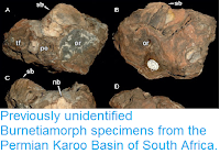
Type A lateral arm plates are elongate with coarsely reticulate stereom on the outer surface and a vertical row of large, freestanding spine articulations on an elevated distal edge. The spine articulations are composed of a single opening encompassed by a pair of arched dorsal and ventral lobes forming a lens-shaped elevation. The inner side has a low and poorly defined vertebral articular knob and a large tentacle notch. The general shape of these plates, as well as the shape of their arm articulations and the presence of coarse reticulation on their outer surfaces is consistent with the modern Ophiuroid order Ophioscolecida, and possibly with the genus Ophioscolex, although Thuy et al. do not go as far as assigning the specimens to genus level.

Type B lateral arm plates are by far the most common in the assemblage, making up about 90% of all the lateral arm plates collected. These are robust and strongly arched, and it appears likely that a pair of them would have been capable of completely enclosing the vertebrae of the arm. The outer surface ornamentation comprises a fine tuberculation, proximally bordered by a band of more coarsely meshed stereom including a central area with a fine horizontal striation and two spurs that establish the position of articulation with the overlapping adjacent lateral arm plate. The inner side of the lateral arm plate has a single, vertical ridge-shaped vertebral articular structure and a deep tentacle notch. The outer distal edge of the lateral arm plates bears a vertical row of small, freestanding spine articulations, each comprising a pair of parallel, horizontal dorsal and ventral lobes encompassing a small nerve opening and a slightly larger muscle opening. The type of spine articulation seen in these plates is exclusively seen in members of the Order Amphilepidida, which again is still extant today.

On the basis of their numeric domination of the sample, other plates found in the Dutton Ranch assemblage can be assumed to have come from the same species as the Type B lateral arm plates. These include ventral arm plates (which show similar ornamentation to the lateral arm plates) and radial shields, although Thuy et al. caution that, although the ventral arm plates appear to be consistent with the lateral arm plates, they would not, in themselves, be sufficient to assign a fossil to the Order Amphilepidida.

The placement of at least two species of Ophiuroids from the Dutton Ranch assemblage within modern orders is surprising, as this implies that these modern orders had emerged considerably before the theoretical date given for the emergence of the crown group Ophiuroids by molecular clock analysis. To test this hypothesis, Thuy et al. performed a Bayesian phylogenetic analysis using a previously established list of significant characters for the group, and using the early Carboniferous stem-group Ophiuroid Aganaster gregarius as an outgroup. This phylogeny confirmed the Type A lateral arm plates as having come from a member of the Order Ophioscolecida, which was close to the modern genera Ophioscolex and Ophiolycus, while the Type B lateral arm plates as having come from a basal member of the Order Amphilepidida.

Evolutionary tree of the crown-group Ophiuroidea, based on Bayesian inference analysis, showing positions of lateral arm plate (LAP) types A and B. Abbreviations: Ceno., Cainozoic; Devon., Devonian; Pennsylv., Pennsylvanian; Guadal., Guadalupian; Lop., Lopingian; L., Lower; Mid., Middle. Thuy et al. (2023).

This phylogenetic analysis confirms that lateral arm plate types A and B represent the earliest known examples of the orders Ophioscolecida and Amphilepidida, as well as (collectively) the oldest known examples of the Superorder  Ophintegrida. This in turn implies that not only had the crown group Ophiuroida arisen by 315 million years ago, it had had time to split into the two superorders, Ophintegrida and Euryophiurida, and that the Superorder Ophintegrida had time to split into the orders Ophioscolecida and Amphilepidida. It has previously generally been assumed that the modern Ophiuroid orders arose during the period of high biotic turnover following the End Permia extinction, which appears to be the case with other Echinoderm groups. Although a pre-End Permian radiation of modern Ophiuroids has been suggested previously, Thuy et al.'s study presents the first direct evidence of this. 

As well as providing evidence for an earlier origin of the crown group Ophiuroids than has previously been expected, Thuy et al.'s study also challenges the generally held assumption that shallow shelf environments are the major driver of biological innovation in the oceans, with some groups subsequently spreading into deep-water environments, and instead adds to a growing body of evidence that deep marine environments can themselves be a source of innovation, producing groups of organisms that go on to invade the shallow seas. 

The assumption that little biological innovation occurs in the deep marine environment may be linked to the paucity of fossil-producing deep-water sediments in the fossil record. This is partly because of the low sedimentation rates in these environments, which leaves the remains of any Animals which die there exposed on the surface for long periods, but is also linked to the recycling of the ocean seafloor, which makes unaltered pre-Mesozoic deep-water deposits extremely rare. Thuy et al.'s study also demonstrates that these obstacles can also be overcome, and that careful sampling of those deposits which have been preserved can uncover ghost-lineages of modern groups, adding to our understanding of evolution in deep time as well as deep marine environments.

See also...

Follow Sciency Thoughts on Facebook.

Follow Sciency Thoughts on Twitter


Monday, 23 January 2023

Fireball over northeast Oklahoma.

The American Meteor Society has received reports of a bright fireball meteor being seen over northeast Oklahoma, slightly after 3.35 am local time (slightly after 9.35 am GMT) on Friday 20 January 2023. People witnesses observing the event from much of Oklahoma, as well as parts of Arkansas, Kansas, Missouri, Mississippi, and Texas, reporting that the meteor moved roughly north to south, to the east of Tulsa. A fireball is defined as a meteor (shooting star) brighter than the planet Venus. These are typically caused by pieces of rock burning up in the atmosphere, but can be the result of man-made space-junk burning up on re-entry. 

Image of the 20 January 2023 Oklahoma Fireball Meteor taken from Tulsa, Oklahoma. Tana Foster/American Meteor Society.

Objects of this size probably enter the Earth's atmosphere several times a year, though unless they do so over populated areas they are unlikely to be noticed. They are officially described as fireballs if they produce a light brighter than the planet Venus. The brightness of a meteor is caused by friction with the Earth's atmosphere, which is typically far greater than that caused by simple falling, due to the initial trajectory of the object. Such objects typically eventually explode in an airburst called by the friction, causing them to vanish as an luminous object. However, this is not the end of the story as such explosions result in the production of a number of smaller objects, which fall to the ground under the influence of gravity (which does not cause the luminescence associated with friction-induced heating).

Heat map showing areas where sightings of the meteor were reported (warmer colours indicate more sightings)and the apparent path of the object (blue arrow). American Meteor Society. 

These 'dark objects' do not continue along the path of the original bolide, but neither do they fall directly to the ground, but rather follow a course determined by the atmospheric currents (winds) through which the objects pass. Scientists are able to calculate potential trajectories for hypothetical dark objects derived from meteors using data from weather monitoring services.

Witness reports can help astronomers to understand these events. If you witness a fireball-type meteor over the US you can report it to the American Meteor Society here. 

See also...


Follow Sciency Thoughts on Facebook.

Follow Sciency Thoughts on Twitter.


Friday, 24 April 2020

Tornadoes and flooding kill at least seven in Texas, Oklahoma, and Louisiana.

At least seven people have died and many more have been injured as tornadoes and severe storms swept across the southern United States on Wednesday 22 and Thursday 23 April 2020. At least three people died and more than twenty were injured when a tornado hit the town of Onalaska in Polk County, Texas, destroying 46 houses and damaging 245 more. Two people died in the city of Madill in Marshall County, Oklahoma, which is reported to have suffered severe damage after being hit by a tornado. In Louisiana a man is reported to have died after falling into a drainage ditch in the city of Mansfield in DeSoto Parish, and a woman is reported to have died in a storm in the town of Lecompte in Rapids Parish. Elsewhere the city of Adel in Cook County, Georgia, is reported to have suffered damage to a number of properties after being hit by a tornado, as is the city of Tallahassee, in Leon County, Florida. In Alabama two emergency workers are reported to have been injured by a falling tree in the city of Anniston in Calhoun County, while attempting to rescue a homeowner trapped by an earlier treefall. Around 150 000 homes and businesses across the southern US are reported to be without electricity due to fallen power lines.

A tornado over the city of Madrill, Oklahoma, at about 5.00 pm local time on Wednesday 22 April 2020. NBC.

Tornadoes are formed by winds within large thunder storms called super cells. Supercells are large masses of warm water-laden air formed by hot weather over the sea, when they encounter winds at high altitudes the air within them begins to rotate. The air pressure will drop within these zones of rotation, causing the air within them so rise, sucking the air beneath them up into the storm, this creates a zone of rotating rising air that appears to extend downwards as it grows; when it hits the ground it is called a tornado.

Tornado damage in Woodworth, Louisiana. USA Today.

Tornadoes can occur anywhere in the world, but are most common, and most severe, in the area of the American mid-west known as 'Tornado Ally', running from Texas to Minnesota, which is fuelled by moist air currents from over the warm enclosed waters of the Gulf of Mexico interacting with cool fast moving jet stream winds from the Rocky Mountains. Many climatologists are concerned that rising temperatures over the Gulf of Mexico will lead to more frequent and more severe tornado events.

Simplified diagram of the air currents that contribute to tornado formation in Tornado Alley. Dan Craggs/Wikimedia Commons/NOAA.

See also...

https://sciencythoughts.blogspot.com/2020/04/tornadoes-kill-at-least-eight-in.htmlhttps://sciencythoughts.blogspot.com/2020/03/homes-destroyed-and-people-treated-for.html
https://sciencythoughts.blogspot.com/2019/12/three-killed-in-tornado-outbreak-in.htmlhttps://sciencythoughts.blogspot.com/2019/11/condominium-complex-evacuated-after.html
https://sciencythoughts.blogspot.com/2019/10/thousands-left-wthout-electricity-after.htmlhttps://sciencythoughts.blogspot.com/2019/10/florida-fish-and-wildlife-conservation.html
Follow Sciency Thoughts on Facebook.

Sunday, 8 March 2020

Homes destroyed and people treated for smoke inhalation as wildfires sweep across the Oklahoma Panhandle.

A number of homes and businesses have been destroyed and several people have been treated for smoke inhalation following a wildfire outbreak in Beaver County in the Oklahoma Panhandle this weekend. The fires were first spotted by a NASA satellite just after 10.00 am on the morning of Saturday 7 March 2020, prompting authorities in the state to order evacuations of the towns of Beaver and Fogan. A number of properties are reported to have been subsequently burned in both towns, with firefighters from Oklahoma and Kansas trying to tackle the blaze, which at the end of Saturday was estimated to have burned 53 km² of vegetation and property and to be only 10% under control.

A house burning in the town of Beaver, Oklahoma, during a wildfire outbreak on 7 March 2020. Cody Rehder/Oklahoma Highway Patrol/Twitter.

Brush fires are common events across much of the southern United States, where long dry periods are often accompanied by high winds, providing the fires with both fuel and the means to spread quickly. As with many other hot-weather related phenomena there is currently serious concern that such fires might become more common in a warming climate, particularly given the recent extreme fire events in Australia. 

See also...

https://sciencythoughts.blogspot.com/2018/04/oklahoma-man-killed-by-wildfire.htmlhttps://sciencythoughts.blogspot.com/2016/11/magnitude-50-earthquake-in-payne-county.html
https://sciencythoughts.blogspot.com/2016/09/magnitude-38-earthquake-beneath-spencer.htmlhttps://sciencythoughts.blogspot.com/2016/09/magnitude-56-earthquake-in-pawnee.html
https://sciencythoughts.blogspot.com/2016/03/magnitude-42-earthquake-in-logan-county.htmlhttps://sciencythoughts.blogspot.com/2015/12/stroms-and-floods-kill-at-least-43.html
Follow Sciency Thoughts on Facebook.

Tuesday, 22 October 2019

Palaeopathology in a Permian Varanopid.

Bone remodelling is an essential physiological process in growth and healing, and deviations from normal bone physiology in the form of pathologies aid in the understanding of normal bone metabolism. The study of such pathologies in the fossil record therefore offers insight into the biology of extinct groups, and the evolutionary history of groups alive today.

In a paper published in the journal PLoS One on 7 August 2019, Yara Haridy and Florian Witzmann of the Museum für Naturkunde at the Leibniz-Institut für Evolutions- und Biodiversitätsforschung, Patrick Asbach of the Institut für Radiologie at the  Charité-Universitätsmedizin Berlin, and Robert  Reisz of the Dinosaur Evolution Research Centre at Jilin University, and the Department of Biology at the University of Toronto Mississauga, discuss an example of a pathological condition in a Varanopid from the Early Permian Richards Spur deposits.

The Early Permian Richards Spur locality at the Dolese Brothers Quarry in Oklahoma preserve one of the most diverse assemblages of terrestrial vertebrates known from any Permian site. The remains are preserved in fissure deposits within the Ordovician Arbuckle Limestone, in a unique depositional environment that has been interpreted as cave systems that underwent periods of flooding not unlike present-day conditions that are commonly associated with monsoonal episodes, preserving an upland biota that is seldom recorded in the fossil record. The fissure fill sediments are early Permian (about 289 million years old) and consist of soft clays and mudstone. They contain articulated specimens or isolated bones of mostly small to medium sized terrestrial Tetrapods.

Varanopids are generally considered to be early Synapsids related to Pelycosaurs (although an alternative suggestion, that they are early Diapsids, has recently been made by Ford and Benson). Varanopids range from the latest Carboniferous to the latest Middle Permian with a broad geographic distribution in Pangea. They were small to medium-sized (1.5 - 2m total body length) predators, superficially resembling extant Varanids (Monitor Lizards) in their habits.

Haridy et al. describe two pathologically fused caudal (tail) vertebrae of an undescribed Varanopid based on outer morphology and the internal microstructure as revealed by X-ray microtomography. The collective anteroposterior length (front to back length) of the fused vertebrae is 23 mm. Taking the midpoint of the ventral fusion zone between the centra (disks) as the boundary between the two elements, the anterior vertebra has a length of 11 mm and the posterior one of 12 mm. The vertebrae can be assigned to Varanopidae based on the proportionally elongate, slender vertebral bodies with a small perforating foramen in the mid-portion of the centrum and the double keelation on the ventral surface. In addition, the neural arches are slender and slightly concave, and the neural spines are delicate, and posterodorsally slanted.

The two vertebral centra are completely fused to one another without any superficial trace of an intervertebral suture, the proximal part of a haemapophyses (part of the bony arch on the ventral side of a tail vertebra) is fused to the posteroventral margin (back) of the anterior centrum and to the anteroventral margin (front) of the posterior centrum. The proximal part of another haemapophysis is fused to the anteroventral margin of the anterior vertebra. The neural arches are completely fused to the centra such that there is no evidence of a neurocentral suture. The fusion zone between the centra protrudes outwardly and forms a large swelling of bone on the right side. In ventral view, the proximal part of the haemapophyses is visible and shows a left/right-asymmetry in the bases of their paired ventral processes; the base of the right process is larger than the left one and extends further anteriorly. In both haemapophyses, the ventral processes are broken off.

On the right dorsolateral surface of the neural arch, a small osseous (bony) growth is present at about the mid-length of the anterior vertebra. The abnormal growth is penetrated by several small foramina (openings) on its dorsal side and has a distinct concavity (indentation) on its posterior part. The pre- and postzygapophysis (articular processes of the neural arch of a vertebra) are fused, but the boundary between them is still discernible. The right postzygapophysis of the anterior vertebra is enlarged as compared to the left one, corresponding to the swelling of the centra in the fusion zone and the larger base of the haemapophyses on the right side. Accordingly, the right prezygapophysis is also larger than the left one in the posterior vertebra. Apart from the zygapophyses, the dorsal side of the neural arch of the posterior vertebra seems to be pathologically unaltered, whereas the dorsal side of the neural arch of the anterior vertebra bears many irregular depressions and crests.

The bone surface is irregular on the left side of the centra, there are irregular grooves, depressions and crests on the right side, sometimes resembling the imprints of large vessels. The bone surface, however, is always smooth with some nutrient foramina penetrating the bone. The right intervertebral foramen for the spinal nerve root between the anterior and posterior vertebra is largely filled with bone; only two smaller openings of unequal size are present in the depression that represents the original foramen. From the larger of these openings, a shallow groove extends in a dorsal direction. In contrast, the intervertebral foramen on the right side is open but slightly constricted by bone growth on the anteroventral margin. A smaller opening is located ventrally on the lateral side of the swollen fusion zone of the centra, also showing a shallow groove running dorsally from the opening. The posterior edge of the posterior vertebra shows that at least the anterior margin of the intervertebral foramen between this and the posteriorly following vertebra was unaltered. The exposed (not fused) anterior and posterior articulation surfaces of the vertebral centra are concave and have a roughened, unfinished surface indicative of a cartilage cover in life. They are round in outline in anterior and posterior view, respectively, and have a centrally located, large notochordal canal. These joint surfaces were not pathologically altered. The opening of the neural canal is well preserved on the anterior face of the anterior vertebra. It is broad ovate in outline, measuring 2.5 mm in width and 1.5 mm in height, and is not constricted by pathological bone growth.

External anatomy of a pathological Varanopid vertebrae. (a) schematic of Varanopid tail with a normal vertebrae used for comparison represented in blue, and the pathological fused vertebrae presented in orange.; (b)-(f) right lateral, left lateral, dorsal, ventral, and anterior views respectively. Abbreviations: as, articular surface; fz, fusion zone; gn, growth nodule; hp, haemapophyses; irg, irregular groove; ivf, intervertebral foramen; na, neural arch; nc, neural canal; ntc, notochordal canal; poz, postzygapophysis fused; vph, ventral processes of haemapophyses. Scale bar is 5mm. Haridy et al. (2019).

The X-ray microtomography scans of the pathological fused vertebrae show that the smooth exterior of the vertebrae did not indicate the extreme pathology that lay beneath the surface. The notochordal canal and the neural canal remain open and unobstructed in longitudinal and cross sections. However, the notochordal canal maintains a consistent diameter throughout both vertebrae, which is unlike the unaffected vertebrae in which the notochordal canal has a wider diameter towards the anterior and posterior ends. The cross-sectional outline of the neural canal is broad-ovate to reniform in the anterior and posterior regions of each vertebra. In the middle of the vertebra, it becomes nearly circular in cross section, as in the normal vertebra. In sagittal section, the neural canal expands at the boundary of the two vertebrae. The intervertebral space is almost absent and restricted to a very thin gap.

The X-ray microtomography scansreveal in great detail the structure of the external cortical compact bone and of the inner trabecular bone of centra and neural arches. The original outer cortex of the vertebrae has been extensively altered through resorption and by addition of bone, and secondary formation of trabecular bone and cortical bone outside the original vertebral cortex has taken place. The cortical bone that surrounds the vertebrae is thickened, and there is a distinguishable difference between the old cortical bone that made up the original surface and the new cortical bone that covers the old cortex. The new cortical bone is denser and thus brighter in the images.

X-ray microtomography internal anatomy of pathological Varanopid vertebrae. (a) Serial cross sections showing the extent of the pathology throughout from most anterior to the posterior the two vertebrae (1)–(12). (b). Closeup of cross section through neural arch showing the old cortical bone overlain by new less dense cortical bone; the wavy line shows the unevenness in the Howship’s lacunae. (c) Closeup of the outward-growth in the fusion zone showing lysis to the old cortical bone and thick erratic trabeculae overlain by a layer of cortical bone. (d) Closeup of the growth node on the second vertebrae’s neural arch, the growth overlays the old cortical bone and has thick trabeculae and large lytic spaces covered by cortical bone. (e) Closeup of the ventral portion of the second vertebrae showing the extent of resorption of old bone and deposition of new pathological bone. Abbreviations: cb, cortical bone; ocb, old cortical bone; nc, neural canal; ntc, notochordal canal. Scale bar is 2mm. Haridy et al. (2019).

The old cortical bone of the centrum and neural arch is covered in Howship’s lacunae, i.e. resorption bays, which are indicative of extensive osteoclastic activity. In some regions of the centrum, the old cortical bone has been so extensively resorbed that it is either a thin remnant covered in resorption bays or has been completely replaced by thickened trabeculae. The trabecular bone consists of thickened trabeculae in both the centra and the neural arch. Most trabeculae are present in about the anterior and posterior thirds of each vertebra, whereas the middle part is nearly devoid of trabeculae and consists of a large hollow space, as seen in the normal vertebra scans. The osseous bump mentioned above on the right side of the neural arch of the anterior vertebra consists of course trabecular bone with a thin external cortex, the trabecular bone is so pervasive that it continues into the neural arch where the cortical bone has been resorbed and replaced with thickened trabecular bone.

X-ray microtomography internal anatomy of pathological Varanopid vertebrae. (a) Off-centre sagittal section showing the internally altered pathological bone. (b) Closeup of the anterior portion of vertebrae 1 showing thickened trabeculae, lytic lesions and cortical bone thickening. (c) Centered sagittal section showing the notochord is still continuous through the pathological vertebrae, also showing the degree of alteration to the neural arch via lytic lesions; (d) Closeup of the ventral region of the fusion zone, showing old cortical bone, large lytic lesions and cortical thickening. Abbreviations: ct, cortical thickening; ocb, old cortical bone; ll, lytic lesion; nc, neural canal; ntc, notochordal canal; tt, thickened trabeculae. Scale bar is 2mm. Haridy et al. (2019).

Haridy et al. consider a number of possible causes of the pathology seen, including infectious arthritis, spondylitis tuberculosa, ankylosing spondylitis, Scheuermann’s disease, spondylitis ankylosans, vertebral tumor, fracture callus causing the growth and subsequent fusion, osteochondrosis intervertebralis, and chronic osteomyelitis, but only two conditions were found to fit the observed symptoms, fibrous dysplasia and Paget’s disease. Of these, Paget’s disease seems the more likely cause as cases of fibrous dysplasia affecting more than one bone are rare.

The cause of Paget’s disease is unclear, though correlations have been suggested between cases of the disease and wood-fired heating, tobacco smoking, consumption of brains, rural life, and especially contact with farm or wildlife animals. This has led to the suggestion that the disease is linked to a zoonotic Virus (Virus typically found in animals but which can infect Humans under some circumstances), with a combination of infection by the Virus and exposure to certain environmental triggers needed to activate the condition. If this theory is correct, and if it also holds true for the infection seen in the Varanopid, then this would represent the oldest known example of a Viral infection in the fossil record.

See also...

https://sciencythoughts.blogspot.com/2018/03/caudal-autotomy-in-early-permian.htmlhttps://sciencythoughts.blogspot.com/2017/07/australopithecus-africanus-deciphering.html
https://sciencythoughts.blogspot.com/2017/02/odontoma-found-in-late-permian.htmlhttps://sciencythoughts.blogspot.com/2016/09/an-osteogenic-tumour-in-198-million.html
https://sciencythoughts.blogspot.com/2016/08/malignant-osteosarcoma-in-17-million.htmlhttps://sciencythoughts.blogspot.com/2014/01/bone-pathology-in-triassic-phytosaur.html
Follow Sciency Thoughts on Facebook.

Saturday, 14 April 2018

Oklahoma man killed by wildfire.

An Oklahoma man has died of injuries sustained while attempting to fight a wildfire in Oklahoma. The unnamed man was injured in Roger Mills County on Thursday 12 April 2018, and died later in a hospital. Two other people have been injured and around 25 houses destroyed in a series of fires sweeping across the state this week, with several hundred more evacuated from homes in Dewey and Woodward Counties. Fires are also burning in Beckham, Caddo an Osage counties.

A burning building in northwest Oklahoma on 12 April 2018. Buck King/Westlake Legal Group.

The fires have a variety of causes and have been feuled by dry winds blowing from the mountains in the northeast of the state, but are essentially due to a prolonged drought in the states of Oklahoma and Texas, with only about 12 mm of rain falling in northwest Oklahoma so far this year. This has dried out vegetation in the area, making it vulnerable to fires. The situation has been made worse this week by high winds in the area, which both fan the fires, bringing them a good suply of oxygen, which can be a limiting factor in still conditions, and help them to spread, by carrying burning debris to new areas.

See also...

http://sciencythoughts.blogspot.co.uk/2016/11/magnitude-50-earthquake-in-payne-county.htmlhttp://sciencythoughts.blogspot.co.uk/2016/09/magnitude-38-earthquake-beneath-spencer.html
http://sciencythoughts.blogspot.co.uk/2016/09/magnitude-56-earthquake-in-pawnee.htmlhttp://sciencythoughts.blogspot.co.uk/2016/03/magnitude-42-earthquake-in-logan-county.html
http://sciencythoughts.blogspot.co.uk/2015/12/stroms-and-floods-kill-at-least-43.htmlhttp://sciencythoughts.blogspot.co.uk/2015/11/flooding-and-winter-storms-thought-to.html
Follow Sciency Thoughts on Facebook.

Friday, 23 March 2018

Caudal autotomy in Early Permian Captorhinids from Oklahoma.

Caudal autotomy is a process by which a vertebrate animal sheds its tail, or a portion of it, in order to escape from a predator. Today it is seen only in Lepidosaurs (Lizards, Snakes and the Turatura), suggesting that it is a unique innovation in this group. Caudal autotomy can occur intravertabrally (through the vertebrae), as seen in the Turatura and most Lizards, or intervertebrally, between the vertebrae, as seen in Agamid Lizards and Snakes.

In a paper published in the journal Scientific Reports on 5 March 2018, Aaron LeBlanc of the Department of Biology at the University of Toronto Mississauga, and the Department of Biological Sciences at the University of Alberta, Mark MacDougall, Yara Haridy, and Diane Scott, also of the Department of Biology at the University of Toronto Mississauga, and Robert Reisz of the Dinosaur Evolution Research Centre at Jilin University, describe evidence for the presence of caudal autotomy in Permian Captorhinids, a Eureptilian group (i.e. animals more closely related to modern Reptiles and Birds than to Mammals, but which split off from the ancestors of these modern animals long before the split between Archosauromorphs (Crocodiles, Birds and Turtles) and Lepidosaurs (Snakes, Lizards and the Turatura).

LeBlanc et al. examined 70 isolated or pairs of Captorhinid caudal (tail) vertebrae from the Royal Ontario Museum collections, obtained from the Early Permian Richards Spur locality at the Dolese Brothers Quarry in Oklahoma. They found that these had a line of weakness, which would apparently allow for caudal autotomy, with a potential line of fracture through the bottom part of the vertebrae. This appeared to be well developed in younger individuals, with the bone becoming more densely mineralised as the animals grew older, suggesting that fully mature individuals lost the ability to autotomise their tails, as is the situation in modern Iguanas.

Fracture planes in Captorhinid caudal vertebrae. (a) Artist’s reconstruction of the Permian Reptile Captorhinus with an autotomous tail (inset showing anterior caudal vertebrae with fracture planes). (b) Image and (c) SEM image of an isolated anterior caudal vertebra with a fracture plane passing through the centrum (black arrow). (d) Ventral view of an anterior, rib bearing caudal vertebra showing the absence of any fracture plane. (e) Ventral view of a caudal vertebra bearing a fracture plane (black arrows) (f) thin-section through the sagittal plane of a caudal vertebra with a fracture plane (black arrow) passing through the ventral portion of the centrum. (g) Close-up of fracture plane (black arrows) in (f) passing into the notochordal canal. Abbreviations: cb, cortical bone; cct, calcified cartilage; ce, centrum; nc, neural canal; ns, neural spine; ntc, notochordal canal. Reconstruction by Danielle Dufault. Anterior is to the left in all of the images. LeBlanc et al. (2018).

Intravertabral caudal autotomy (breaking off of the tail through the vertebrae) is generally associated with animals that can regrow their tails. No sign of any such regrowth was seen in any Captorhinid examined, though this does not necessarily imply that they were not capable of such recovery, as all of the specimens from the Richards Spur locality are isolated vertebrae rather than entire skeletons, which would make such preservation unlikely, and even were this not the case, the proportion of animals with recovering tails in the population would presumably be quite low, making the likelihood of such an animal being preserved equally low.

See also...

http://sciencythoughts.blogspot.co.uk/2017/08/moschops-capensis-synchrotron-scanning.htmlhttp://sciencythoughts.blogspot.co.uk/2017/03/vertebrate-remains-from-late-permian-of.html
http://sciencythoughts.blogspot.co.uk/2017/02/odontoma-found-in-late-permian.htmlhttp://sciencythoughts.blogspot.co.uk/2016/09/unidentified-burnetiamorph-specimens.html
http://sciencythoughts.blogspot.co.uk/2014/06/pareiasaurs-from-late-permian-of-niger.htmlhttp://sciencythoughts.blogspot.co.uk/2014/04/scavenging-on-body-of-dicynodont.html
Follow Sciency Thoughts on Facebook.