Showing posts with label Echinoderms. Show all posts

Sunday, 12 February 2023

Ophiuroids (Brittle Stars) from the Carboniferous of Oklahoma.

Determining when groups of organisms first originated is one of the great challenges faced by evolutionary biologists. Traditionally, the fossil record has been the only way to determine when such groups first appeared, although since the 1960s molecular clock techniques, which use mutation rates to determine when groups of organisms diverged, have also been employed. Modern studies typically use a combination of the two techniques to achieve better results, but the accuracy of any study is still dependent on the quality of the available data.

The fossil record is not complete. Organisms stand a far better chance of being preserved in some environments that others, and organisms themselves differ greatly in their potential to leave fossil remains. Furthermore, Humans are often selective in both where they look for fossils, and in the types of fossils they collect, concentrating on larger, more eye-catching fossils, and often overlooking unconventional deposits which do not produce abundant, obvious fossils completely. Fossil groups which have skeletons made up of large numbers of small sections, such as small Vertebrates and Echinoderms, are often overlooked in environments which do not produce articulated skeletons, leaving gaps in our knowledge of their histories, even though their fossils are both numerous and widespread. Deposits from environments where articulated skeletons are unlikely to be preserved, such as the deep marine seafloor (where sedimentation rates are very low, and storm events never rapidly cover organisms) are often overlooked in palaeontological studies. These environments, however, often contain numerous disarticulated skeletal elements, which can potentially tell us a great deal about the history of traditionally undersampled groups.

Ophiuroids, or Brittle Stars, are the most specious of the five extant classes of Echinoderms, and a well resolved phylogeny, based upon molecular data. However, the skeleton of Ophiuroids typically disaggregates into a large number of sand-sized particles within a few hours of death, with the result that fossils of entire Ophiuroids are very rare. The plates which make up the skeletons of Ophiuroids are actually very distinctive, making it possible to identify individual plates to the species level, and a great deal of work has been done on Mesozoic and Cainozoic Ophiuroids using individual plates, but the Palaeozoic Ophiuroid fossil record has been largely overlooked to date.

In a paper published in the journal Geology on 3 January 2023, Ben Thuy of the Department of Palaeontology at the Natural History Museum Luxembourg, Larry Knox of Earth Sciences at  Tennessee Tech University, Lea Numberger-Thuy, also of the Department of Palaeontology at the Natural History Museum Luxembourg, and Nicholas Smith and Colin Sumrall of the Department of Earth and Planetary Sciences at the University of Tennessee, present the results of a study of Ophiuroid elements from deep water sediments from the Carboniferous of Oklahoma.

Thuy et al. examined 81 sieved micropalaeontological assemblages, obtained from bulk samples collected at Dutton Ranch in central southern Oklahoma for Ophiuroid samples. One of these, collected in 1981 from a blue-to-olive-grey shale identified as Unit 9BC, which is considered to be roughly equivalent to the Bostwick Member of the Lake Murray Formation, which dates to the latest Bashkirian (about 315 million years ago) proved to be particularly rich in Ophiuroid fossils. This unit was deposited in the northern Ardmore Basin, a northwestern extension of the Panthalassic Ocean. Based upon grain size, mineralogy, and Ostracod fauna, this deposit is thought to have been laid down in a deep slope or upper continental shelf environment.

Paleogeographic map of the southern United States during the Early Pennsylvanian, with sampled locality marked by the star.  Deep Time Maps in Thuy et al. (2023). 

In total, the samples produced about 400 Ophiuroid microfossils, including various types of arm plates (single ambulacrals and pairs fused into vertebrae; and lateral, ventral, and dorsal arm plates) as well as disc plates (radial shields and oral plates). From these lateral arm plates were chosen for further examination, as these are considered to show the greatest complexity, and are therefore the most useful taxonomically. Five distinct types of lateral arm plate were present in the sample, which Thuy et al. believe represent five separate species. Furthermore, two of these five species appear to be crown group Ophiuroids (the 'crown-group' of a group of organisms includes all living species within that group, their most recent common ancestor, and everything descended from that ancestor), assignable to extant orders, which Thuy et al. refer to as types A and B.

Brittle Star microfossils from the latest Bashkirian (Atokan, Upper Carboniferous), of Dutton Ranch, Oklahoma, USA. (A)–(G) Lateral arm plate type B (A)–(C) and associated skeletal remains (E)–(G), corresponding to unnamed Amphilepidid, with median (A) and proximal (B) lateral arm plates in external (A1), (B1) and internal (A2), (B2) views and with details of spine articulations (C), proximal vertebra (D) in lateral view, proximal ventral arm plate (E) in external view, radial shield (F) in external view, and oral plate in adradial view (G). All plates are shown with their respective position in a typical modern ophiuroid skeleton. (H) Lateral arm plate type A, corresponding to an unnamed Ophioscolecid, with proximal lateral arm plate shown in external (H1) and internal (H2) views. Abbreviations: di, distal; do, dorsal. Thuy et al. (2023).

Type A lateral arm plates are elongate with coarsely reticulate stereom on the outer surface and a vertical row of large, freestanding spine articulations on an elevated distal edge. The spine articulations are composed of a single opening encompassed by a pair of arched dorsal and ventral lobes forming a lens-shaped elevation. The inner side has a low and poorly defined vertebral articular knob and a large tentacle notch. The general shape of these plates, as well as the shape of their arm articulations and the presence of coarse reticulation on their outer surfaces is consistent with the modern Ophiuroid order Ophioscolecida, and possibly with the genus Ophioscolex, although Thuy et al. do not go as far as assigning the specimens to genus level.

Type B lateral arm plates are by far the most common in the assemblage, making up about 90% of all the lateral arm plates collected. These are robust and strongly arched, and it appears likely that a pair of them would have been capable of completely enclosing the vertebrae of the arm. The outer surface ornamentation comprises a fine tuberculation, proximally bordered by a band of more coarsely meshed stereom including a central area with a fine horizontal striation and two spurs that establish the position of articulation with the overlapping adjacent lateral arm plate. The inner side of the lateral arm plate has a single, vertical ridge-shaped vertebral articular structure and a deep tentacle notch. The outer distal edge of the lateral arm plates bears a vertical row of small, freestanding spine articulations, each comprising a pair of parallel, horizontal dorsal and ventral lobes encompassing a small nerve opening and a slightly larger muscle opening. The type of spine articulation seen in these plates is exclusively seen in members of the Order Amphilepidida, which again is still extant today.

On the basis of their numeric domination of the sample, other plates found in the Dutton Ranch assemblage can be assumed to have come from the same species as the Type B lateral arm plates. These include ventral arm plates (which show similar ornamentation to the lateral arm plates) and radial shields, although Thuy et al. caution that, although the ventral arm plates appear to be consistent with the lateral arm plates, they would not, in themselves, be sufficient to assign a fossil to the Order Amphilepidida.

The placement of at least two species of Ophiuroids from the Dutton Ranch assemblage within modern orders is surprising, as this implies that these modern orders had emerged considerably before the theoretical date given for the emergence of the crown group Ophiuroids by molecular clock analysis. To test this hypothesis, Thuy et al. performed a Bayesian phylogenetic analysis using a previously established list of significant characters for the group, and using the early Carboniferous stem-group Ophiuroid Aganaster gregarius as an outgroup. This phylogeny confirmed the Type A lateral arm plates as having come from a member of the Order Ophioscolecida, which was close to the modern genera Ophioscolex and Ophiolycus, while the Type B lateral arm plates as having come from a basal member of the Order Amphilepidida.

Evolutionary tree of the crown-group Ophiuroidea, based on Bayesian inference analysis, showing positions of lateral arm plate (LAP) types A and B. Abbreviations: Ceno., Cainozoic; Devon., Devonian; Pennsylv., Pennsylvanian; Guadal., Guadalupian; Lop., Lopingian; L., Lower; Mid., Middle. Thuy et al. (2023).

This phylogenetic analysis confirms that lateral arm plate types A and B represent the earliest known examples of the orders Ophioscolecida and Amphilepidida, as well as (collectively) the oldest known examples of the Superorder  Ophintegrida. This in turn implies that not only had the crown group Ophiuroida arisen by 315 million years ago, it had had time to split into the two superorders, Ophintegrida and Euryophiurida, and that the Superorder Ophintegrida had time to split into the orders Ophioscolecida and Amphilepidida. It has previously generally been assumed that the modern Ophiuroid orders arose during the period of high biotic turnover following the End Permia extinction, which appears to be the case with other Echinoderm groups. Although a pre-End Permian radiation of modern Ophiuroids has been suggested previously, Thuy et al.'s study presents the first direct evidence of this. 

As well as providing evidence for an earlier origin of the crown group Ophiuroids than has previously been expected, Thuy et al.'s study also challenges the generally held assumption that shallow shelf environments are the major driver of biological innovation in the oceans, with some groups subsequently spreading into deep-water environments, and instead adds to a growing body of evidence that deep marine environments can themselves be a source of innovation, producing groups of organisms that go on to invade the shallow seas. 

The assumption that little biological innovation occurs in the deep marine environment may be linked to the paucity of fossil-producing deep-water sediments in the fossil record. This is partly because of the low sedimentation rates in these environments, which leaves the remains of any Animals which die there exposed on the surface for long periods, but is also linked to the recycling of the ocean seafloor, which makes unaltered pre-Mesozoic deep-water deposits extremely rare. Thuy et al.'s study also demonstrates that these obstacles can also be overcome, and that careful sampling of those deposits which have been preserved can uncover ghost-lineages of modern groups, adding to our understanding of evolution in deep time as well as deep marine environments.

See also...

Follow Sciency Thoughts on Facebook.

Follow Sciency Thoughts on Twitter


Thursday, 24 November 2022

Acanthaster benziei: A new species of Crown-of-thorns Starfish from the Red Sea.

Crown-of-thorns Starfish, Acanthaster spp., are highly distinctive Starfish found across the tropical Indo-Pacific region from the east coast of Africa to the west coast of Mexico, which get their popular name from the covering of long, venomous spines found in most species. They are typically corallivorous, feeding on Coral Polyps by extruding their stomachs and digesting them externally. Notably, Crown-of-thorns Starfish can undergo sudden rapid population increases, known as outbreaks, which can lead to large areas of Coral Reefs being denuded of their living Polyps, something of great concern to conservationists at a time when Coral Reefs are facing a range of other threats, which has led to them being one of the most extensively studied groups of Marine Invertebrates.

Crown-of-thorns Starfish were first described by the German naturalist Georg Eberhard Rumphius in 1705, and given their own generic name, Acanthaster, by the French palaeontologist François Louis Paul Gervais  in 1841. For a long while, only two species were described within the genus, Acanthaster planci, the typical, long-spined, venomous, corallovorous form, and Acanthaster brevispinus, a shorter-spined, non-venomous form, which does not feed on Corals. However, genetic studies carried out within the past three decades have shown that Acanthaster planci is in fact a species cluster, made up of a number of physically very similar species (cryptospecies), which are nevertheless genetically distinct, which often appear to have diverged from one-another a long time ago. 

Based upon this, it was suggested that the original species should be split into four different species, each inhabiting a different geographical area; the Pacific, the Southern Indian Ocean, the Northern Indian Ocean and the Red Sea, which each of these species probably needing further division into several subspecies. Subsequent studies have indeed confirmed that the Pacific, North Indian Ocean, and South Indian Ocean populations are in fact separate species, although genetic material from the Red Sea population has not, until now, been available.

In a paper published in the journal Zootaxa on 17 November 2022, Gert Wörheide of the Department of Earth and Environmental Sciences Palaeontology and Geobiology, and the GeoBio-Center at Ludwig-Maximilians-Universität München, and the Bavarian State Collection of Palaeontology and Geology, Emilie Kaltenbacher and Zara-Louise Cowan, also of the Department of Earth and Environmental Sciences Palaeontology and Geobiology at Ludwig-Maximilians-Universität München, and Gerhard Haszprunar, also of the GeoBio-Center at Ludwig-Maximilians-Universität München, and of the Bavarian Zoological State Collections, describe the Red Sea population of Crown-of-thorns Starfish as a new population.

The new species is named Acanthaster benziei in honour of marine biologist John Benzie, for his extensive work on Crown-of-thorns Starfish. The description is based upon four specimens collected from species within the territorial waters of Saudi Arabia by  Sara Campana and OliverVoigt in 2017.

Typical colouration of Acanthaster benziei. (A) GW4081 (Paratype, hiding during the day under a crevice), Al-Lith, Saudi Arabia, (B)–(D) Thuwal Reefs, Saudi Arabia. Approximate diameter of specimens is 25–30 cm. Oliver Voigt & Gert Wörheide in Wörheide (2022).

Acanthaster benziei is a large Starfish with a convex disk and 11-14 arms (the range for the genus being 10-25), of uneven lengths, and tapering to a point. Each arm has two rows of ambulacral tube feet, which have flattened tips and lack suckers. The central disk of the species is 28-65 mm across, with an aboral (upper surface) covered in papulae (pimples) arranged in an apparently random manner. Both surfaces are covered in calcareous ossicles (plates) and spines. These Starfish are grey-green to grey-purple in colour, although the aboral spines are orange or red. The papulae on the aboral surface of the central disk can form darker patterns, giving this surface a 'bulls-eye' appearance.

See also...

Follow Sciency Thoughts on Facebook.

Follow Sciency Thoughts on Twitter.


Sunday, 3 April 2022

Benthodytes jiaolongi: A new species of deep-sea Sea Cucumber from the Western Pacific.

Sea Cucumbers (Holothuroidea) are a class of Echinoderms that have become elongated and worm-like, effectively becoming secondarily bilaterally symmetrical. Like all Echinoderms they have a calcareous exoskeleton, but this is greatly reduced, typically only occurring as ossicles within the epidermis. The muscle structure of Sea Cucumbers is instead supported by a layer of modified collagen that can be stiffened or relaxed at will, allowing for a more flexible body than that of other Echinoderms. The Family Psychropotidae currently comprises 37 species of deep-sea Sea Cucumbers, which are elongated and flattened to facilitate swiming, and typically have a large tail-appendage of uncertain purpose. Although the Psychropotidae were first discovered during the H.M.S. Challenger Expedition of 1872–1876, the group are relatively understudied, and phylogenetic relationships within the family poorly understood.

In a paper published in the journal ZooKeys on 9 March 2022, Chuan Yu of the School of Oceanography at Shanghai Jiao Tong University, and the Key Laboratory of Marine Ecosystem Dynamics at the Second Institute of Oceanography of the Ministry of Natural Resources, Dongsheng Zhang, also of the School of Oceanography at Shanghai Jiao Tong University, and the Key Laboratory of Marine Ecosystem Dynamics at the Second Institute of Oceanography of the Ministry of Natural Resources, and of the Southern Marine Science and Engineering Guangdong Laboratory, Ruiyan Zhang, again of the School of Oceanography at Shanghai Jiao Tong University, and Chunsheng Wang, once again of the School of Oceanography at Shanghai Jiao Tong University, and the Key Laboratory of Marine Ecosystem Dynamics at the Second Institute of Oceanography of the Ministry of Natural Resources, the Southern Marine Science and Engineering Guangdong Laboratory, and of the State Key Laboratory of Satellite Ocean Environment Dynamics of the Ministry of Natural Resources, describe a new species of Psychropotid Sea Cucumber from the Western Pacific.

The new species is described from four specimens collected from the Weijia Guyot and Kyushu-Palau Ridge by the Jiaolong Human Operated Vehicle. The new species is placed in the genus Benthodytes , and given the specific name jiaolongi, in reference to the vehicle used to recover the specimens.

 
Benthodytes jiaolongi specimen (RSIO6017101, paratype) in situ on the Kyushu-Palau Ridge. Yu et al. (2022).

Benthodytes jiaolongi is a red-to-violet elongated and subcylindrical Sea Cucumber reaching about 25 cm in length. It has eighteen tentacles surrounding its mouth, and two rows of midventral tube feet. 

Benthodytes jiaolongi. (A) Dorsal view, (B) ventral view. Yu et al. (2022).

See also...


Follow Sciency Thoughts on Facebook.

Follow Sciency Thoughts on Twitter


Tuesday, 16 February 2021

Cantabrigiaster fezouataensis: A new Somasteroid Echinoderm from the Early Ordovician Fezouata Lagerstätte in Morocco.

Asterozoans, whose most familiar members include Starfish and Brittle Stars, are the dominant group of extant Echinoderms based on their diversity, abundance and biogeographic distribution. Despite their ecological success and a fossil record spanning more than 480 million years, the origin and early evolution of Asterozoans, and those of crown-group Echinoderms more generally, remain uncertain given the difficulty of comparing the organisation of the calcified endoskeleton in diverse Lower Palaeozoic groups, such as the Edrioasteroids and Blastozoans. The extraxial–axial theory, which supports the homology of the biserial ambulacral ossicles of pentaradial and non-pentaradial Echinoderms based on embryonic and ontogenetic data, has been proposed as a developmentally informed model that facilitates comparisons among groups with disparate morphologies. Although the extraxial–axial theory can potentially clarify the early evolution of crown-group Echinodermata, the broad implications of this hypothesis have never been examined under a comprehensive quantitative phylogenetic framework. Consequently, the main phylogenetic predictions of the extraxial–axial theory, pertaining to the evolutionary relationships of Cambrian and Ordovician Echinoderms, such as the origin of the crown group from Edrioasteroid-like ancestors, although analysed with other homology schemes, have yet to be critically tested using the extraxial–axial theory.

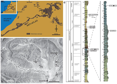
In a paper published in the journal Biology Letters on 20 January 2021, Aaron Hunter of the Department of Earth Sciences at the University of Cambridge and the School of Earth Sciences, at the University of Western Australia, and Javier Ortega-Hernández of the Museum of Comparative Zoology and Department of Organismic and Evolutionary Biology at Harvard University, and the Department of Zoology at the University of Cambridge, describe a new Somasteroid Echinoderm from the Early Ordovician Fezouata Lagerstätte of Zagora, in thecentral Anti-Atlas of Morocco.

 
Locality and stratigraphic occurrence of Cantabrigiaster at Lower Ordovician Echinoderm sites from the Anti-Atlas, Morocco. (a) Location of the Anti-Atlas range in northwestern Africa. (b) Map of the Anti-Atlas showing the distribution of Ordovician outcrops and the location of the Zagora area. (c) Landsat view of the Zagora area showing the location of all main Early Ordovician echinoderm localities that contain Somasteroids photograph courtesy of the U.S. Geological Survey. (d) Synthetic, composite stratigraphic column showing the Lower Ordovician succession in the Zagora area, central Anti-Atlas, Morocco. With detailed, stratigraphic columns showing the late Tremadocian interval (Araneograptus murrayi Zone and part of the Hunnegraptus copiosus Zone) showing the position of the main Somasteroid fossils. Hunter & Ortega-Hernádez (2021).

The exceptionally preserved morphology of the specimens reveals a unique plate organisation among Somasteroids, and allows us to test the phylogenetic implications of this taxon for the origin of total-group Asterozoa. Central to Hunter and Ortega-Hernádez's phylogenetic hypothesis is the presence of an imperforate extraxial body capsule on the aboral surface of the Somasteroids which is then lost in derived Asterozoans so that the aboral surface is entirely composed of perforate extraxial body wall, for example, carinals in Asteroids, and ventral, dorsal and lateral arm plates in Ophiuroids.

The new species is named Cantabrigiaster fezouataensis, where 'Cantabrigiaster' derives from Genus name derived from ‘Cantabrigia’, after the cities of Cambridge in the UK and USA, which were home to the influential Asterozoan workers John William Salter (University of Cambridge), Juliet Shackleton (neé Dean) (University of Cambridge) and Howard Barraclough ‘Barry’ Fell (Harvard University). No explanation is given for 'fezouataensis', although it appears to mean 'coming from Fezouata'.

Cantabrigiaster fezouataensis is a Somasteroid typified by biserial and offset ambulacrals with thin transverse bar, wide perradial groove, multiple interconnected virgal ossicles and aboral carinal region with network of spicule-like ossicles. Adambulacral ossicle series lacking along abaxial body margins (perpendiculars (virgals), structures 90° to the axial ambulacrals).

The designated holotype of the species (i.e. the specimen to which all other specimens are compared in order to determine whether they are the same species) is UCBL-FSL 424961, an articulated  specimen and latex moulds deposited at the University of Lyon 1. This is derived from the primarily Stylophoran-dominated beds in the upper part of the Araneograptus murrayi Zone, late Tremadocian, Z-F2 (Jbel Tizagzaouine), Z-F4 (Bou Izargane) and Z-F9 (Bou Glef), in the lower part of the Fezouata Shale Formation, Lower Ordovician, Zagora area (central Anti-Atlas), Morocco. The 70 m thick interval yields assemblages typical of the Fezouata Biota at about 260–330 m above the base of the Ordovician.

Also referred to Cantabrigiaster fezouataensis are another 31 specimens, including: specimens housed at the Yale Peabody Museum, Yale University, Hotchkiss collection (YPM IP 535545535559); the collections of Vizcaïno (UCBL-FSL 424962) and Lefebvre (UCBL-FSL 711938 and 711939) housed at the University of Lyon 1; and the Catto collection deposited in the Natural History Museum of Nantes (MHNN.P.045596).

Cantabrigiaster fezouataensis from the Lower Ordovician (?late Tremadocian) of Morocco. All body fossils. (a) YPM 535547, oral view. (b) Detail of dashed area in a. (c) YPM IP 535557-535558, oral and aboral view. (d) YPM IP 535559, aboral view. (e) Detail of dashed area in d. (f) YPM 535552, aboral view. (g) Detail of dashed area in (f). Abbreviations: am, ambulacral ossicles; cr, carinal region ossicles (preserved on the aboral surface); mc, mouth cavity; pb, podial basins; vr, virgal ossicles. Hunter & Ortega-Hernádez (2021).

The arms of Cantabrigiaster fezouataensis are broad, petaloid and arranged in a pentagonal outline The aboral skeleton (carinal region) is composed of randomly scattered spicule-like ossicles arranged into an irregular network. On the oral side, the ambulacrals consist of flattened ossicles with a subquadrate outline. These ossicles abut each other following the orientation of the perradial axis. The perradial suture is straight, and the ambulacrals at either side are stepped out of phase by approximately half an ossicle. The abaxial organisation of the ambulacrals consists of an elevated perradial ridge, less than a quarter in width relative to the ambulacral, and bears a thin transverse bar that occupies a central position, conferring a T-shape in oral view. The perradial ridges of the ambulacral ossicles at either side of the perradial suture are substantially separated from each other, forming a wide oral groove. The podial basins are shared equally between adjacent ambulacrals. Abaxially, the following ossicle series consist of the perpendiculars, also known as virgals in Somasteroids. The perpendicular series is composed of interconnected and robust rod-like virgal ossicles without spines. These ossicles follow a perpendicular orientation relative to the perradial suture. The virgal ossicles close to the ambulacrals are the largest, becoming smaller in length and width towards the abaxial body margins. Likewise, adjacent perpendicular series are in direct contact with each other adaxially relative to the perradial suture, whereas it is possible to observe open gaps between them towards the abaxial body margins. Proximal (relative to the mouth) perpendicular series consist of up to nine virgal ossicles, which gradually decrease in number towards the tips of the arms. The circumoral ossicles are enlarged relative to ambulacral ossicles, and the first podial pore is shared equally with the small and sub-triangular mouth angle plates. The madreporite is not preserved.

 
Cantabrigiaster fezouataensis from the Lower Ordovician (Tremadocian) of Morocco. Holotype UCBL-FSL 424961 (Van Roy coll.). (a) Oral view (body fossil). (b) Interpretative diagram of (a). (c) Close-up of extended arm (latex mould). (d) Interpretative diagram of (c). (e) Close-up of oral region (latex mould). ( f ) Interpretative diagram of (e). am, ambulacral ossicles; co, circumoral ossicles; cr, carinal region ossicles (preserved on the aboral surface); map, mouth angle plates; mc, mouth cavity; pb, podial basins; ps, podial suture; vr, virgal ossicles. Hunter & Ortega-Hernádez (2021).

The presence of virgal ossicles in Cantabrigiaster strongly supports its affinities with Somasteroids. Cantabrigiaster bears the greatest similarity to the Tremadocian taxa Chinianaster, Thoralaster and Villebrunaster,  but is unique among somasteroids in lacking ossicles along the abaxial lateral margins of the arms. The arm construction of Cantabrigiaster consists of flattened and offset biserial ambulacrals, each of which articulates with an abaxially oriented perpendicular series composed of simple virgal ossicles. In addition to these features, the arms of all other Somasteroids also possess a series of axially oriented ossicles along the lateral margins that vary from small and bead-like, albeit with occasional spikes, in Tremadocian taxa, to robust and block-like in the stratigraphically younger (Floian) Ophioxenikos and Darriwilian) Archegonaster. The absence of this key character and the results of  Hunter and Ortega-Hernádez's phylogeny demonstrate that Cantabrigiaster embodies the ancestral condition by virtue of lacking ossicles defining the lateral arm margins, whereas other Somasteroids record the first appearance of these structures along the edges of the arms, and their subsequent changes in size and shape. Based on this sequence,we propose that the origin of new axially oriented ossicle series in early Asterozoans required their formation on the abaxial edges of the arms. Our hypothesis implies that the proximity of axially oriented ossicle series relative to the perradial axis reflects the order of their evolutionary appearance since virgals are abaxially oriented, they are not directly comparable with any of the axially oriented ossicle series observed in Palaeozoic Asterozoans. In this context, Cantabrigiaster specifically lacks the adambulacral ossicle series present in more derived Somasteroids, Ophiuroids, Asteroids and Stenuroids (a group considered intermediate between Somasteroids and Ophiuroids/Asteroids), highlighting its profound significance for understanding the evolution of the Asterozoan body plan.

 
Phylogeny of total-group Echinodermata. Strict consensus topology based on the Bayesian-inference analysis of 38 taxa and 74 morphological characters informed by the extraxial–axial theory. The Asterozoan/Crinoid clade represented does not imply a sister group relationship; Echinozoan/Asterozoan monophyly has been established using molecular data. Wen., Wenlock; Lud., Ludlow; Prid., Přídolí. Hunter & Ortega-Hernádez (2021).

The extraxial–axial theory supports the homology of the ambulacrals across pentaradial total-group Echinoderms based on their developmental origin and postembryonic ontogeny, and allows comparison of the skeletal organization of Cantabrigiaster on a broader phylogenetic scale. Outside Asterozoa, the absence of adambulacrals in Cantabrigiaster draws parallels with Tremadocian Crinoids (e.g. Protocrinoids, Apektocrinus, Eknomocrinus), whose arm construction incorporates flattened and offset biserial ambulacrals articulated to an abaxially oriented (perpendicular) series of simple ossicles, here expressed as the cover plates. A similar axial skeletal organisation is also observed among Cambrian forms, most notably Edrioasteroids,  which also possess flattened and offset biserial ambulacrals but lack feeding appendages, and to a lesser extent Blastozoans, which have feeding appendages formed by modified ambulacrals known as brachioles. The widespread occurrence of these characters among non-asterozoan groups suggests that their presence in Cantabrigiaster is symplesiomorphic.

Cantabrigiaster fezouataensis from the Lower Ordovician (Tremadocian) of Morocco. All latex molds. (a) Holotype UCBL-FSL 424961 (Van Roy coll.), oral view. (b) UCBL-FSL 711938 (Lefebvre coll.), oral view. (c) UCBL-FSL 424961 (Van Roy coll.), oral view. (d) UCBL-FSL 711939 (Lefebvre coll.), aboral view. (e) MHNN.P.045596 (Catto coll.), oral view. (f) UCBL-FSL 424962a (Vizcaïno coll.), aboral view. (g) UCBL-FSL 424962b (Vizcaïno coll.). Abbreviations: am, ambulacral ossicles; co, circumoral ossicles; cr, carinal region ossicles (preserved on the aboral surface); map, mouth angle plates; mc, mouth cavity; tb, transverse bar; pb, podial basins; pr, perradial ridge; ps, perradial suture; vr, virgal ossicles. Hunter & Ortega-Hernádez (2021).

Hunter and Ortega-Hernádez's phylogenetic analysis of representative Lower Palaeozoic total-group Echinoderms tests the significance of Cantabrigiaster for the origin of Asterozoa. The dataset reflects the ambulacral homology proposed by the extraxial–axial theory, the oral symmetry model proposed by Universal Element Homology and Hunter and Ortega-Hernádez's hypothesis for the correspondence of axially oriented ossicle series in early Asterozoans. Bayesian and parsimony-based analyses recover practically identical topologies, despite a loss in tree resolution in the earliest divergent representatives that can be expected from the former methodology, indicating a robust phylogenetic signal within Asterozoa. Cantabrigiaster occupies the earliest diverging position within total-group Asterozoa, supporting our hypothesis that the absence of adambulacrals is an ancestral condition, rather than a case of secondary reduction. Tremadocian Somasteroids are resolved as a paraphyletic grade of stem-group Asterozoans, whereas the Floian Ophioxenikos and Darriwilian Archegonaster consistently occupy a more derived position as members of crown-group Asterozoa. The analyses argue against the monophyly of Stenuroids, but corroborate their close phylogenetic relationship to Ophiuroids, specifically as their earliest diverging stem-group representatives. These findings indicate that the evolution of a well-developed adambulacral ossicle series constitutes a critical step in the origin of crown-group Asterozoa, and suggest that the abaxially oriented virgals of Somasteroids became independently reduced, and ultimately lost, within the stem lineages of Ophiuroidea and Asteroidea.

 
Morphological reconstruction of Cantabrigiaster fezouataensis. (a) Aboral view. (b) Oral view. (c) Cross section of isolated arm in oblique view. (d) Cross section of main body cavity lateral view. (e) Isolated virgal ossicle series and ambulacrals in oral view. (f) Life reconstruction of Cantabrigiaster fezouataensis. Abbreviations: am, ambulacral ossicles; cr, carinal region ossicles (preserved on the aboral surface); map, mouth angle plates; pr, perradial ridge; ps, podial suture; tb, transverse bar; vr, virgal ossicles. Marguerite Lardanchet in Hunter & Ortega-Hernádez (2021).

See also...


Online courses in Palaeontology. 

Follow Sciency Thoughts on Facebook.

Follow Sciency Thoughts on Twitter.


Wednesday, 6 January 2021

Ichnological evidence for limb-autotomy in Triassic Holocrinids.

Many Animals, including Echinoderms, are able to autotomize parts of their body usually as a defense strategy against predators. It has been argued that shed appendages, which sometimes display vigorous post-autotomy movements, reduce the animal’s mortality in two major ways: (1) they enable the animal to break away from predators that have grasped it, and (2) divert the attention of the predators away from the vulnerable body parts. Crinoids, commonly referred to as Sea Lilies, are a group of Echinoderms that is subject to a high predation pressure, have remarkable ability to autotomize and regenerate their appendages. It has been shown that arm autotomy in these echinoderms is achieved through the nervously mediated (L-Glutamate invoked) destabilisation of collagenous ligamentary fibres at specialized autotomy planes, namely (crypto)syzygial articulations. 

Although considerable effort has been devoted to study autotomy in Crinoids, little attention has been paid to post-autotomy thrashing behaviour. A 2010 study by Iain Wilkie, Alice Barbaglio, William Maclaren, and Maria Candia Carnevali, on living stalkless Comatulids (Feather Stars) only briefly reported that: 'after autotomy induced in both intact Animals and isolated arms, the detached distal portion of the arm showed rhythmical cycles of flexion and extension in the oro–aboral plane'.

In a paper published in the journal Scientific Reports on 15 September 2020, Przemysław Gorzelak of the Institute of Paleobiology of the Polish Academy of Sciences, Mariusz Salamon and Krzysztof Brom of the Faculty of Natural Sciences at the University of Silesia in Katowice, Tatsuo Oji of the University Museum at Nagoya University, Kazumasa Oguri of the Japan Agency for Marine-Earth Science and Technology, Dorota Kołbuk, also of the Institute of Paleobiology of the Polish Academy of Sciences, Marek Dec of the Polish Geological Institute, Tomasz Brachaniec, also of the Faculty of Natural Sciences at the University of Silesia in Katowice, and Thomas Saucède of Biogéosciences at the Université Bourgogne Franche-Comté, further examine thrashing behaviour in extant Sea Lilies, and explore its ichnological potential by conducting neoichnological aquarium experiments using the Stalked Crinoid Metacrinus rotundus, and then analyzed samples with articulated arm fragments of the oldest (Early Triassic) stem-group Isocrinids (Holocrinids) in order to identify evidence of this behaviour in the fossil record.

Specimens of living Stalked Crinoids of Metacrinus rotundus were dredged from a depth of about 140 m in Suruga Bay (near the town of Numazu in Shizuoka Prefecture, Japan) using a 90-cm wide naturalist dredge with a net. Then, the living specimens were transferred to an experimental seawater tank in the Nagoya University Museum. The aquarium was maintained at a constant seawater temperature (roughly 16°C) in darkness and under circulation provided by a water pump. After few weeks of acclimatising the Crinoids, a box (approximately 40 × 30 cm) floored with fine-grained sand was placed, which was smoothed before an autotomised arm was introduced. Neoichnological experiments were repeated several times using different arms from different individuals.

For comparison purposes, a series of aquarium experiments was made on production of transport-induced sole markings left on the sediment surface (fine-grained sand) by isolated dead arms being dragged (with the pinnules facing upstream or downstream) by a current (0.35 cm per second) adjusted by a water pump that was placed at the bottom of the aquarium. At the lower or higher velocity, the arms (which were placed at a distance of about 25 cm from the pump) were not moving or were lifted above the sediment surface, respectively, leaving no traces. These experiments were conducted at the Laboratory of Experimental Taphonomy at the Faculty of Natural Sciences of the University of Silesia in Katowice, Sosnowiec, Poland. Gypsum casts from both experiments are deposited at the Institute of Paleobiology of the Polish Academy of Sciences (ZPALV.42ICH).

A Lower Triassic (lowest Spathian) slab of thin-bedded silty limestone of the Thaynes Group (west of Paris, Idaho, USA) preserving five isolated fragments of arms belonging to one of the oldest (if not the oldest) post-Palaeozoic holocrinid taxon (Holocrinus sp.) deposited in the collection of the Université de Bourgogne, Géologie Dijon, France (UBGD 30564) was investigated. These arm fragments are of different lengths and lack distal arm tips and some distal pinnules.

Following autotomy of the arm, within a few seconds, its activity increased, i.e. it started to thrash mostly in the oral–aboral plane. Both frequency and style of flexions (sluggish writhing and violent flicks) and the duration of movement varied (several hours to up to about 7 days). However, only in the first hours after autotomy, detached arms showed the highest activity. The frequency and amplitude of motions decreased with time; i.e. after several hours autotomised arms remained in place; their activity could still be recorded but they did not produce any traces. In the first hours after autotomy, each autotomized arm produced two major types of traces on the sediment surface, which can be closely associated with each other.

 
Traces produced by autotomised arms of Metacrinus rotundus. (a)–(c) Straight deep grooves arranged in radiating group; (d)–(f) a few sets of straight parallel grooves and furrows inclined at different angles to each other; (g)–(i) two large arched grooves; (j)–(l) small short parallel furrows. (a), (d), (g), (j) Photographs of sediment surface; (b), (e), (h), (k) photographs of gypsum casts; (c), (f), (i), (l) false-colour depth maps of gypsum casts. (m) colour scale of elevation. Large and small arrows indicate deep grooves left by the arm and shallow furrows made by pulling the pinnules along the substrate, respectively. Scale bars are 1 cm. Gorzelak et al. (2020).

The first type comprises straight to slightly arched grooves, 1.5–3 cm long (mean: 2.1 cm), 0.2–0.6 cm wide (mean: 0.3 cm) and about 0.1 cm deep, usually arranged in radiating groups; the grooves may be inclined at different angles to each other (the angles between adjacent grooves ranging from 9° to 28°) and are separated from each other by a distance of 0.9–2.8 cm (measured in the widest distance between two ends of neighbouring grooves). The length-to-width ratio of these grooves ranges from 3.8 to 10.9 (mean: 6.8). These traces were left by a rotating and more or less rhythmic movement of the flicking arm that placed the most pressure on the substrate with its median-distal arm part.

The second type comprises sets of thin and parallel (locally curving) furrows, 0.3–1.7 cm long (mean: 1.1 cm), 0.04–0.17 cm wide (mean: 0.1 cm),  and about 0.03 cm deep; these sets may be inclined at different angles to each other, locally forming a herringbone pattern. The length-to-width ratio of these furrows ranges from 4.1 to 33.6 (mean: 11.4). These traces were made by pulling the pinnules along the substrate.

Current-produced sole markings, resulting from the more or less continuous contact of the arm with the sediment surface, display a very different morphology. These grooves are continuous, long (4–6 cm), generally linear, deep (~ 0.2 to 0.4 cm) and run parallel to the flow direction.

 
Current-induced sole markings (arrows) left on the sediment surface. (a) Photograph of sediment surface; (b) photograph of gypsum cast. Gorzelak et al. (2020).

Close examination of Lower Triassic slabs from the Thaynes Group (Spathian, west of the city of Paris, Idaho, USA) revealed similar traces associated with isolated articulated fragment of arm of Holocrinus sp. Given their size, morphology and close association with the Crinoid arm, it appears that these traces could have been produced by thrashing movements of shed arm. However, the scarcity and imperfect state of preservation make the erection of the new ichnospecies impossible.

 
Possible trace fossils associated with articulated piece of arm of Holocrinus sp. (UBGD 30564; Thaynes Group, Lower Triassic; collection of Université de Bourgogne, Géologie Dijon, France). (a) Slab preserving articulated fragment of arm and associated traces (arrows); a photograph taken using a camera fixed to a tripod. (b) Slab preserving articulated fragment of arm and arched grooves (curved red arrows) arranged in radiating group and a few sets of indistinct straight parallel furrows (straight small red arrows with a question mark) and analogical traces (in dotted circle) produced by autotomised arms of Recent isocrinid; a photograph taken using a camera attached to an optical microscope. (c) False-colour depth map of a slab; (d) false-colour depth map of a slab acquired from photographs taken from binocular microscope (elevation has been increased (× 2) to enhance depth contrast); (e) colour scale of elevation. Scale bars are 1 mm; scale bar in dotted circle is 1 cm. Gorzelak et al. (2020).

One arm fragment is associated with slightly arched grooves, about 1.9 to 3.2 mm long (mean: 2.3), 0.5 to 0.8 mm wide (mean: 0.55), arranged in a radiating group (the angles between adjacent grooves range from 12° to 24°). These grooves are separated from each other by a distance of 1.9–2.6 mm (measured in the widest distance between two ends of neighbouring grooves). Dimensionally, the mean length-to-width ratio of these grooves (4.3) falls well within the range of this ratio for the grooves produced by the median-distal arm part during our neoichnological experiments. These grooves are associated with a few sets of indistinct thin and parallel (locally slightly curving) furrows, 1.8–3.2 mm long (mean: 2.4), 0.07–0.13 mm wide (mean: 0.1) of uncertain origin. Notably, however, the mean length-to-width ratio of these furrows (25.5) falls within the range of this ratio reported in Gorzelak et al.'s neoichnological experiments.

As no ethological term exists for the trace fossils produced by post-autotomy thrashing movements of shed appendages Gorzelak et al. propose a new ethological category, autotomichnia (from the Greek auto- 'self-' and tome 'severing', αὐτοτομία). Although thrashing behaviour occurs in a wide range of Vertebrate and Invertebrate taxa, autotomichnia are probably rare in the fossil record, and its identification is difficult if the traces are not found in association with the 'producer'. Such cases, however, are not likely to be common given that shed appendages displaying movements attract attention of the predators which attempt to consume them. The only ichnospecies ascribed to activity of Crinoids described so far is Krinodromos bentou. This ichnotaxon, however, in contrast to the presently described traces, refers to unusual locomotion trail, and shows different morphology in the form of two bordering grooves combined with pushed sediment piles, and a central flat area or a narrow winding furrow. Possible explanation for the fossil traces reported herein is that they simply represent sole markings produced by a dead arm moved along by a current. However, these Triassic traces bear no morphological resemblance to the experimentally induced abiotic sedimentary structures.

It has been argued that ichnology can provide valuable insights on the evolutionary origins of behaviors and on many of their functional and adaptive aspects. Gorzelak et al.'s experiments showed that autotomised arms of Stalked Crinoids (isocrinids) display vigorous movements similar to that observed in shed arms of Brittlestars. They produce distinct traces on the sediment surface, which may have some potential to be preserved as trace fossils. This finding opened new perspectives for tracing the origin of thrashing behaviour in crinoids. Indeed, Gorzelak et al.'s examination of fossil material from the Thaynes Group suggests that similar traces are associated with isolated articulated fragment of arm of the oldest (approximately 250.6 million years old) stem-group Isocrinids (Holocrinus). The fact that this arm fragment lacks distal arm tip and some distal pinnules is suggestive that it was autotomised due to predatory attack prior to rapid burial rather than another type of disturbance (e.g. storm). It must be emphasised that although abiotic factors (such as high current velocities induced by storms or other environmental trauma) may lead to arm loss, most autotomy in natural settings results from biotic interactions. This is consistent with recent data which showed that the thruster-produced extreme flow or suction force did not lead to arm loss or any other type of injury in deep-water, Stalked Crinoid Democrinus sp.

Trace fossils described by Gorzelak et al. suggest that the origin of thrashing behaviour in Crinoids could be traced back to at least the Early Triassic. The question of whether this behaviour could have appeared in the Palaeozoic Crinoids (independently in several clades or in one lineage and was then inherited by the post-Palaeozoic descendants) is presently unclear. However, it should be emphasised that, in contrast to the highly flexible muscular arms of recent and post-Palaeozoic Crinoids in which syzygial or cryptosyzygial ligamentary articulations specifically designed for autotomy are localised, many Palaeozoic Crinoids possessed primitive arms with limited flexibility. Although regeneration is frequently documented in Palaeozoic Crinoids, most of them, if not all, have not yet developed autotomy planes. This suggests that they lacked autotomy abilities, or at least that they were less specialised in that regard.

The end-Permian mass extinction profoundly influenced the evolutionary history of Crinoids, not only through the demise of major Palaeozoic crinoid groups but also through changes in their functional morphology. Notably, post-Palaeozoic crinoids are considered to have descended from only a single survivor (i.e. a Cladid ancestor, Ampelocrinids). In the Middle Triassic, however, they rebounded and underwent a major radiation resulting in the appearance of several motile taxa showing many anti-predatory morphological and behavioral innovations to increased predation pressure during the Mesozoic Marine Revolution. Holocrinids are among the first Crinoids to appear in the aftermath of the end-Permian Mass Extinction. These Crinoids display many adaptations to benthic and nektonic predators. For instance, they developed specialised rupture points at the distal nodal facets in their stalk, allowing them to free themselves of the sea bottom, crawl with the aid of highly flexible muscular arms and re-attach. Furthermore, they elaborated localised autotomy planes (cryptosyzygies) in the arms, which in Recent Isocrinid descendants greatly reduce mortality and arm damage. Gorzelak et al. suggest that Holocrinids might have also displayed post-autotomy arm thrashing. Interestingly, in some Lizards, it has been observed that their wildly thrashing autotomised tails (in contrast to taxa having a much lower rate of tail thrashing) consistently distract attention of predator away from the escaping Lizard and increase predator-handling time, providing additional opportunity to escape. Intriguingly, however, in some Lacertid Lizards, duration of thrashing between different species, shows little variation, either because it is less costly, or because the underlying physiological pathways are evolutionary conservative. Notwithstanding, the high numbers of shed Lizard tails are commonly found in predator stomachs. All these indicate that caudal autotomy and thrashing behaviour in these Vertebrates is an effective antipredatory strategy. Accordingly, Iain Wilkie argued that thrashing behaviour in Ophiuroids is also likely to be an effective decoy against Fish predators. Consequently, Gorzelak et al. hypothesise that this behaviour in crinoids also probably acts as a defense strategy to distract predator attention, and/or to increase predator handling time, providing additional getaway chances to Crinoids. A moving autotomized arm might itself be sufficient attraction as a food source. Accordingly, acquisition of thrashing behaviour in the Early Triassic Crinoids may underscore the magnitude of the anti-predatory traits that occurred during the Mesozoic Marine Revolution, which had already started soon after the end-Permian extinction. The thrashing behaviour along with the specific arm construction consisting of two types of articulations (muscular ones playing a great role in locomotion and postural changes by maximising arm flexibility, and strictly ligamentary (crypto-)syzygial ones, specifically designed for autotomy) have been maintained in Isocrinid/Comatulid descendants up to the present.

Given the scarcity and imperfect state of preservation of the trace fossils described herein, further ichnological findings are needed to fully test the above hypotheses. Results of neoichnological experiment presented herein highlight the preservation potential of thrashing behaviour of Crinoid arms, thus rock slabs preserving detached Crinoid arms are worthy of in-depth investigation.

See also...


Online courses in Palaeontology. 

Follow Sciency Thoughts on Facebook.

Follow Sciency Thoughts on Twitter.


Tuesday, 30 June 2020

Ophiambix kagutsuchi & Ophiambix macrodonta: Two new species of Brittle Stars from Japanese hot vents and cold seeps.

Brittle Stars, Ophiuroidea, contains the largest number of species within the Echinodermata and occurs in a great variety of marine habitats, such as muddy substrates, living infaunally in sediments, under rocks, in the interstices of Sponges and Hard Corals, and on surfaces of various Animals such as Soft Corals. They are globally one of the dominant deep-sea megafaunal groups, but diversity in chemosynthetic ecosystems such as hydrothermal vents and hydrocarbon seeps remains poorly understood. Since 1985, 10 species of Ophiuroids, including Ophiacantha longispina, Ophiactis tyleri, Ophienigma spinilimbatum, Ophiocten centobi, Ophioctenella acies, Ophiolamina epraewas, Ophiomitra spinea, Ophioplinthaca chelys, Ophiotreta valenciennesi rufescens, and Spinophiura jolliveti, have been collected from chemosynthetic ecosystems. As our knowledge grows, we have gained better understanding of Ophiuroids within these settings, and it appears Ophiuroid diversity has likely been underestimated. In Japanese waters, surveys of Ophiuroids have recorded 346 species, which represent approximately three-quarters of the North Pacific Ocean Ophiuroid fauna. However, no Ophiuroid species have been recorded from chemosynthetic habitats around Japan to date.

In a paper published in the journal Raffles Bulletin of Zoology on 17 April 2020, Masanori Okanishi of the Misaki Marine Biological Station of the University of Tokyo, Moe Kato of the School of Natural System at Kanazawa University, Hiromi Kayama Watanabe and Chong Chen of the Japan Agency for Marine-Earth Science and Technology, and Toshihiko Fujita of the National Museum of Nature and Science, describe two new species of Ophiuroids from chemosynthetic ecosystems in Japanese waters.

Both new species are placed in the enigmatic Ophiuroid genus Ophiambix, which was originally erected by Edward Lyman in 1880 for the monotypic Ophiambix aculeatus, and is known from the deep waters (146–5,315 m) of the Pacific and Atlantic Oceans. Out of the four species of Ophiambix currently considered to be valid, only Ophiambix aculeatus has been recorded from Japanese waters. Ophiambix has a characteristically flat body and their arms are well differentiated from the disc in a manner superficially similar to that of Asteroids.

The first new species is named Ophiambix kagutsuchi, in reference to Kagutsuchi, the god of fire in ancient Japanese mythology, referring to the hot-vent habit of the new species. The species was found at a series of hydrothermal vents in the Okinawa Trough, as well as on sunken wood found in the Ryukyu Trench, southwest of Japan, at a depth range of 276–1,979 m.

Ophiambix kagutsuchi, holotype (NSMT E-13071). (A) Aboral body; (B) aboral disc and proximal portion of arm; (C) oral disc and proximal portion of arms, arrow heads indicate oral papillae; (D) proximal portion of aboral surface of arm; (E) proximal portion of oral surface of arm; (F) distal portion of aboral surface of arm; (G) distal portion of oral surface of arm. Abbreviations: ASh, adoral shield; ASp, arm spine; D, dorsal arm plate; GS, genital slit; L, lateral arm plate; OP, oral plate; OS, oral shield; RS, radial shield; SD, supplementary dorsal plate; Te, tentacle scale; V, ventral arm plate. Scale bars 1 mm. Okanishi et al. (2020).

The disk of Ophiambix kagutsuchi is pentagonal, and about 4 mm in diameter. The aboral surface covered by small granules of almost uniform size, separated from each other, approximately 50–70 μm in diameter. Removal of granules shows underlying plates are scalar, circular in outline, and imbricating, each approximately 250–350 μm in diameter. Radial shields are triangular, distally wider, 350 μm in width, and 350 μm in length, sharpen towards the centre, and completely concealed by the granules. On the oral surface, the adoral shields are parallelogram-shaped, wider than long, approximately 250 μm in width, and 120 μm in length, one overlapping the other. The oral plates are trapezoidal, approximately 250 μm in width, and 180 μm in length at the radial edge, 250 μm in length at the abradial edge, and in contact with each other. The oral shields are pentagonal, slightly rounded, with a convex distal edge, approximately 360 μm in width, and 60–260 μm in length. The interradial oral disc area is narrow, covered by scales under thick skin, and approximately 200–300 μm in length. The genital slits are narrow, almost extending from the edge of the oral shield to two-thirds the height of the oral interradial disc, and 0.1 mm in width. The oral papillae and teeth are rudimentary, very thin, narrow and flat ossicles, approximately 10 μm in length, forming a continuous horizontal row on oral edge of dental and oral plates. The teeth and oral papillae quite similar in shape but for descriptive purposes, the ossicles on top of the dental plate are called teeth and ossicles on oral edge of oral plate are called oral papillae. With the exception of the oral-most row of papillae, there are 6 to 7 thin and fan-shaped teeth forming vertical row on the dental plate. There is a second tentacle pore inside the mouth slit.

Ophiambix kagutsuchi, paratype (NSMT E-13070), scaning electron microscope images of ossicles. (A)–(D) Vertebrae from proximal portion of arm, distal view (A), proximal view (B), oral view (C), aboral view, green illustration indicates 'T'-shaped groove (D); (E)–(G) lateral arm plates from proximal portion of arms, oral view (E), adoral view (F), inner view (G) an arrow head indicates a perforation; (H) ventral arm plates from proximal portion of arm, inner view; (I), (J) arm spines from distal (I) and proximal (J) portion of arm, arrow heads indicate secondary teeth. Orientations: ab, aboral side; ba, basal side; dis, distal side; ex, external side; in, inner side; o, oral side; pro, proximal side. Abbreviations: AF, aboral muscle fossae; DL, dorsal lobe; ES, elongated structure; LAC, lateral ambulacral canal; MO, muscle opening; NO, nerve opening; OF, oral muscle fossae; VL, ventral lobe. Okanishi et al. (2020).

On the proximally arm, theaboral surface is covered by small granules the same as those on the aboral disc, these decrease in number and disappearing on arm tip. After removal of the granules, the exposed dorsal arm plates are oblong, longer than wide, the proximal edge is slightly wider than the distal edge. There is a pair of fan-shaped supplementary dorsal arm plates on both lateral sides of each dorsal arm plate and irregularly shaped smaller supplementary plates on the distal side of the dorsal arm plates separating them from each other. On the middle portion of the arm, these supplementary plates disappear and the dorsal arm plates are in contact, gradually decreasing in size toward the arm tip. The lateral arm plates are thin, and widely separated from each other throughout the arm. The tentacle pore forms a large hole. On the proximal portion of the arm, the ventral arm plates are almost square with a concave distal edge, and toward the arm tip, become oblong, longer than wide, with a distal concave edge, contiguous throughout the arms. There are three flat and pointed arm spines on the proximal portion of each arm, the middle one is longest, the same length as the corresponding arm segment, the aboral-most half length of the middle spine, and the oral-most spine is shortest, approximately one-fifth length of the middle spine. The arm spines decrease in number to one toward the arm tip, transforming into a hook-shape, approximately the same length as the corresponding arm segment. There is one small, rudimentary tentacle scale at each tentacle pore, although small, tentacle scales present on the distal portion of each arm.

Ophiambix kagutsuchi, paratype (NSMT E-13050), scanning electron microscope images of ossicles. (A), (B) oral plates of abradial side (A) and adradial side (B), partly cracking, arrows indicate neural grooves; (C), (D) adradial genital plates, aboral view (C) and oral view, partly cracking (D); (E) inner view of a radial shield; (F) external view of a dental plate. Orientations: ab, aboral side; cen, centre of disc; o, oral side; per, periphery of disc. Abbreviations: AbMA, abradial muscle attachment area; AdMA, adradial muscle attachment area; AG, articulation for genital plate; DAT, depression for aboral tentacle; DOT, depression for oral tentacle; DW, presumable depression for water ring canal. Scale bars 100 μm. Okanishi et al. (2020).

The second new species is named Ophiambix macrodonta, which is a Latin adjective which means to have large teeth, referring to the flat, wide teeth of the species. The species is known only from a hydrocarbon seep site on Kuroshima Knoll, southeast of the Yaeyama Islands, part of the Ryukyu Island chain, southwest of Japan, at a depth range of 638–644 m.

Ophiambix macrodonta, holotype (NSMT E-13059). (A) Aboral body; (B) oral body; (C) aboral disc and proximal portion of arm; (D) aboral periphery of disc and proximal portion of arm; (E) oral disc and proximal portion of arms; (F) oral periphery of disc and proximal portion of arm; (G) a jaw; (H) top of a jaw, arrow heads indicate teeth and oral papillae. Abbreviations: ASh, adoral shield; DP, dental plate; GS, genital slit; L, lateral arm plate; OP, oral plate; OS, oral shield; RS, radial shield; Te, tentacle scale; V, ventral arm plate. Scale bars 1 mm. Okanishi et al. (2020).

The disc of Ophiambix macrodonta is pentagonal, and 6 mm in diameter. The aboral surface is covered by polygonal scales, approximately 250–350 μm in diameter, arranged in a mosaic pattern. Each scale is covered by small granules of uniform size, approximately 40–60 μm in diameter, in contact with each other and forming two or three circular rows on the periphery of each scale. The radial shields are oval, 850 μm in length and 500 μm in width, and almost completely concealed by granules except on the margins. The adoral shields are curved, bar-like, wider than long, approximately 500 μm in length and 150 μm in width, and separated from each other. The visible part of the jaws is trapezoid, approximately 500 μm in length and 250 μm in width, and contiguous. The oral shields are elliptical in shape, longer than wide, slightly acute on both adradial edges, and approximately 300 μm in length and 650 μm in width. The interradial oral disc area is narrow, covered by polygonal and mosaic scales, approximately 200–300 μm in length as those on aboral disc. The genital slits are long and wide, almost extending to the disc edge from distal edge of the oral shield, and 0.3 mm in width. The oral papillae and teeth are rudimentary, very thin, narrow and flat ossicles, approximately 10 μm in width, forming a horizontal row on the oral edge of the dental plate and oral plate. The second tentacle pore is inside the mouth slit.

Ophiambix macrodonta, holotype (NSMT E-13059) (A)–(E), (G), (H) and a paratype (NSMT E-13060) (F), scanning electron microscope images. (A) Aboral disc and proximal portion of arm; (B) aboral periphery of disc and proximal portion of arm; (C) granules (arrow heads) on aboral disc; (D) oral disc and proximal portion of arms; (E) jaws, an arrow head indicates teeth or oral papillae; (F) a jaw from lateral view, arrow heads indicate teeth and oral papillae; (G) proximal portion of aboral surface of arm, arrow heads indicate supplementary plates; (H) proximal portion of oral surface of arm. Abbreviations: ASh, adoral shield; ASp, arm spine; D, dorsal arm plate; GS, genital slit; RS, radial shield; V, ventral arm plate. Okanishi et al. (2020).

The hollowtype of Ophiambix macrodonta has two complete arms 25 mm and 26 mm, while the other three arms have lost their tips. The proximal portion of the arms is 1.5 mm wide and 1 mm high, and eliptical in cross section. The arms taper gradually towards the tip. The aboral surface is covered by dorsal arm plates and supplementary dorsal arm plates, with no granules. On the proximal portion of the arm, the dorsal arm plates are semicircular, and wider than long; the proximal edge is straight. There is one large, fan-shaped supplementary dorsal arm plate on both lateral sides of the dorsal arm plate. Three smaller, polygonal plates are at the distal edges of the dorsal arm plates and the two larger supplementary dorsal plates. Toward the arm tip, these supplementary plates decrease in size and gradually disappear with the dorsal arm plates becoming contiguous. The lateral arm plates are thin, and widely separated throughout the arm. On the proximal portion of the arm, the ventral arm plates are almost square, with slightly concave lateral edges and oblong. On the distal portion of the arm, the ventral arm plates are longer than wide, concave, and the lateral edges more pronounced. The ventral arm plates are contiguous throughout the arms. There are three flat arm spines on the proximal portion of arms, the aboral-most and middlevspines are flat and leaf-like, and the oral-most spine is cylindrical, narrow, and pointed. All three spines are equal in length to the corresponding arm segment. Toward the tip, the aboral-most and middle arm spines transform to a cylindrical and pointed shape, and the oral-most one transforms into a hook, approximately the same length as the corresponding arm segment. There is one small, triangular tentacle scale at each tentacle pore, although small, tentacle scales are present on distal portion of the arm.

Ophiambix macrodonta, holotype (NSMT E-13059). (A) Proximal portion of aboral surface of arm, a part enlarged in (B), arrow heads indicate supplementary plates; (C) distal portion of aboral surface of arm; (D) proximal portion of oral surface of arm; (E) distal portion of oral surface of arm; (F) distal portion of lateral surface of arm. Abbreviations: ASp, arm spine; D, dorsal arm plate; L, lateral arm plate; Te, tentacle scale; V, ventral arm plate. Scale bars 1 mm. Okanishi et al. (2020).

The difference in body size of the two new species Ophiambix macrodonta (3.3 to 7.0 mm in disc diameter) and Ophiambix kagutsuchi (0.8 to 3.2 mm in disc diameter) suggests that these could be interpreted as different sizes of the same species. However, the smallest specimen of Ophiambix macrodonta (3.3 mm in disc diameter), and the largest specimen of Ophiambix kagutsuchi (3.2 mm in disc diameter), were similar in size but exhibited the full set of respective diagnostic characters. All examined specimens of Ophiambix kagutsuchi, were collected from hydrothermal vents or sunken wood environments, whereas those of Ophiambix macrodonta, new species, were collected only from hydrocarbon seeps. Therefore, Okanishi et al. consider that these two taxa are indeed separate new species that can be distinguished from each other by morphological characters and by different environmental preferences.

See also...

https://sciencythoughts.blogspot.com/2019/10/ophiopsila-xmasilluminans.htmlhttps://sciencythoughts.blogspot.com/2019/08/ophiacantha-scissionis-fissiparous-six.html
https://sciencythoughts.blogspot.com/2014/12/a-new-species-of-brittle-star-from.htmlhttps://sciencythoughts.blogspot.com/2014/06/a-new-species-of-brittle-star-from.html
https://sciencythoughts.blogspot.com/2012/06/brittle-star-from-late-jurassic-of.html
Follow Sciency Thoughts on Facebook.