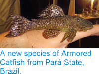
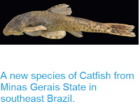
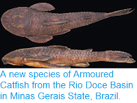
The genus Trichogenes was described by Heraldo Britski and Herman Ortega in 1983 to include a single new species, Trichogenes longipinnis. The taxon was discovered in hydrographically isolated systems in the Atlantic rainforest near the border of the states of Rio de Janeiro and São Paulo in southeastern Brazil, still its known geographical range. The discovery of Trichogenes was most important in understanding the evolution of Trichomycterids (Pencil Catfish) and Loricarioids (members of the superfamily that includes all Catfish except the Diplomystidae, or Velvet Catfish), because it bears plesiomorphic character conditions then unknown elsewhere in the family. Since its discovery, it was obvious that Trichogenes occupied a markedly basal position among Trichomycterids, a circumstance that in combination with other character-state conditions, had significant impact on the phylogeny of lower Loricarioid Catfish. Given the large morphological gap between Trichogenes longipinnis and other Trichomycterids, the eventual discovery of intermediate forms was considered likely. The subfamily Copionodontinae came in part to fulfill that expectation, as a clade that shares most of the putatively primitive features seen in Trichogenes. But copionodontines, although also basal in trichomycterids, are a rather divergent clade, and not a variation of the Trichogenes lineage. A second species in the genus came only 27 years after the description of Trichogenes longipinnis, with the discovery of Trichogenes claviger from the Rio Itapemirim drainage in the State of Espírito Santo, southeastern Brazil.
In a paper published in the American Museum Novitates on 23 April 2020, Mário de Pinna, Vinícius Reis, and Heraldo Britski of the Museu de Zoologia da Universidade de São Paulo, describe a third species of Trichogenes.
The discovery of this taxon was made under peculiar circumstances. Three preserved specimens in good condition were located without any associated data. Exhaustive searches and inquiries about the possible provenance of the material did not disclose any relevant information. Also, field trips to possible localities based on indirect inference failed to locate any specimens and no additional material is known. Despite such shortcomings, the new form is highly distinctive, there being no doubt of its taxonomic differentiation. In itself, it is of special phylogenetic importance, and it represents a most relevant addition to a highly relictual genus whose two species described to date are endangered. The unexpected existence of this new Trichogenes is in itself remarkable and significantly adds to our knowledge of the biodiversity of neotropical freshwater Fish. Having the new taxon reported and described will make it more widely known, encouraging the search for its provenance and, it is hoped, the discovery of existing populations.
The new species is clearly related to other species of Trichogenes, but differs from them in several traits of pigmentation, external and internal anatomy. The new species is the sister group of Trichogenes claviger. Some of its character states shed light on the homology and evolution of the antorbital and the opercle in trichomycterids, providing corroborative evidence for the transition between the generalised condition in other Loricarioids and the highly specialised situation in most other Trichomycterids.
Field activities related to the search for the locality of the new species resulted in the discovery of Trichogenes claviger in a new locality. Data on such specimens is reported, including photographs and video of live specimens and ecological information not previously available.
The new species is named Trichogenes beagle, in honour of the Beagle Laboratory of Molecular Systematics of the Department of Animal Biology at the Universidade Federal de Viçosa, the lab where the only specimens of the new species were found preserved. The three known specimens were found by Vinícius Reis during a routine search for molecular samples of Trichomycterids in a freezer at the Beagle Laboratory. The specimens were in a plastic centrifuge vial, in good overall condition and clearly had been fixed in formalin before their preservation in alcohol, as evidenced by the transparent aspect of their eye lenses. No label was associated with the vial, which had 'Cachoeira do Cobra' inscribed by hand in pen on the outside. Extensive and repeated inquiries with lab members revealed no memory of who might have collected the fish or placed the vial in that freezer. The equipment is used by lab students, faculty, and visiting professionals, and is intended as a convenience facility for temporary storage, although some material is occasionally abandoned, as was apparently the case with the T. beagle material. As a result, the name 'Cachoeira do Cobra' was the only clue as to the provenance of the material, even though vials of that sort are often repeatedly reused and inscriptions on their outside may bear no relation with their current content.

Trichogenes beagle is distinguished from its two congeners, Trichogenes longipinnis and Trichogenes claviger, by: (1) the colour pattern composed of small uniform-sized dark markings irregularly distributed on body, not forming any pattern (compared to presence of both small and large round spots, with the latter aligned in parallel to base of anal fin); (2) the lack of dark concentrations around latero-sensory pores on head (as opposed to pores outlined in dark); (3) the presence of a faint white vertical band along the margin of the hypural plate, delimited anteriorly and posteriorly by darker areas (as opposed to equivalent dark band or vertical series of dark spots in same position); (4) the absence of differential dark concentration along the base of the dorsal fin (instead of well-defined dark band present); (5) the presence of a wide (about 25% of the depth of the anal fin) white band parallel to the base of the anal fin ( band absent in Trichogenes longipinnis and narrower than 20% of the depth of the anal fin in Trichogenes claviger); (6) the lack of dark pigment concentration on the ventral area of opercular odontodes (as opposed to no differential dark pigmentation along area of opercular odontodes); (7) the barbular bone articulating with the distal portion of the antorbital process of the lateral ethmoid (this barbular bone is absent in Trichogenes claviger and articulates with the midlength of the antorbital process in Trichogenes longipinnis); (8) the opercular odontodes continuously distributed along a large portion of the margin of the opercle (as opposed to opercular odontodes distributed in two separate patches); (9) the presence of some tricuspidate teeth on jaws (as opposed to none). Further distinguished from Trichogenes claviger by having 8+9 principal caudal fin rays (instead of 7+8); I+8 dorsal-fin rays (Trichogenes claviger has I+7); and by the presence of a barbular bone. Further distinguished from Trichogenes longipinnis by the terminal mouth; the presence of ossification on the second hypobranchial (compared to second hypobranchial entirely cartilaginous); the presence of an anterodorsal clawlike process on the dorsal surface of the neural arch of each of the anterior four free vertebrae; the interopercular odontodes extending onto the posterodorsal margin of the interopercle (as oposed to mostly restricted to ventral and posterior margins of the bone); by the posterior naris broader than long (rather than round); by the presence of an entirely differentiated fleshy lobe laterally on the lower lip (compared to a fleshy lobe mostly continuous with the lower lip); by the lack of any branched anal-fin rays (compared to most anal-fin rays branched in adults); the deeper head (head depth 70.4–71.5% head length; compared to 50.3–62.8% head length); by the deep coronoid process of the lower jaw (in Trichogenes longipinnis the coronoid process is comparatively less deep); by the presence of 36 free vertebrae (rather than 38 or 39); by the presence of 6 branchiostegal rays(rather than 7); by the absence of a pelvic splint; by the presence of 9 pleural ribs (rather than 10 or 11); by a well-defined thin dark line along base of anal fin, formed by a regular row of slanted elongate spots on the distal portion of each pterygiophore; by the lack of dark spots on the sides of head; by the dark spots on body not extending onto base of caudal fin; and by having the deepest part of the body at the middle of the abdomen, then continuously less deep posteriorly to the base of the caudal fin.

The body of Trichogenes beagle is blunt, deeper than broad anteriorly and gradually more compressed posteriorly to caudal fin. Anterior part of body and head, from snout to dorsal-fin origin, straight or in broad continuous convex arc. Dorsal profile of posterior region of body, from endpoint of dorsal fin to caudal-fin origin, mostly straight. Ventral profile variably convex (due to abdominal distension) broadly convex from snout to origin of pelvic fins, then straight along entire length of anal-fin base. Deepest part of body at middle of abdomen immediately anterior to pelvic-fin origin, then gradually less deep to base of caudal fin.
Head approximately as wide as deep, its depth approximately three-quarters or more of the head length. Mouth terminal, positioned at middepth of head, with jaws equally long or upper jaw slightly longer than lower one. Upper and lower lips narrow. Upper lip continuous laterally with maxillary barbel. Lower lip smaller and narrower than upper lip, laterally differentiated into well-defined elongate fleshy lobe attached anteriorly on lip, separating lip from base of rictal barbel. Dentary teeth 15–17, disposed in three rows. Outer row with 6 or 7 large, distally expanded, compressed and slightly tan colored, with bicuspid and tricuspid teeth interspersed, with round cusp edges, gradually smaller laterally, with lateralmost one or two teeth conical. Teeth on inner row smaller, less markedly expanded and less deeply bilobed than on outer one. Two rows mixing up laterally, with 2–4 lateral teeth conical. Premaxillary teeth similar to those on dentary, 15–18 in number and also disposed in two rows, with 6–7 teeth in outer row. Difference in tooth morphology between rows similar to that described for dentary. Two or three lateralmost premaxillary teeth of inner row conical. Many replacement tooth cusps alongside inner tooth row on both dentary and premaxilla.

Center of eye located anterior to middle of head length, closer to lateral margin of head than to dorsal midline in dorsal view. Skin over eye thin and transparent, orbital margin free. Infraorbital laterosensory canal complete, with five ossicles plus lacrimal-antorbital anteriorly. Five infraorbital pores, first one positioned posterodorsally to eye, second one posteroventrally to it, third and fourth ones along ventral margin of eye and fifth one directly anterior to that, close to dorsal margin of maxillary barbel base. Anterior naris surrounded by short anterolateral integument tube, continuous posterolaterally with nasal barbel. Posterior naris large and wide, broader than long in shape, located directly posterior to anterior one and partly occluded by two partly continuous flaps of skin. Three pairs of barbels, maxillary one maximally reaching base of first pectoral-fin ray or slightly beyond. Rictal barbel inserted ventrally to maxillary one, reaching posterior tip of interopercular patch of odontodes. Nasal barbel originating on posterolateral region of anterior naris, reaching anterior margin of eye. Opercular odontodes disposed along posteroventral margin of opercle with concentration of 6–7 small odontodes at well-defined distal opercular process with round expanded tip. Remaining 4–5 odontodes more or less evenly disposed in single irregular file. Interopercle with well-developed patch of odontodes, visible in lateral and ventral aspect of head. Odontodes extending onto dorsal margin of bone posteriorly. Approximately 20 interopercular odontodes distributed along entire ventral, posterior, and part of dorsal margin of interopercle. Dorsal odontodes four in number, forming well-defined roundish sawlike arrangement of erect small odontodes. Odontodes along ventral portion of interopercle disposed in two series, with those on outer row larger than those on inner row.

Branchial arches with ossified basibranchials 2 and 3, and cartilaginous basibranchial 4, plus anterior cartilaginous body putatively representing vestigial basibranchial 1 (otherwise absent in siluriforms). Hypobranchials mostly cartilaginous, except for ossified anterolateral tips of hypobranchials 1 and 2 (with former produced into differentiated anterolateral process). Gill rakers present on all ceratobranchials and on short sector of first epibranchial. Accessory cartilaginous element of ceratobranchial 4-segmented and connected with elongated posterior cartilage of ceratobranchial 5 by membrane. Epibranchials 1–3 bearing well-defined uncinated processes directed posteromesially, largest on third element. Upper pharyngeal toothplate only slightly larger than associated ossified portion of pharyngobranchial 4.

Pectoral fin large, with convex distal profile, its base immediately posterior to vertical through tip of interopercle, shorter than head length. Pectoral-fin rays I+9 (in all 3 specimens). Pelvic fin with round distal profile, its origin slightly posterior to vertical through tip of pectoral fin. Tip of pelvics extending posteriorly beyond origin of anal fin. Pelvic-fin rays I+6 (in all 3 specimens). First pelvic-fin ray (unbranched) shorter than others. Pelvic splint absent. Dorsal fin smaller than pectoral fin, its distal profile convex. Dorsal-fin origin closer to base of caudal fin than to tip of snout, and anterior to middle of anal fin. Dorsal-fin rays I+7 (1 specimen with malformed posterior ray), plus single large procurrent ray anteriorly. Anal fin long, its distal profile straight or gently convex, with round posterior end. Last anal-fin ray adnate for 80% of its length. Origin of anal fin slightly anterior to middle of standard, its base longer than 50% of body length (excluding head). All anal-fin rays unbranched, numbering 31 (in 1 specimen) or 32 (in the remaining 2 specimens), plus 2 small procurrent rays anteriorly to the first segmented and unbranched ray. Caudal fin strongly bilobed, with 8+9 principal rays. Upper lobe longer than lower lobe. Procurrent caudal-fin rays 7–9 dorsally and 5–6 ventrally, plus one accessory segmented unbranched ray on each lobe (in all 3 specimens).

Lateral line extending in straight path along mid-lateral line, from posterior part of head to vertical through middle of pelvic fin, almost reaching vertical through origin of anal fin, internally corresponding to position of 10th free vertebral centrum. Lateral line continuous for anterior two-thirds, then prolonged as series of four short independent tubules. Total of 16 lateral-line pores, eight of which in anterior continuous portion and eight at interrupted portion (each tubule with two terminal pores). Eleven short lateral-line ossicles positioned between pore pair. Vertebrae 36 (in all three specimens). First anal-fin pterygiophore inserted posterior to hemal spine of 12th free vertebra (in all three specimens). First dorsal-fin pterygiophore inserted posterior to neural spine of 17th (two specimens) or 18th (one specime) free vertebra. Dorsal-fin pterygiophores 7, anal-fin pterygiophores 31. Pleural ribs 9. Branchiostegal rays 6.
Dorsal and lateral sides of body and head with uniform background scattering of small dark chromatophores, denser dorsally. Dark background abruptly ending near base of anal fin, forming broad white band parallel to base of anal fin, equivalent to approximately 25% of fin depth. Concentration of melanophores forming well-defined round or oblong spots scattered irregularly on dorsum and sides of body, not arranged in any specific pattern. Spots and background covering not entering caudal fin. Abdominal region white. Well-defined dark line on body along base of anal fin, formed by series of partly coalescent, elongate, and anteriorly slanted dark markings on distal portion of each anal-fin pterygiophore. Such a dark line underlies broad white band mentioned above, forming striking contrast with it. Narrow vertical white bar over distal portion of hypural plate, formed by abrupt and well-defined lack of background melanophores. Head with uniform covering of fine chromatophores, similar to background color of body but lacking larger spots of latter. Skin at dorsal platform of opercular odontodes darker than surrounding areas, forming dark spot. Ventral patch of opercular odontodes white, lacking any concentration of dark chromatphores. Interopercular odontode patch white. Posterior naris outlined in narrow white ring, then surrounded posteromesially by dark field. Laterosensory pores white, without any dark concentrations. Nasal, maxillary, and rictal barbels with fine scattering of dark chromatophores, densest on their dorsal surfaces and posterior margins. Rictal barbel lightest, with sparse dark chromatophores at base and white distally. Ventral part of head with dark fields on region of lower jaw and margin of lower lip. Dorsal, pectoral, and caudal fins with sparse dark chromatophores alongside individual rays, most pronouncedly along first pectoral-fin ray. Base of dorsal fin without any differential dark concentration. Anal and pelvic fins lacking dark pigment.
The degree of development of the posterior opercular process in all three specimens available of Trichogenes beagle conforms to the female morphology described for Trichogenes claviger. Sexing of one paratype confirms that it is a female. More specimens are needed before additional inferences can be made about the possible presence of sexual dimorphism in Trichogenes beagle.
The geographical distribution of Trichogenes beagle is unknown at present, a result of the unlikely circumstances of its discovery. Search in gazetteers and Web resources revealed two potential candidates for a locality with the name 'Cachoeira do Cobra', one in the Rio São José at the town São Gabriel da Palha and the other in the Rio Guandú at the town of Afonso Claudio, both tributaries of the Rio Doce basin, State of Espírito Santo. Field trips were made by de Pinna et al. to the two localities in January 2019, but did not locate any specimens of Trichogenes. The former locality, a relatively large river with a varied community of Fish, did not seem likely as a Trichogenes habitat. The second site was a more fitting location, a relatively isolated mountain watercourse, although it too had many resident species, including one of Trichomycterus, and the river had recently been heavily impacted by recreational dam construction at the time of the visit. While the presence of Trichogenes cannot be ruled out in the yet-unsampled upper course, the specific locality called Cachoeira da Cobra did not have any Trichogenes on the occasion of de Pinna et al.'s sampling. Thus, the geographical provenance of Trichogenes beagle remains a mystery. Most of the ichthyological material that finds its way to or through the Beagle Laboratory is derived from field activities in the broadly surrounding area, roughly covering the adjacent portions of southeastern State of Minas Gerais, southern State of Espírito Santo, and northern State of Rio de Janeiro. That area is drained by the basins of rivers Doce, Itapemirim, Itabapoana and Paraíba do Sul. This encompasses a large range of possibilities for future prospection, especially considering the narrowly restricted geographical ranges of other species of Trichogenes.
The variety and degree of distinguishing characteristics in external morphology and internal anatomy between Trichogenes beagle and Trichogenes claviger, suggest a relatively long history of divergence. Such differences are still larger between Trichogenes beagle and Trichogenes longipinnis. Matching the relative degree of similarity, some of the conditions shared between Trichogenes beagle and Trichogenes claviger are apomorphic and indicate that the two species are sister groups. Those include the presence of the median clawlike process on the anterior neural arches, the terminal mouth, the dark line and associated white band along the base of the anal fin, the lack of branched anal-fin rays and the broader than long posterior nostrils. Other unusual similarities between the two species, such as the lack of a pelvic splint and the compressed tooth cusps, are present also in Copionodontinae and thus are likely apomorphic at a broader level including the latter subfamily plus Trichogeninae, with reversals in Trichogenes longipinnis. Some of the differential characters among the three species of Trichogenes warrant further discussion. Vertebral number seems to be a consistent meristic difference between the species in the genus, because it displays surprisingly little variation. Trichogenes longipinnis has 38 (nine specimens including the holotype) or 39 (five specimens) vertebrae and all eighteen known specimens of Trichogenes claviger have 35. Vertebral number in Trichogenes beagle (36 in all specimens) is unique in the genus and falls between the values for the two other species, but the limited number of specimens available does not allow an estimate of variation. The same happens with pleural rib number (10 or 11 in Trichogenes longipinnis, 9 in Trichogenes beagle, and 8 in Trichogenes claviger). The pattern of integumentary pigmentation also consistently differs among the three species of Trichogenes. In Trichogenes longipinnis, much variation is seen among populations, but it never matches that in Trichogenes claviger or Trichogenes beagle, with some pronounced qualitative differences. For example, the dark line along the base of the anal fin of the two latter species is never present in Trichogenes longipinnis. Uniquely in the genus, Trichogenes beagle lacks differentially large dark spots forming a series along the ventral part of the body and all its spots are small throughout their position in the body. Additionally, its spots are not disposed in a row anywhere on the body.
The field activities related to the search for Trichogenes beagle led our team to attempt to sample material representative of Trichogenes claviger as well. Much information from that experience is new and, considering the rarity of the species, considered by de Pinna et al. to be worth recording. Sampling in the type locality was impossible because it is located on private land belonging to a large corporation to which access has been repeatedly denied. So de Pinna et al. directed our efforts to the surrounding areas, finally locating specimens in a different tributary to the Rio Itapemirim, also on private property (Fazenda Santa Clara) but with access granted by the owners. The new locality is near the type locality, approximately 3 km apart in straight line, in a different tributary to the Picada Comprida Creek. Still, it is an important record because it shows that Trichogenes claviger is composed of more than a single population and may in fact be spottily distributed in the region. Previous field efforts in the region have not been successful in locating records additional to the original type locality, confirming that the species is not readily found.

The creek is a small body of water (about 1 m wide and 10 cm deep for most of its course) with limited flow. The sector visited had gentle declivity and no major waterfalls and was shaded with secondary-growth gallery forest. Specimens of Trichogenes claviger were observed in two spots. One was a deep pool, about 1 m in diameter and 1 m deep, heavily covered by gallery thicket, with debris on the sides and bottom and no detectable current within its perimeter, although water movement was evident by constant outflow in one of the edges. Only two specimens were found at that site. The other spot was about 100 m downstream from the first one and consisted of a small depression, roughly 25 cm deep, in the main channel of the creek, immediately after a short (about 50 cm) shallow rapid sector. Substrate in that spot was sand interspersed with rocks, covered with a thin layer of mud. Margins were muddy. Water was clear, slightly cloudy, but quickly turned turbid by disturbance. The time of de Pinna et al.'s visit was the peak of the dry season, although evidence of former water levels in the marginal vegetation indicated that the creek can be much larger at other times. Most of the extension of the creek visited by us had no specimens of Trichogenes claviger, which appears to concentrate in a few widely spaced preferred spots, at least during the low water period. Location of such spots and detection of specimens requires careful inspection of relatively long stretches of the creek. No other Fish species was captured in the stream sector surveyed for Trichogenes claviger.

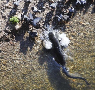
Observation of live specimens in the field and immediately after captureshows that Trichogenes claviger is a midwater swimmer with constant swimming mode, much like Trichogenes longipinnis. Several specimens were seen gulping air from the surface and others expelling a bubble of air, showing that Trichogenes claviger has accessory air breathing, like many other trichomycterids and loricarioids. Specimens with air in their stomachs swim at an oblique angle, with the head pointing down, again like Trichogenes longipinnis. When captured in the net, specimens elbowed their way up the net walls with the aid of their interopercula, in fashion similar to that of Trichogenes longipinnis and most other Trichomycterids. The sexual dimorphism in opercle morphology previously reported for the species is clearly visible in live specimens.
The general habits observed for Trichogenes claviger therefore are similar to those reported for Trichogenes longipinnis. Their habitat, on the other hand, is markedly different. Trichogenes longipinnis occupies highly oligotrophic mountain streams with mostly rocky substrate, no mud at margins or bottom, steep altitudinal declivity, and mostly high-energy water current (although specimens concentrate on quiet deep pools, usually immediately downstream from waterfalls). As noted above, the kind of stream inhabited by Trichogenes claviger has pronouncedly divergent characteristics. This shows that the target habitats for searching new localities of Trichogenes species (including that of Trichogenes beagle) must be kept considerably broad. Despite the dissimilarities, however, some parallels exist in the ecology of the two species, such as their preference for deeper sectors following waterfalls or rapids sectors (on a tiny scale in the case of Trichogenes claviger), the spotty distribution within water bodies and the absence or paucity of cooccurring Fish species.
Trichogenes beagle stands out as a remarkable species among Trichomycterids due to unique morphological character states, which bring to light broader issues in the evolution of certain morphological complexes. One of them is the presence of the bone normally called antorbital, supraorbital, or frontolacrimal tendon bone in trichomycterids. The structure, called the barbular bone by de Pinna et al., has been known for a long time in the family, and was first illustrated by Carl Eigenmann in 1908 in Hatcheria, Scleronema, and Eremophilus, misidentified as the nasal. The bone is present in the form of an elongated rod or splint, lacking any laterosensory canal association, extending from the margin of the frontal towards the lacrimal in all Trichomycterinae, and at least in basal members of the Sarcoglanidinae and Glanapteryginae. It is absent in Copionodontinae, Tridentinae (except in Potamoglanis), Stegophilinae, and Vandelliinae. Within Trichogenes, the bone is present in Trichogenes beagle and Trichogenes longipinnis, and absent in Trichogenes claviger. The condition of the structure in species of Trichogenes differs from that in all other Trichomycterid taxa, having the shape of a flat, discoidal, or roughly triangular bone, dorsally concave, located at about midlength of the lateral process (sometimes called antorbital process) of the lateral ethmoid. In Trichogenes longipinnis it lacks contact with the margin of the neurocraniun and is positioned rather distant from the eye, not shielding the orbit. In T. beagle, the bone is proportionally larger and articulates directly with an expansion of the lateral ethmoid, shielding the orbit anterodorsally. The situation Trichogenes beagle thus matches topologically the supraorbital of Characiforms and many other Bony Fish, more so than in any other Trichomycterid. This invites a more detailed analysis of that bone, its potential homologies, and phylogenetic significance.

It is likely that the discoidal structure in species of Trichogenes is homologous to the rod-shaped bone in other Trichomycterids, a hypothesis first proposed in the description of Trichogenes longipinnis. In both cases, the elements are noncanal bones located at the margin of the neurocranium, positioned at the space between the frontal and the lacrimal, overlaying the antorbital process of the lateral ethmoid (in Trichogenes) or equivalent space when that process is reduced or absent (other trichomycterid taxa) and connected anteriorly with the lacrimal-antorbital by a ligament or sheet of connective tissue. De Pinna et al.'s observations reveal that in both cases the bone develops as an independent ossification in the middle of connective tissue or ligament between the margin of the neurocranium and the lacrimal-antorbital. There is thus little question that all such structures are homologous, be they disc or rod shaped. De Pinna et al. propose the name barbular bone as the anatomical term to refer to them. The previously employed 'fronto-lacrimal tendon bone' correctly implies non-homology with other known bones in the Catfish skull, but it is a descriptive term and not a proper anatomical name.
Homology of the barbular bone with similar structures in other fishes is more controversial. Previous proposals have called it an antorbital, supraorbital, or a neomorphic structure. Suggestions of a supraorbital or antorbital homology face considerable difficulties in the context of Siluriform relationships. No other Siluriforms have a supraorbital ossification, which is also absent in their sister taxa, the Gymnotiformes. Phylogenetically, it is highly unlikely that a supraorbitallike element in taxa phylogenetically deeply internested within siluriforms, such as the one in Trichomycterids, is actually homologous to the supraorbital of other teleosts. This would imply numerous parallel losses of the bone in related taxa, a highly unparsimonious scenario. So, despite the remarkable topological similarity between the discoid bone in Trichogenes beagle and the supraorbital in other ostariophysans, such a homology is most unlikely.
The situation is more complicated with the antorbital hypothesis. The antorbital in Siluriforms is considered fused with the lacrimal, forming the compound lacrimal-antorbital, which is the anteriormost element in the infraorbital branch of the laterosensory canal system. Such a fusion is common in many lower Teleostean Fish. In Catfish, the compound nature of the element is obvious by its complex structure, partly associated with a canal but with divergent processes in the adult condition. The composite nature of the lacrimal-antorbital in Siluriforms is an old hypothesis, first suggested by the observations of James Playfair McMurrich in 1884. The idea has never been refuted, and continues to be accepted today. However, direct evidence of ontogenetic fusion remains scarce or nonexistent, so that final demonstration of the compound nature of the structure is still missing.
In most Trichomycterids the lacrimal-antorbital, when present, is markedly reduced to a short cylinder associated with a tiny sensory-canal branch and lacking the complex processes and expansions of most other Siluriforms. This has normally been interpreted as a simplification of the lacrimal-antorbital of other Catfish. Still, such simplicity raisesthe possibility that the splintlike bone posteriorly might be a secondarily separated and highly modified antorbital and that the anterior canal-bearing portion is solely the lacrimal. The situation in Trichogenes and Copionodontinae may resolve the question. In both taxa the lacrimal-antorbital has retained a more complex structure typical of catfishes, with a central expanded canal portion and two processes anteriorly. Furthermore, juvenile specimens of Trichogenes longipinnis show direct evidence of fusion. In a 20.3 mm standard length specimen (MZUSP 83452), the lacrimal-antorbital clearly shows two elements in the process of fusion, one of which is a short conical basal ossification prolonged as an anterolateral process. This element ossifies independently of surrounding canal elements and is separated from the rest of the complex by a small cartilage layer (in the stage observed it is connected with the canal element via a narrow bony strut). This element corresponds putatively to the antorbital. The other element is mostly composed of a canal ossification, putatively corresponding to the lacrimal. The latter element bears an anteromesial process that is clearly a simple expansion of the canal element with no evidence of independent ossification. These observations make it clear that the adult structure is actually formed by fusion of separate elements, not only directly demonstrating the hypothesis of a compound lacrimal-antorbital in catfishes but also that trichomycterids primitively conform to that pattern. A corollary of this conclusion is that the discoidal frontolacrimal bone of Trichogenes species cannot be homologous with the antorbital, because the latter is already incorporated into their lacrimal-antorbital. By extrapolation, the same set of conclusions can be extended to the homologous rodlike structure in other Trichomycterids.

Thus, the logical deduction is that the fronto-lacrimal bone in trichomycterids is a neomorphic structure, as first proposed by Jonathan Baskin in 1973, who proposed it as a synapomorphy for the so-called 'Trichomycterinae-Group' comprising subfamilies Trichomycterinae, Glanapteryginae, and Sarcoglanidinae. De Pinna et al.'s observations confirm that the barbular bone is present in all members of the Trichomycterinae, in most Sarcoglanidinae and in basal Glanapteryginae (Listrura spp. in reduced or vestigial condition). Concomitantly, it is consistently absent in all distal trichomycterids, namely Tridentinae, Stegophilinae, and Vandelliinae. It is present, however, in species of Potamoglanis. In case the proposed relationship of that genus with Tridentinae is corroborated, then the barbular bone is also present in that clade.
Among Catfish, the only structure potentially homologous to the barbular bone of Trichomycterids occurs in the Scoloplacidae, Loricariidae, and Astroblepidae, a monophyletic subgroup of Loricarioids. In the two latter families there is an elongate bone extending along the lateral margin of the palatine, posteriorly splint- or threadlike in Loricariids and rodlike in Astroblepids. In Scoloplacids the putatively homologous bone has a complex, wishbone morphology. The shape and position of that bone, as well as the phylogenetic proximity of the taxa involved, invite comparisons with the barbular bone of Trichomycterids. Scott Allen Schaefer originally considered the bone in scoloplacids, astroblepids, and loricariids as a neomorph sesamoid ossification. Later however, he noticed nasal capsule association of the bone in scoloplacids and thus concluded that it was more likely homologous to the lacrimal (or lacrimal-antorbital) of other Catfish, not a neomorph. Schaefer extended and elaborated on that hypothesis, convincingly concluding that the bone in Scoloplacids, Astroblepids, and Loricariids is homologous among the three families and represents a modified lacrimal-antorbital, a view with which de Pinna et al. concur. Schaefer´s conclusion, along with de Pinna et al.'s own argument above that the lacrimal-antorbital is primitively present in Trichomycterids and entirely distinct from the barbular bone, demonstrate that the latter is not homologous with the modified lacrimal-antorbital of Scoloplacids, Astroblepids, and Loricariids.
De Pinna et al. conclude that the discoidal bone in Trichogenes longipinnis and Trichogenes beagle is homologous to the rod-shaped bone present in many other Trichomycterids. They are different states of a neomorph structure named the barbular bone by De Pinna et al., probably a synapomorphy for Trichomycteridae that has been secondarily lost in more distal lineages of the family. Concomitantly, the antorbital is ontogenetically fused with the lacrimal in Trichogenes longipinnis, confirming the broader hypothesis of a composite lacrimal-antorbital in Catfish.
Another feature in which Trichogenes beagle brings insights into the evolution of typical Trichomycterid especialisations is the opercle. That bone in Trichomycterids has some of the most extreme modifications among Siluriforms, usually with a compact shape with a specialised posterior platform bearing distal odontodes and a complex ventral articulation with the dorsal part of the interopercle. Opercular odontodes in Trichomycterids are present only on the posterior platform, a situation contrasting with that in other Loricarioids whose odontodes are distributed more evenly along the margin of the bone. Species of Trichogenes and Copionodontinae retain some degree of laminar structure of the opercle, which itself provides some intermediacy in the evolution of the typical compact trichomycterid opercle. In terms of odontode distribution, Trichogenes longipinnis and Trichogenes claviger have two sets of opercular odontodes, one homologous to the posterior platform of other trichomycterids and another anterior patch unique to them. Between the two patches there are no odontodes. On the other hand, Trichogenes beagle is unique in that its opercle bears odontodes more or less evenly dispersed along its posterior margin, in addition to a concentration on the posterior platform. This condition is far more similar to that in other Loricarioids with opercular odontodes evenly distributed (Callichthyidae, Loricariidae) and provides an interesting intermediate stage for understanding the evolution of the trichomycterid operculum. A morphology such as that in Trichogenes beagle might be the ancestral state, most similar to loricarioid outgroups. The two-patch morphology of Trichogenes longipinnis and Trichogenes claviger results from the loss of odontodes in the middle range of their distribution along the edge of the opercle. Finally, the state in most other Trichomycterids is reached via loss of the ventral odontode patch. A parallel, partial or total loss of opercular odontodes occurs in most Copionodontinae and some other Trichomycterid subgroups such as the Glanapterygines Glanapteryx, Pygidianops, and Typhlobelus, and the Stegophiline Megalocentor.
See also...


Follow Sciency Thoughts on Facebook.