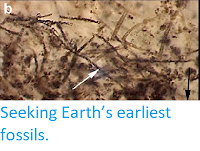
The earliest evidence for life on Earth comes in two forms; chemical evidence, i.e. compounds believed to have been derived from the activities of living organisms and isotopic signals thought to be indicative of biological activity, and through the presence of structures believed to be microfossils in ancient sedimentary deposits. In both cases, identifying these with confidence requires considerable skill, as there remains a danger that abiotic processes might have achieved the same results. One of these dangers is the potential formation of pseudofossils (structures which resemble fossils but are of non-biological origin). The most ancient structures interpreted as being microfossils take two forms, microbial filamentous and spherical cells, but structures morphologically similar to both of these have been synthesised on the lab by non-biological means, raising the possibility that none of these ancient 'fossils' are actually of biological origin. The researchers were able to recreate these strucutures, also known as organic biomorphs, by oxidising sulphides in the presence of organic materials, under which circumstances they formed spontaneously. This has been shown to be possible under a wide range of conditions likely to have been present on the ancient Earth, and with a wide range of organic compounds serving as precursors.
However, the fact that such biomorphs can be formed in the laboratory does not automatically prove that all, or indeed any, of the various Archaean and Palaeoproterozoic fossil deposits are in fact pseudofossils; indeed some of these have been studied for many years and their status as being of biological origin is not really in doubt. Nevertheless, some caution is clearly needed when establishing the nature of any apparent fossils in these ancient deposits, particularly if their host rocks are sulphide-rich.
In the oceans of the Proterozoic, euxinic (low oxygen, high sulphur) conditions are thought to have been fairly common, and this may have also have sometimes been the case in the Archaean, at least at a local level, although ocean sulphide levels appear to have been low for the most part. Many of the microfossils known from these deposits are associated with pyrite, which indicates the deposits which produced them did indeed have raised sulphur levels. However, there is not currently any clear data on the likelihood of biomorphs being preserved in these deposits.
In a paper published in the journal Geology on 28 January 2021, Christine Nims and Julia LaFond of the Department of Geosciences at Pennsylvania State University, Julien Alleon of the Institut des Sciences de la Terre at the Université de Lausanne, Alexis Templeton of the Department of Geological Sciences at the University of Colorado, Boulder, and Julie Cosmidis, also of the Department of Geosciences at Pennsylvania State University, describe the results of an experiment in which they performed experimental silicification of organic biomorphs along with the Sulphur Bacterium Thiothrix, in order to assess the likelihood of their becoming preserved in the fossil record as pseudofossils.

As the majority of putative Precambrian microfossils are preserved in chert (fine-grained sedimentary rock composed of microcrystalline crystals of quartz), Nims et al. decided to investigate the preservational potential of organic biomorphs through silicification. They also carried out the same experimental procedures on mats of the sulphur-oxidising Bacterium Thiothrix as a control measure; experiments on the silicification of colonial micro-organisms have been undertaken before, but the majority of these have been performed on Cyanobacteria, despite many putative Precambrian microfossils being interpreted as most likely being sulphur-cycling organisms. Nims et al. felt that Thiothrix would be a good analogue for these organisms, as it forms intracellular sulphur globules.
Nims et al. obtained organic biomorphs by reacting dissolved sulphides with yeast extract (which contains a variety of complex organic compounds) in a sterile environment. Both the biomorphs and the Thiothrix mats were then placed into a supersaturated sodium-metasilicate solution, then stored for up to five months at room temperature. Samples were taken from these experiments at regular intervals, and examined using scanning electron microscopy and transmission electron microscopy coupled with energy-dispersive X-ray spectroscopy, Raman spectromicroscopy, attenuated total reflectance–Fourier transform infrared spectroscopy, X-ray absorption near-edge structure at the sulphur K-edge, and scanning transmission X-ray microscopy at the carbon K-edge, nitrogen K-edge, and sulphur L-edge.
The biomorphs produced were a mixture of spheres (0.5–3 μm in diameter) and filaments (0.1–1 μm thick). These generally retained their shape during the silicification process; whereas the spherical Thiothrix cells rapidly lost their shape, first becoming elongate, then deforming into flattened ribbons. As non-silicified Thiothrix cells retained their shape. Nims et al. presume this flattening to be a result of the silicification process, rather than the microscopy preparation. In contrast, the silicification process appeared to have little impact upon the shape of the organic biomorphs, other than a small degree of fragmentation in some of the filamentous specimens. In fact, the biomorphs were preserved very rapidly, by the precipitation of nano-colloidal silica on their surfaces, which formed a thin crust around each specimen, preserving it from any further degredation. The Thiothrix cells, in contrast, quickly became covered in a thick silica-gel, possibly due to the presence of extracellular polymeric substances around the Bacterial cells, which may have caused the silica to behave in a different way, or possibly due to the metabolic activity of the cells, which initially reduced the pH of the surrounding media, enabling an increase in the silica saturation, whereas the pH around the biomorphs remained constant at about 7.

During the silification process, the discrete globules of elemental sulphur present within the cells of Thiothrix broke up, with the sulphur becoming diffused out of the cells into the surrounding medium. Sulphur also diffused out of the spherical biomorphs, leaving empty organic vesicles, but here it subsequently re-precipitated along the envelope of the silicified biomorphs, as both sulphur and oxidised sulphur forms (such as sulphate, thiosulphate, and/or sulphones and ester sulphates).
Nims et al. suggest it is likely that the diffusion of sulphur from both the Thiothrix cells and the organic biomorphs was caused by solubilisation as polysulphides, which are highly reactive toward organics, causing rapid organic-matter sulphurisation. This may be the cause of the formation of the sulphur-rich organic envelopes around the organic biomorphs. Attenuated total reflectance–Fourier transform infrared spectroscopy showed that sulphur-bearing groups such as sulphates and sulphones were forming during silification, although it was not possible to confirm the incorporation of intramolecular sulphur into the biomorphs. Early digenetic sulphurisation would favour the preservation of microstructures in the rock record, so establishing whether this actually happens with the organic biomorphs would be a major step in establishing their preservation potential.

If sulphur was lost from the organic biomorphs during silicification, then the result would be organic microstructures in the chert which did not contain sulphur-bearing minerals. However, if they were being preserved in an iron- and sulphur-rich environment, we would expect to see the formation of pyrites close to the preserved biomorph structures, something which is commonly observed in cherts which host ancient organic microfossils.
Prior to silicification, the composition of the biomorphs was dominated by carboxylic groups and unsaturated carbon, along with aliphatics, alcohols, and carbon-oxygen groups. During the silicification process, the proportions of aliphatic, aromatic, and unsaturated carbon compounds rose, while those of other organic compounds fell. In Thiothrix cells the composition was dominated by amide groups (the major component of proteins) both prior to and during silicification. It has previously been established that if peptides are present in the synthesis medium, then amides can also be incorporated into organic biomorphs. Nims et al. did not detect any amide groups in the biomorphs, despite these having been formed using yeast extract, which contains peptides. However, nitrogen was found in the envelopes of the biomorphs, in an unidentified inorganic or organic form. Thus, the carbon and nitrogen species present in the biomorphs and Thiothrix were quite different.
The initial carbon/nitrogen ratio of the biomorphs was 0.27, which rose to 0.40 during the silicification process, whereas that of the Thiothrix cells started at 0.16 and rose to 0.42, i.e. a similar value to that of the biomorphs. It is unclear how this proportion would be affected by subsequent high-temperature and pressure diagenesis, although it is possible that this might result in some detectable difference in the carbon/nitrogen ratio of organic biomorphs and Bacterial cells developing.

Nims et al. conclude that organic biomorphs can form via the reaction of sulphides with organic compounds, and are highly likely to be preserved as pseudofossils in chert by the process of rapid silica encrustation, possibly in combination with the sulphurisation of organic matter. Such pseudofossils would not only be extremely similar to fossils produced by Bacteria, or similar Prokaryotes, they would also have similar chemical characteristics. Indeed, such organic biomorphs might actually have better preservational potential than actual microbes.
None of this proves that any Precambrian microfossil assemblage is in fact made up of pseudofossils, but it certainly suggests that there is a possibility for pseudofossil assemblages to exist in these ancient rocks, and indicates that a degree of caution must be used before such assemblages are accepted as being of biological origin. In particular, specimens with an apparently Bacterial morphology and chemical composition will need to be viewed with some caution, particularly if they originate from deposits interpreted as having been laid down in high-sulphur environments. Nims et al. strongly feel that more work must be done to identify possible geochemical signatures which might indicate specimens are of non-biological origin.
See also...


Online courses in Palaeontology.
Follow Sciency Thoughts on Facebook.
Follow Sciency Thoughts on Twitter.