Showing posts with label Nematodes. Show all posts

Tuesday, 9 October 2018

Caenorhabditis inopinata: A new species of Nematode from Japan, and a long sought-after sister species to the highly studied Caenorhabditis elegans.

The Nematode Caenorhabditis elegans is one of the most closely studied organism in modern biology, due to its small size and rapid reproduction, which makes it an ideal laboratory subject. The genome of Caenorhabditis elegans has been comprehensively mapped, and its development fully documented, allowing manipulation of its genetic code to be carefully studied, which has led to many discoveries with implications for other animals, including Humans. As such there has been a great deal of interest in discovering a sister species to Caenorhabditis elegans (that is to say a very closely related species, with a similar genetic history), with the species currently used as a sister in genetic studied, Caenorhabditis briggsae, being about as closely related to Caenorhabditis elegans as Humans are to Mice.

In a paper published in the journal Nature Communications on 10 August 2018, a team of scientists led by Natsumi Kanzaki of the Kansai Research Center of the Forestry and Forest Products Research Institute, Isheng Tsai of the Biodiversity Research Center at Academia Sinica, and Ryusei Tanaka and Vicky Hunt of the Faculty of Medicine at the University of Miyazaki, describe a sister species of Caenorhabditis elegans from Ishigaki Island, Japan.

The new species is named Caenorhabditis inopinata, where ‘inopinata’ means ‘surprising’ as the species was discovered unexpectedly by scientists who were not looking for a sister species to Caenorhabditis elegans, and only realised their discovery when they ran a genetic comparison of the new species to other members of the genus. Caenorhabditis inopinata was found living in Figs of the species Ficus septica, with the only members of the species found outside of these fruit being larvae attached to the abdomen of the Fig Wasp Ceratosolen bisulcatus, the pollinator of Ficus septica, making it highly likely that the Nematodes are carried from Fig-to-Fig during pollination. This is completely different to the life-cycle of Caenorhabditis elegans, which is a free-living Nematode found in soil and rotting plant matter. This different life-style is reflected in the anatomy of Caenorhabditis inopinata, which is longer and thinner than Caenorhabditis elegans, and has short stomatal flaps, a short tail and rounded appendage on the anterior cloacal lip in females and tongue-like postcloacal appendage in males.

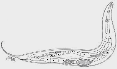
Caenorhabditis inopinata biology compared with the sibling species Caenorhabditis elegans. (a) Differential interference contrast microscopic views of Caenorhabditis inopinata (adult female; bottom) with Caenorhabditis elegans (hermaphrodite; top). Scale bar: 100 μm. (b) Key morphological characters of Caenorhabditis inopinata. (a) Right lateral view of stomatal part in different focal planes showing dorsal tooth (left) and right subventral tooth (right) with arrow heads. (b) Ventral view of male tail. (c) Female tail in right lateral (left) and ventral (right) view showing phasmids in arrowheads. (d) Face view of stomatal part of adult. (e) Face view of stomatal part of dauer-like juvenile. (f) Left lateral view of male tail. (g) Ventral view of male cloacal opening. Scale bars: 20 μm for (a)–(c), 2 μm for (d)–(g). (c) Life cycle of Caenorhabditis inopinata. The nematodes multiply in fresh syconia of Ficus septica, which has a mutualistic association with the pollinating wasp Ceratosolen bisulcatus. When the syconia mature nematodes in the dauer form disperse to new young syconia using the wasps as a vector. Kanzaki et al. (2018).

See also...

https://sciencythoughts.blogspot.com/2017/09/oncholaimus-zhangi-new-species-of.htmlhttp://sciencythoughts.blogspot.co.uk/2017/07/strongyloides-stercoralis-determining.html
http://sciencythoughts.blogspot.co.uk/2015/04/tripius-gyraloura-sphaerularid-nematode.htmlhttp://sciencythoughts.blogspot.co.uk/2015/04/philometrid-nematodes-from-perciform.html
http://sciencythoughts.blogspot.co.uk/2015/03/a-new-species-of-capillariid-nematode.htmlhttp://sciencythoughts.blogspot.co.uk/2014/12/two-new-species-of-pinworms-from.html
Follow Sciency Thoughts on Facebook.

Sunday, 3 September 2017

Oncholaimus zhangi: A new species of Oncholaimid Nematode from the East China Sea.

Oncholaimid Nematodes are free-living (as oposed to parasitic) Nematodes found in marine, brackish and freshwater aquatic environments around the world. Members of the genus Oncholaimus are sediment dwelling Nematodes, distinguished by a large buccal cavity with three teeth and a short tail.

In a paper published in the Chinese Journal of Oceanology and Limnology on 25 October 2017, Gao Qun and Huang Yong of the College of Life Sciences at Liaocheng University, describe a new species of Oncholaimus from intertidal sediments on Dongshan Island in Fujian Province, China.

The new species is named Oncholaimus zhangi, in honour of Zhang Zhinan of the College of Marine Life Science at the Ocean University of China, in recognition of his work on Nematodes. The species is described from four male and three female specimens collected from sandy soils at deths of up to 8 cm. The male measures 3718–3934 μm in length, while the females were 3 228–3 783 μm; the females have only a single ovary while the males have paired gonads.

Oncholaimus zhangi, (a) lateral view of male anterior end; (b) lateral view of female body; (c) lateral view of the male tail end; (d) lateral view of the male anterior end showing amphidial fovea; (e) lateral view of the female anterior end; (f) lateral view of spicule. Gao & Huang (2017).

See also...

http://sciencythoughts.blogspot.co.uk/2017/07/strongyloides-stercoralis-determining.htmlhttp://sciencythoughts.blogspot.co.uk/2015/04/tripius-gyraloura-sphaerularid-nematode.html
http://sciencythoughts.blogspot.co.uk/2015/04/philometrid-nematodes-from-perciform.htmlhttp://sciencythoughts.blogspot.co.uk/2015/03/a-new-species-of-capillariid-nematode.html
http://sciencythoughts.blogspot.co.uk/2014/12/two-new-species-of-pinworms-from.htmlhttp://sciencythoughts.blogspot.co.uk/2014/11/a-parasitic-heterakid-nematode-from.html
Follow Sciency Thoughts on Facebook.

Wednesday, 26 July 2017

Strongyloides stercoralis: Determining the origin of Human infection in a parasitic Nematode.

Parasitic Nematodes of the genus Strongyloides infect the guts of a wide range of Vertebrate hosts, including Birds, Reptiles, Amphibians, and Mammals, as well as Humans, livestock, and companion animals. Two species are known to infect Humans, Strongyloides fuelleborni, which has two subspecies, one found in tropical Africa and the other in Papua New Guinea, and Strongyloides stercoralis, which has a cosmopolitan distribution, though most infections occur in the tropics and subtropics, and which is also found in Dogs and Chimpanzees.

In a paper published in the journal Scientific Reports on 7 July 2017 a team of scientists led by Eiji Nagayasu of the Division of Parasitology at the University of Miyazaki published the result of a phylogenetic study of Strongyloides stercoralis, using four gene sets plus morphometric analysis. Nematodes were isolated from Humans from Japan, Myanmar, Thailand, Laos, Uganda, the Central African Republic and Tanzania, as well as Dogs from Japan and Myanmar and Chimpanzees from Tanzania, with a view to establishing a phylogenetic tree and potentially determining the origin of infection in Humans.

 
A Strongyloides stercoralis nematode. Oklahoma State University.

Nagayasu et al. found that the Nematodes could be divided into two main clades (groups with common ancestry), one of which contained all the Human derived worms, plus the Chimpanzee derived worms and all worms from Dog infections in Japan, and some of the worms from Dogs in Myanmar. The second clade was contained entirely worms obtained from Dogs in Myanmar. They interpret this data as indicating that Strongyloides stercoralis infection originated in Dogs and has spread to Humans during the two species’ long association. The first clade has apparently made the jump between Dogs and Humans (and subsequently to Chimpanzees, which are very similar to Humans in immunological terms), while the second clade not made this jump. 

Median-joining haplotype network for the mitochondrial Cox1 gene. Each circle represents one haplotype. The size of the circle represents the number of hosts that harbour the given haplotype. The colour inside the circle indicates the host species/geographical origin (country). Numbers beside the branches indicate the number of mutational steps between haplotypes (no number is shown in case of a single-step difference). Branch lengths are roughly proportional to the number of mutational steps. Nagayasu et al. (2017).

Interestingly, a previous study of Strongyloides stercoralis, published in 2014 that looked as a smaller number of gene-sets and which concentrated on infections in Cambodia, had also identified two major clades within the total population of Nematodes, one of which was associated principally with Dogs, but which was also found in some Humans. However, re-examination of this data reveals that all of the Humans infected with the second strain were immunocompromised individuals the HIV from Myanmar; HIV is known for its destruction of the immune system, and often individuals infected with it are infected by organisms which do not usually infect Humans, or even which are not usually pathogenic at all, suggesting that such individuals would be vulnerable to a strain of Nematodes which normally attacks only Dogs.

See also...

http://sciencythoughts.blogspot.co.uk/2015/04/tripius-gyraloura-sphaerularid-nematode.htmlhttp://sciencythoughts.blogspot.co.uk/2015/04/philometrid-nematodes-from-perciform.html
http://sciencythoughts.blogspot.co.uk/2015/03/a-new-species-of-capillariid-nematode.htmlhttp://sciencythoughts.blogspot.co.uk/2014/12/two-new-species-of-pinworms-from.html
http://sciencythoughts.blogspot.co.uk/2014/11/a-parasitic-heterakid-nematode-from.htmlhttp://sciencythoughts.blogspot.co.uk/2014/10/parasite-infections-in-german-soldiers.html
Follow Sciency Thoughts on Facebook.

Sunday, 6 September 2015

Exceptional preservation in Early Ordovician fossils from Hubei Province, China.


Fossil Lagerstätten are deposits which produce exceptional fossils, providing us with valuable insights into vanished ecosystems. There are a number of Ordovician Fossil Lagerstätten known which produce invertebrate fossils with soft tissues, providing information on organisms otherwise not seen in the fossil record. These are the Early Ordovician Fezouata Formation of Morocco, the Middle Ordovician Winneshiek Lagerstätte in northeast Iowa and the Late Ordovician Beecher’s Trilobite Bed of New York, Llanfawr Mudstone of Wales, Soom Shale of South Africa and Late Ordovician deposits from Manitoba.

In a paper published in the Chinese Science Bulletin on 17 March 2015, Andrzej Baliński of the Instytut Paleobiologii and Yuanlin Sun of the Key Laboratory of Orogenic Belts and Crustal Evolution at Peking University discuss a new Early Ordovician Fossil Lagerstätten from Hubei Province in China. These fossils come from the Fenxiang Formation, a deepwater shale, and show fossils phosphatised in a similar manner to the Burgess Shale and Chengjiang Cambrian Fossils, a form of preservation extremely rare in post-Cambrian deposits (so rare that until quite recently such preservation was not thought to be seen in post-Cambrian deposits at all). The fossils were deposited in a deepwater environment, but many are of shallow-water organisms, thought to have been moved there by a submarine landslip, in a similar way to the shallow water fossils seen in the Burgess Shale. They were deposited about 470 million years ago, slightly before the Great Ordovician Biodiversification Event, in which the Cambrian Evolutionary Fauna (dominated organisms such as Trilobites, Polychaete Worms, Monoplacophorans and Inarticulate Brachiopods) was largely replaced by the Palaeozoic Evolutionary Fuana (dominated by organisms such as Articulate Brachiopods, Crinoids, Cephalopod Molluscs and Anthozoan Corals), and contains organisms from both faunas.

The first fossils described are Linguloid Brachiopods assigned to the genus Leontiella with soft tissues preserved. Linguloids are considered to have been the first Brachiopods to have appeared, and are documented with soft tissue preservation from the Burgess Shale, but reliably interpreted soft-body preservation in post-Cambrian Linguloid Brachiopods is extremely rare, with the earliest unquestionable examples coming from the Early Lower Devonian Hunsrück Slate of Germany, and possible examples from the Devonian of England, Middle Ordovician of New York State and Late Ordovician of South Africa. The Fenxiang Brachiopods show three-dimensional preservation of the pedicle, showing details of the external morphology. Leontiella has a streamlined shell with radial ornamentation, characteristics associated with true burrowing behaviour in Brachiopods (unlike the simpler shells of the Cambrian Linguloids); the Fenxiang specimens have long, vermiform pedicles, which appears to support this, suggesting that Linguloid Brachiopods had indeed evolved true burrowing behaviour by this point.

Linguloid brachiopod Leontiella sp. with preserved pyritized vermiform pedicle. Balinski & Sun (2015).

The Fenxiang Biota also contains a variety of trace fossils, which also show evidence of burrowing behaviour not seen in Cambrian deposits. These include numerous sinusoidal horizontally-orientated cylindrical burrows 20–60 μm in diameter, tightly packed with framboid structures, thought to be replacement minerals occupying spaces which formerly contained pyrite crystals, which themselves would have been formed by bacterial decomposition of an organic infilling of the burrows, such as fecal pellets. Balinski and Sun interpret these as the burrows of free-living Nematodes, which form very similar burrows in modern environments. These traces considerably predate the first Nematode body-fossils, which are found in the Early Devonian Rhynie Chert of Scotland, and the earliest trace fossils previously ascribed to Nematodes, from the Middle Triassic of Germany. Nematodes are thought to have diversified from a common ancestor before the appearance of the earliest Arthropods and Velvet Worms, both groups which appear in the Early Cambrian, so the presence of such organisms in the Early Ordovician is not surprising, however Nematodes are not thought to have been able to adopt a burrowing lifestyle until larger burrowing and bioturbating organisms had broken up a layer of microbial matting which covered most of the seafloor in the Cambrian and prevented much oxygen from penetrating to the sediments bellow.

Sinusoidal trace fossil from the Fenxiang Formation interpreted as a Nematode burrow. Balinski & Sun (2015).

The Fenxiang Biota has also produced an upright colonial Hydroid, possibly belonging to the Haleciidae, preserved as part and counterpart on a split rock. Hydras and Medusas (Jellyfish) are thought to have been the earliest Cnidarians to appear, due to their simple body-plans and placement on genetically-based phylogenetic trees, and are expected in the fossil record very early. However numerous fossils assigned to these groups from Ediacaran, Cambrian, Ordovician and Silurian deposits have subsequently been re-assigned to different groups, leaving the earliest confidently assigned Medusas coming from the Late Carboniferous Mazon Creek Formation and the earliest known Hydras those from the Late Ordvician of Kentucky and (possibly) Wales. The Fenxiang specimen represents an advanced colonial Hydroid, the earliest known example of such an organism.

Pyritized colony of Hydroid. Balinski & Sun (2015).

The Fenxiang Biota also includes a variety of other Cnidarians, including Conulariids, an extinct group of Palaeozoic Scyphozoans, and numerous Black Corals (Antipatharia), an important group of modern Corals, particularly in deeper waters, but until know not known from the fossil record at all (though they have been predicted to have been an ancient group from genetic studies of their phylogenetic position). These Fenxiang fossils have been processed in acetic acid revealing extremely well preserved details of their anatomy, which leave little doubt on their assignment to the group.

Phosphatic coralla of Antipatharian Coral  showing basal part of a colony with crowded spines (l), spinose branch (m), and partly preserved spinose basal part of colony and erect stem (n). Balinski & Sun (2015).

The deposits also contain numerous fossils which have yet to be identified or studied in detail, including branching feather-like fossils, which may prove to be another form of colonial Hydroid, numerous Arthropod fragments, and a possible Scalidophoran Worm embryo similar to those known from Cambrian deposits in China, Russia and Australia and earliest Ordovician of the United States.

See also…

Reworked Late Ordovician Sponges have been collected from Miocene to Pleistocene across a wide area of northern Europe for over two centuries. These are associated with the course of the Baltic River System, which drained much of northern Europe for...


The enigmatic fossil Furca bohemica was first recorded from Late Ordovician deposits at Veselá Gorge, in what is now the Czech Republic, by the French Palaeontologist Joachim Barrande in 1846, though he never formally described the specimens. A formal description was provided by Antonin Fritsch in...


Brachiopods (or Lampshells) superficially resemble Bivalve Molluscs, though they are not closely related. They have a filter feeding apparatus called a lophophore, unlike anything found in any Mollusc, but also found in Bryozoans and Phoronid Worms. This is encased with in a shell with two valves, each symmetrical about a...


Follow Sciency Thoughts on Facebook.

Tuesday, 14 April 2015

Tripius gyraloura, a Sphaerularid Nematode infecting the Arundo Gall Midge.


Sphaerularid Nematodes are highly virulent parasites of Insect hosts. While most parasites seek to keep their host alive for as long as possible, enabling them to live and produce offspring for as long as possible, Sphaerularids begging to reproduce at a high rate shortly after entering a new host,  producing a large number of young which quickly overcome the host. While some of these young may exit the host while it still lives and start seeking out new hosts, the majority will exit the host after its death.

In a paper published in the journal Systematic Parasitology on 1 November 2014, George Poinar of the Department of Zoology at Oregon State University and Donald Thomas of the United States Department of Agriculture-Agricultural Research Service Cattle Fever Tick Research Laboratory, describe a new species of Sphaerularid Nematode infecting the Arundo Gall Midge, Lasioptera donacis.

Gall Midges lay their eggs within the tissues of plants, where they cause the plant to form a gall, in which the larval Midge lives, surviving on the plant’s natural fluids and protected from predators by the gall. The larval stage of the Insect lives its entire life within the gall, emerging from the egg, and growing till it is ready to pupate, which it does within the gall. Only the adult stage emerges to mate and lay eggs on new hosts. High levels of Gall Midge infections are harmful to plants and may even kill them, and species of Gall Midge are often capable of infecting only a single species of plant, making them of great interest to biologists looking for control species for invasive plants (plants which have been introduced to new environments by human activities, and lacking natural predators in these environments, become to dominate them and exclude native flora and fauna).

The Arundo Gall Midge, Lasioptera donacis, is a natural parasite of the Old World Giant Reed, Arundo donax, a species native to waterways of the Mediterranean Basin, but introduced to North America by European settlers in the sixteenth century, where it has become a serious invasive pest, as it lacks natural predators or parasites in the New World, and has an ability to modify and dominate aquatic ecosystems. As such the Arundo Gall Midge is considered to be a potential biological control for the Giant Reed, and is being studied at a quarantined laboratory run by the United States Department of Agriculture at Moore Air Base at Edinburg, Texas, with a view to releasing it into American ecosystems.

Midges were imported to Moore Air Base inside the stems of infected Giant Reeds harvested in France, Italy and Greece. Many of these Midges were found to be infected with a virulent Sphaerularid Nematode, which suppressed the reproductive ability of, and eventually killed its hosts. Such parasites have important implications when trying to establish control species for invasive pests. On the plus side, if the biological agent proves capable of switching to unintended hosts and itself becomes an invasive species, the parasite can be used to control it, but on the minus side, if the parasite is accidentally releases with the control species, it can prevent it from becoming established, thereby undermining the control effort, or worse still, the parasite may itself prove capable of switching to a new host (or hosts) in its new home, further damaging an already vulnerable ecosystem (and eradicating a new parasitic Nematode, once released, is almost impossible).

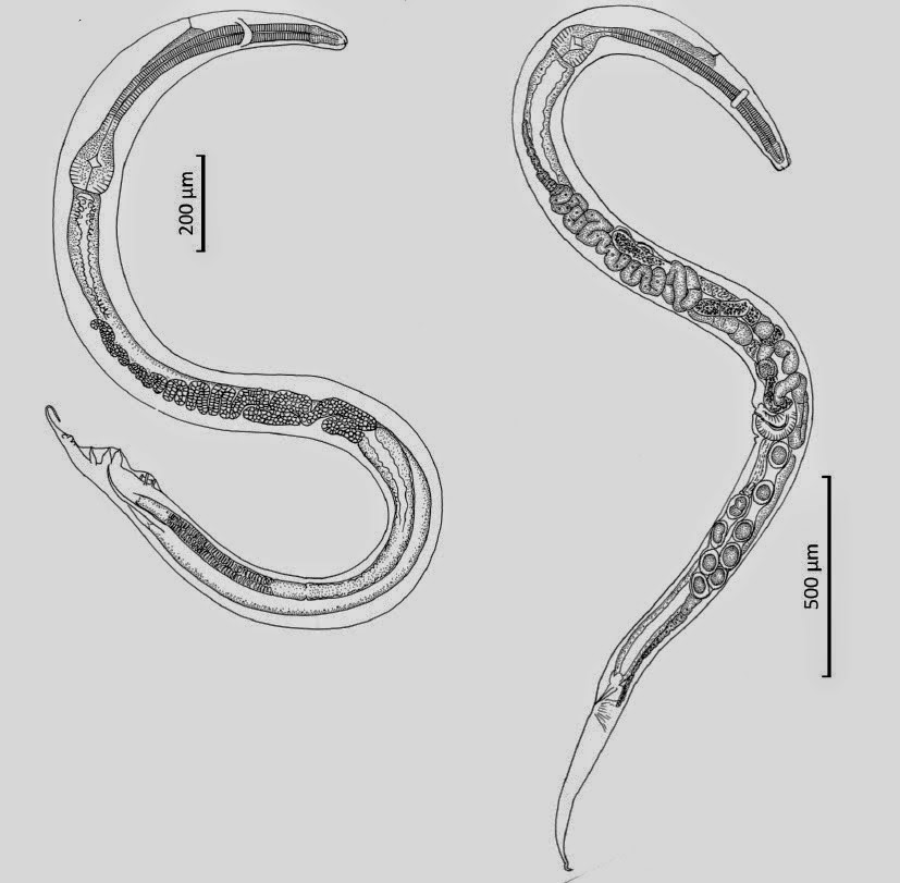
The new Nematode is placed in the genus Tripius, and given the specific name gyraloura, meaning ‘round-tail’. The species has a free-living sexual stage, which seeks out new hosts to infect, but does not feed independently or infect a secondary host (many parasites have more than one host in their life-cycle, and thus have primary and secondary, and sometimes even tertiary hosts). The free-living females reach 297–360μm in length and have a well-developed stylet (a hollow mouthpart similar to a hyperdermic needle), while the males reach 415–428μm and have an underdeveloped stylet. These mate in the environment, whereupon the males die and the females seek out a new host, using secretions to penetrate the body wall of juvenile Midges shortly before pupation. The infective female everts her uterine cells into the host’s haemocoel, giving birth to secondary, parasitic females, which can reach 0.6-2.0 mm in length, and which produce asexually, giving birth to larvae 290-430 μm in length. Sexual reproduction in infected Midges emerging form their pupas appeared to be supressed, and the Midges soon after, revealing body cavities packed with Nematodes. Some of these may have escaped through the ovipositors of female hosts, but most are thought to have escaped after the host’s death.

Tripius gyraloura. (1) Cluster of parasitic juveniles (arrow) in a dissected female Lasioptera donacis (arrowhead shows anterior end of a mature parasitic female); (2) free-living infective stage female. Scale-bars: (1) 144 μm; (2) 39 μm. Poinar & Thomas (2014).

As well as being of interest due to its potential impact on the biological control program for the Old World Giant Reed, the discovery of Tripius gyraloura is interesting because it isonly the second such infection in a living Gall Midge discovered, one other member of the same genus Tripius gibbosus infects the Gall Midge Cecidomyia pini, while a third member of the genus infects Sciarid Flies. A Gall Midde infected with parasitic Nematodes is also known from Eocene Baltic Amber.

See also…

http://sciencythoughts.blogspot.co.uk/2015/04/philometrid-nematodes-from-perciform.htmlPhilometrid Nematodes from Perciform Fish off the north Australian coast.                 Philometrids are large Nematodes parasitizing Fish . They show a high degree of sexual dimorphism, with males typically only a few mm in length, while females may reach tens of...

Capillariid Nematodes are parasitic worms infecting a variety of different Vertebrate hosts. The group is split into 22 genera, of which nine are parasites of Fish. Members of the genus Capillaria cause infections in...


Pinworms, Oxyuridae, are parasitic Nematodes infecting the digestive tracts of Mammals. They have short life cycles, typically undergoing several generations in a year, with eggs being released in the host’s faecal matter to infect new hosts. Some species of Pinworm appear to be quite cosmopolitan, infecting...


Follow Sciency Thoughts on Facebook.

Friday, 10 April 2015

Philometrid Nematodes from Perciform Fish off the north Australian coast.


Philometrids are large Nematodes parasitizing Fish . They show a high degree of sexual dimorphism, with males typically only a few mm in length, while females may reach tens of centimetres. The females give birth to live young.

In a paper published in the journal Parasite on 6 February 2015, František Moravec of the Institute of Parasitology of the Biology Centre of the Academy of Sciences of the Czech Republic and Diane Barton of Fisheries Research at the Department of Primary Industries & Fisheries of the Northern Territory and Aquatic Ecology & Management at the Research Institute for the Environment and Livelihoods at Charles Darwin University, describe two species of Philometrid Nematodes from Perciform Fish off the north Australian coast.

The first species is placed in the genus Philometra, and given the specific name protonibeae, in reference to its host species, the Blackspotted Croaker, Protonibea diacanthus. The males of this species reach 3.37–3.90 mm in length; no gravid (i.e. fully mature and pregnant) female was recovered intact, but the largest partial specimen was 427 mm (42.7 cm) in length. The nematodes were found within the ovaries of female Blackspotted Croakers caught at Camdem Sound in Western Australia and Fenton Patches and Ruby Island in the Northern Territory. About 25% of Fish examined were infected, and since the host species has a broad Indo-West Pacific distribution, it is likely that the parasite has a similar distribution.

Philometra protonibeae, left: (A) Anterior end of gravid female, lateral view. (B) Cephalic end of gravid female, apical view. (C) Caudal end of male, apical view. (D) Anterior end of male, lateral view. (E) Anterior end of gravid female (another specimen), lateral view. (F) Larva from uterus, lateral view. (G) Posterior end of gravid female, lateral view. (H) Anterior end of nongravid female, lateral view. (I, J) Posterior end of male, ventral and lateral views. (K, L) Distal end of gubernaculum, dorsal and lateral views. (M) Distal end of spicule, lateral view. (N) Caudal end of gravid female, lateral view. (O) Posterior end of nongravid female, lateral view. Right: (A, B) cephalic end of subgravid female, subapical and apical views. (C) Caudal end of male with protruded spicules and gubernaculum, lateral view. (D) Protruded distal ends of spicule and gubernaculum, lateral view. (E) Distal end of gubernaculum, dorsal view (arrow indicates elevated triangular structure). (F) Caudal end of male, apical view (arrow indicates phasmid). Abbreviations: a, group of four flat caudal papillae; s, spicule. Moravec & Barton (2015).

The second species is also placed within the genus Philometra, but is not assigned to a species as only the females were discovered, and female members of this genus can be very hard to distinguish. Nevertheless it is thought that this is a new species, as it is the first member of the genus described from the John’s Snapper, Lutjanus johnii, and species of Philometra tend to be host specific. The species was found infecting the ovaries of female Fish caught at Bynoe Harbour, Lee Point and Nicol Island, all in the Northern Territory; 45.5% of inspected Fish were infected. One complete gravid female was recovered, this being yellowish in colour and 47 mm in length, as was a subgravid female, 32 mm in length, and two nongravid females, 11.97 and 2.49 mm in length.

Philometra sp. from Lutjanus johnii. (A) Anterior end of largest gravid female, lateral view. (B, C) Posterior end of smaller gravid female, lateral and dorsoventral views. (D) Larva from uterus, lateral view. Moravec & Barton (2015).

See also…

Capillariid Nematodes are parasitic worms infecting a variety of different Vertebrate hosts. The group is split into 22 genera, of which nine are parasites of Fish. Members of the genus Capillaria cause infections in...


Pinworms, Oxyuridae, are parasitic Nematodes infecting the digestive tracts of Mammals. They have short life cycles, typically undergoing several generations in a year, with eggs being released in the host’s faecal matter to infect new hosts. Some species of Pinworm appear to be quite cosmopolitan, infecting...


Parasitic Nematodes of the superfamily Heterakoidea are typified by having three lips, an esophagus with a valved bulb, thick shelled eggs and a pre-anal sucker on the males. They are typically parasites of the digestive tracts of small vertebrates, which do not require an intermediate host (i.e. the species only needs to infect one species of hosts, rather than...


Follow Sciency Thoughts on Facebook.

Sunday, 29 March 2015

A new species of Capillariid Nematode from New Caledonia.


Capillariid Nematodes are parasitic worms infecting a variety of different Vertebrate hosts. The group is split into 22 genera, of which nine are parasites of Fish. Members of the genus Capillaria cause infections in a wide range of Mammals, Birds, Amphibians, Fish and Sharks, though those infecting marine Fish are poorly known.

In a paper published in the journal Parasite on 23 December 2014, František Moravec of the Institute of Parasitology at the Biology Centre of the Academy of Sciences of the Czech Republic and Jean-Lou Justine of the Institut Systématique, Évolution, Biodiversité at the Muséum National d’Histoire Naturelle describe a new species of Capillaria from a Leopard Coral Grouper, Plectropomus leopardus, caught off Baie de Koutio, on Nouméa Island, New Caledonia.

The new species is named Capillaria plectropomi, in reference to the host species. Nineteen specimens of both sexes were collected from the intestine of a single Fish; 23 other Leopard Coral Grouper’s inspected yielded no further specimens. The males ranged from 7.52-10.00 mm in length, the females from 9.57–14.24 mm. The cuticle of the Worms were finely striated. The distribution of the species is unknown, but the host is found in the Western Pacific from southern Japan to Australia and eastwards to the Caroline Islands, Fiji and Tonga.

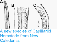
Capillaria plectropomi from Plectropomus leopardus. (A) Anterior end of male, lateral view. (B) Stichocyte in middle part of stichosome. (C) posterior end of male, lateral view. Moravec & Justine (2015).

See also…

Pinworms, Oxyuridae, are parasitic Nematodes infecting the digestive tracts of Mammals. They have short life cycles, typically undergoing several generations in a year, with eggs being released in the host’s faecal matter to infect new hosts. Some species of Pinworm appear to be quite cosmopolitan, infecting...


Parasitic Nematodes of the superfamily Heterakoidea are typified by having three lips, an esophagus with a valved bulb, thick shelled eggs and a pre-anal sucker on the males. They are typically parasites of the digestive tracts of small vertebrates, which do not require an intermediate host (i.e. the species only needs to infect one species of hosts, rather than...

Parasite infections in German soldiers from the Kilianstollen First World War archaeological site.
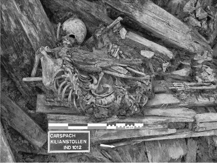
The science of palaeoparasitology involves the study of parasite remains from palaeontological and archaeological sites. This rarely involves the recovery...


Follow Sciency Thoughts on Facebook.

Tuesday, 30 December 2014

Two new species of Pinworms from Sulawesi Soft-furred Rats.


Pinworms, Oxyuridae, are parasitic Nematodes infecting the digestive tracts of Mammals. They have short life cycles, typically undergoing several generations in a year, with eggs being released in the host’s faecal matter to infect new hosts. Some species of Pinworm appear to be quite cosmopolitan, infecting a range of hosts, but most species have a close relationship with a specific host species and rarely cross-infect other species.

In a paper published in the Raffles Bulletin of Zoology on 18 September 2014, Kartika Dewi of the Zoology Division at Museum Zoologicum Bogoriense, Hideo Hasegawa of the Department of Biology at OitaUniversity and Mitsuhiko Asakawa of the Department of Parasitology at Rakuno Gakuen University describe two new species of Pinworms from Sulawesi Soft-furred Rats, Eropeplus canus, trapped at Lambanan in the South Sulawesi Administrative District. The Sulawesi Soft-furred Rat is an indigenous species to Sulawesi (i.e. found nowhere else), known only from a few mountainous localities (altitudes between 1800  and 2300 m) and considered to be Vulnerable under the terms of the International Union for the Conservation of Nature’s Red List of Threatened Species. Both of the new species are placed in the genus Syphacia, seven species of which have previously been recorded from Indonesia; the cosmopolitan Syphaciamuris, which infects a variety of Rats (Rattus spp.), plus four species infecting indigenous Rodents on Sulawesi and one species each infecting Rodents on Halmahera and Papua.

The first new species is placed in a new subgenus, Rumbaisyphacia, which is a combination of ‘rumbai’, an Indonesian word meaning ‘fringe’ and the name of the genus (i.e. ‘fringed Syphacia’) and given the specific name kumis, meaning ‘moustache’ in Indonesian; both names refer to a fringe of papillae around the mouthparts. The species is described from twenty two specimens, eleven of each species. The females are considerably larger than the males, at 3.21–4.12 mm as opposed to 1.51–1.72 mm.

Syphacia (Rumbaisyphacia) kumis, from Eropeplus canus in south Sulawesi, Indonesia. (1) male, lateral view; (2) cephalic end of male, apical view. Dewiet al. (2014).

The second new species is also placed in a new subgenus, Segienamsyphacia, from ‘Segienam’, which means ‘hexagonal’ in Indonesian, a reference to the mouthparts of the female, and given the specific name yuniae in honour of Yuni Apriyanti, who prepared the specimens for observation by SEM. This species is also described from eleven male and eleven female specimens. The females of this species are also bigger than the males (though not as much so as in Syphacia (Rumbaisyphacia) kumis) at 2.34–2.56 mm in the females and 1.00–1.25 mm in the males.

Syphacia (Segienamsyphacia) yuniae, from Eropeplus canus in south Sulawesi, Indonesia. (11) male, lateral view; (12) cephalic end of male, apical view. (18)Female, lateral view; (19) cephalic end of female, apical view. Dewi et al. (2014).

Dual infections of Pinworms are rare in Rodents, with most species having a single Pinworm parasite, and where more than one species is present this is generally thought to be an exceptional situation in which unhealthy animals have been opportunistically infected by non-specific parasites. However all of the Sulawesi Soft-furred Rats inspected were suffering from infections of both Pinworm species, and did not appear to be particularly unhealthy. Dewiet al. suggest that as additional resource partition by the parasites, with the Pinworms probably infecting different microhabitats of utilizing different nutrients.

SEM of mouth opening Syphacia (Rumbaisyphacia) kumis showing setiferous apical margin of pharynx. Dewi et al. (2014).

See also…

Parasitic Nematodes of the superfamily Heterakoidea are typified by having three lips, an esophagus with a valved bulb, thick shelled eggs and a pre-anal sucker on the males. They are typically parasites of the digestive tracts of small vertebrates, which do not require an intermediate host (i.e. the species only needs to infect one species of hosts, rather than infecting different...
The science of palaeoparasitology involves the study of parasite remains from palaeontological and archaeological sites. This rarely involves the recovery...
 Nematodes are probably the most numerous and specious group of animals on Earth, though relatively few species have actually been described. The title of...


Follow Sciency Thoughts on Facebook.