Showing posts with label Anhui Province. Show all posts

Friday, 8 December 2023

Mesechinus orientalis: A new species of Hedgehog from eastern China.

Hedgehogs, Erinaceinae, are a distinctive group of spiney insectivorous Mammals found across Eurasia and Africa, but absent from Australia and the Americas. Their most notable features are their spines, which are enlarged hollow hairs, and ability to role into a ball, presenting only these spines to the outside world. There are five extant genera of Hedgehogs (a number of extinct genera are also known in the fossil record, including one from North America). The genus Atelerix is found exclusively in Africa, Paraechinus in North Africa, the Middle East and South Asia, Hemiechinus in Central and South Asia, Erinaceus across Europe, the Middle East and much of Asia, and Mesechinus is restricted to eastern Asia.

Hedgehogs of the genus Mesechinus, known as Forest Hedgehogs, are found in China, Mongolia, and the Russian Far East, where they are found in semiarid and dry steppes, semideserts, cold-temperate deciduous and temperate deserts, warm-temperate deserts, grasslands, deciduous broad-leaf forests, and subalpine and low-elevation coniferous forests. Formerly, all known species were restricted to the cold habitats of northeastern China, Mongolia, and the Russian Far East, but in 2007 a new species Mesechinus wangi, was described from the tropical and subtropical rainforests of Yunnan Province, in the southwest of China, opening the posibility that other species in the genus might be found in warmer climates.

In a paper published in the journal ZooKeys on 28 November 2023, Zifan Shi and Hongfeng Yao of the Collaborative Innovation Center of Recovery and Reconstruction of Degraded Ecosystem in Wanjiang Basin at Anhui Normal University, Kai He of the Key Laboratory of Conservation and Application in Biodiversity of South China at Guangzhou University, Weipeng Bai of the Institute of Nihewan Archaeology at Hebei Normal University, Jiajun Zhou of the Zhejiang Forest Resources Monitoring Center, Jingyi Fan, also of the Collaborative Innovation Center of Recovery and Reconstruction of Degraded Ecosystem in Wanjiang Basin at Anhui Normal University, Weiting Su and Wenhui Nie of the State Key Laboratory of Genetic Resources and Evolution & Yunnan Key Laboratory of Biodiversity and Ecological Security of Gaoligong Mountain at the Kunming Institute of Zoology, Shuzhen Yang of the National Nature Reserve of Mount Tianmu, Kenneth Onditi and Xuelong Jiang, also of the State Key Laboratory of Genetic Resources and Evolution & Yunnan Key Laboratory of Biodiversity and Ecological Security of Gaoligong Mountain at the Kunming Institute of Zoology, and Zhongzheng Chen again of the Collaborative Innovation Center of Recovery and Reconstruction of Degraded Ecosystem in Wanjiang Basin at Anhui Normal University and the State Key Laboratory of Genetic Resources and Evolution & Yunnan Key Laboratory of Biodiversity and Ecological Security of Gaoligong Mountain at the Kunming Institute of Zoology, describe a new species of Mesechinus from Anhui and Zhejiang provinces in eastern China. 

The new species is named Mesechinus orientalis, where 'orientalis' means 'of the east', in reference to it having been discovered in eastern China. The new species is described on the basis of seven specimens collected in southeast Anhui and northwest Zhejiang provinces. They are small-bodied Hedgehogs, reaching slightly under 190 mm in length. Their spines have four colour rings, two-thirds of the length is white at the base, followed by a 3–4 mm black ring, a narrow light ring, and a black tip. The species appears to be sexually dimorphic with regard to colour, with all known females having reddish fur, while all known males are greyish, although Shi et al. are cautious about asserting this with any confidence, due to the small number of known specimens.

A living Mesechinus orientalis from Xuancheng, Anhui. Shi et al. (2023).

A genetic analysis found that Mesechinus orientalis is the sister group to the pairing of Mesechinus hughi (found in central China) and Mesechinus wangi (from southwest China). It also found that the most recent common ancestor of all members of the genus Mesechinus was likely to have lived about 1.71 million years in the Early Pleistocene, with Mesechinus orientalis probably diverged from the common ancestor of Mesechinus hughi and Mesechinus wangi about 1.10 million years ago, with those two species having diverged about 740 000 years ago.

Divergence times estimated using BEAST based on mitogenome data. Branch lengths represent time. Numbers above branches refer to divergence time in millions of years. Asterisks indicate the location of correction points. Shi et al. (2023).

See also...

Follow Sciency Thoughts on Facebook.

Follow Sciency Thoughts on Twitter.


Monday, 29 August 2022

Guangdedendron micrum: A Lycopsid Tree from the Late Devonian of Anhui Province, China.

The first major evolutionary radiation of the Vascular Plants occurred in the Devonian, and was a vital step on the path towards the formation of almost all modern terrestrial ecosystems. During this time, three distinct groups of Vascular Plants began to produce Trees, the Pseudosporochnaleans, Fern-like Plants which may have been related to the true Ferns, Archaeopteridaleans, Fern-like Plants thought to have been ancestral to the Gynosperms, and Lycopsids, or 'Giant Club Mosses'. The Lycopsids are thought to have been the first of these groups to appear, and played an important role in early terrestrial ecosystems, forming the first forests in the Devonian, and dominating the forests of the Carboniferous, providing the majority of the biomass for the great Carboniferous coal deposits. Unlike the Carboniferous forests, the forests of the Devonian are relatively poorly known, with most examples coming from Europe and North America, and only a single example on Svalbard Island having produced in situ Lycopsid trees before 2019.

South China is known to have been a significant centre for the evolution of Lycopsids, and may have been the place where the first forests appeared. In 2019 a Devonian Lycopsid forest was reported at Xinhang in Anhui Province, where trees of the Lycopsid Guangdedendron micrum were uncovered at a working clay pit, yielding the oldest known examples of stigmarian roots.

Because this site is being actively worked for commercial reasons, further fossils are being continuously uncovered, adding to our knowledge of this early forest-forming Lycopsid Tree. In a paper published in the journal BMC Ecology and Evolution on 23 May 2022, Xue Gao of the Key Laboratory of Orogenic Belts and Crustal Evolution at Peking University,  Le Liu of the School of Geoscience and Surveying Engineering at the China University of Mining and Technology-Beijing, Min Qin of the Institute of Geology and Paleontology at Linyi University, Yi Zhou, also of the Key Laboratory of Orogenic Belts and Crustal Evolution at Peking University, Lei Mao of the Anhui Geological Museum, and De‑Ming Wang, once again of the Key Laboratory of Orogenic Belts and Crustal Evolution at Peking University, present an updated description of Guangdedendron micrum, based upon new material that has become available over the last three years.

Guangdedendron micrum was a small Lycopsid Tree, with separate male and female plants, in which adult Plants produced seeds only once before dying. It produced stigmaria-type rhizomous roots, which divided bilaterally on four different axes, and produced helical rootlets. The step branched dichotomously at its terminus, producing a single large strobilus ('cone'). Leaves were narrow with entire margins, and arranged spirally on the stems. The Stobili were typically singular, but in some cases branched dichotomously once. They were roughly cylindrical in shape, with helically arranged megasporophylls (spore bearing leaves), each of which had a keeled pedicel (leaf stem) and an upturned lamina (leaf); spores were born on the upper surface of these laminae.

The rhizomatous roots reached down to about 27 cm beneath the soil surface, with each axes being 8.3-31 cm long, and arranged at angles of 19-60° to the ground surface. The rootlets are up to 27.2 cm long and 7 cm wide. The largest preserved stems are 88 cm high and about 18.7 cm in diameter. The vegetative leaves 2.0-9.2 cm long and 0.12-0.90 cm wide, each having a single vein. The largest stobili reach about 23.4 cm long and 3.0 cm wide. The sporangia laminae reach about 18 mm long and 5.8 mm wide. The megasporangia (spore producing bodies on the leaves are 4.0-7.4 mm long.

The new material presented by Gao et al. shows that the  stigmarian rhizomorph root of Guangdedendron micrum has four evenly distributed axes, which grew to about 31 cm in length, before dichotomously branching once. These rhizomorph roots angle downwards between 19-51° to the ground surface, covering an area up to 41.1 cm wide and 27.0 cm deep. The first order branches (which arise from the initial dichotomous branching) grow to about 7.2-7.8 cm in length, before dividing again to produce third order branches, which reach 2.8-6.5 cm in length. Rootlets are arranged helically along the roots and reach up to 12 mm in length and 5 mm in width. These are generally unbranched, and do not produce root hairs.

In‑situ rooting systems of Guangdedendron micrum from Yongchuan mine. (A) Stem and connected rhizomorph axes. (B) Rooting system with branched rhizomorph axes. PKUB21015. (C) Stems with connected rhizomorph axes bearing rootlets. (D) Top view of the specimen shown in (A), after removing the stem and surrounding rocks partially peeling off. A rooting system with four once‑dichotomized rhizomorph axes. Arrows indicating 8 second‑order branches. (E) Stem base and connected two rhizomorphic axes. (F), (G) Rhizomorphic axes connected to moulds of stem bases and bearing rootlets. Scale bars: (B), (D), (G) 5 cm; diameter of the coin for scale: 2 cm (A), (C), (E), (F). Gao et al. (2022).

Stems have been found preserved as compressions and erect casts. These reach up to 71.3 cm high and 1.1-12.2 cm in diameter, excluding the expanded bases which form the connection to the root system. Dichotomous branching is rare, but does occur, happening no more than twice on a single plant, with the branches angled at 13-43°. 

Stems and vegetative leaves of Guangdedendron micrum, from Yongchuan (A), (B), (D), (G)–(M), (O)–(Q), (S), (U), (V) and Jianchuan (C), (E), (F), (N), (R), (W) mines. (A), (B) In‑situ stems with expanded bases. PKUB21000, 21014. (C) Two adjacent in‑situ stems with expanded bases. (D) An in‑situ stem with an expanded base. (E), (F) Two sides of a stem displaying basal expansion and helically arranged oval fissures of broken leaf cushions after leaf abscission. PKUB21004. (G)–(J) Stems perpendicular to the bedding plane. (H) YC‑103. (K) An in‑situ stem with leaf cushions. (L) A once‑dichotomised stem. YC‑101. (M), (N) Twice‑dichotomised stems. (M) YC‑102. (O) A once‑dichotomised leafy stem with leaf bases. (P), (Q) Part and counterpart of helically arranged leaf cushions. YC‑105, 104. (R) A stem with helically arranged leaf cushions. (S) Fusiform leaf cushions. PKUB21013. (T) Line illustration of a leaf cushion based on arrowed portion in (Q). (U) Helically arranged leaf bases. Arrow indicating portion enlarged in (V). PKUB21007. (V) Enlargement of arrowed portion in (U), indicating fusiform leaf bases with middle vertical grooves in the lower part. (W) Helically arranged leaf bases, each showing a vertical groove in the middle. PKUB16052a. Scale bars: (A), (E), (F) 2 cm, (D), (G) 5 cm, (P)–(R), (U), (W) 1 cm, (S), (T), (V) 5 mm; diameter of the coin for scale: 2 cm (B), (C), (H), (J), (K), (M)–(O); length of the hammer for scale: (I) 28.6 cm, (L) 27.3 cm. Gao et al. (2022).

Vegetative leaves are 3.5-8.5 cm long and 0.29 to 0.90 cm wide, and spindle-shaped with entire margins and a single vein running the entire length of the leaf. These are arranged spirally around the stems, departing at an angle of 59-98° to the stem. Slender twigs, interpreted as the stems of juvenile plants, have leaves to their tips arranged in a similar way.

Vegetative leaves of Guangdedendron micrum from Jianchuan mine. (A), (B) Terminal parts of vegetative axes bearing leaves. PKUB21001, 16067. (C), (D) Part and counterpart of a terminal vegetative axis. PKUB21017a, 21017b. (E), (F) Tapering vegetative axes with persistent linear leaves. Arrow indicating portion in (F) enlarged in (G). PKUB21018, 16144. (G) Enlargement of arrowed portion in (F), showing veins of vegetative leaves. Scale bars: (A)–(E) 2 cm, (F) 10 cm, (G) 1 cm. Gao et al. (2022).

The fertile stems are up to 6.4 cm long and 0.21-0.55 cm wide, and tipped by strobili. One fertile stem found was 17.6 cm wide, and branched dichotomously, producing two daughter stems angled at 60° to one-another. Vegetative leaves on the lower part of the fertile stems are angled at 70-85° to the stems, and are curved at their tips.

Fertile axes and strobili of Guangdedendron micrum from Jianchuan (A)–(E), (H)–(J), (L), (M) and Yongchuan (F), (G), (K) mines. (A), (B) Part and counterpart of a once‑dichotomized axis bearing linear leaves and a single strobilus. (C) A strobilus without basal fertile axis preserved. (D), (E) Part and counterpart of a strobilus terminating a fertile axis. PKUB16001a, 16001b. (F), (G) Part and counterpart of terminal strobili in pairs, with sporophylls along central strobilar axis and persistent vegetative leaves on fertile axis. PKUB21002a, 21002b. (H) At least eight strobili (arrows 1–8) preserved in the same direction (1 and 2, 7 and 8 possible paired, respectively). PKUB16047. (I) A dichotomized strobilus. PKUB21011. (J) Terminal strobili in pairs. PKUB16035. (K) A single and a pair of strobili perpendicular to the layers. (L) A short strobilus may partially preserved. PKUB16065. (M) Strobilus displaying central strobilar axis. PKUB16020a. Scale bars: (A), (B) 5 cm, (C), (J), (L), (M) 1 cm, (D)–(I) 2 cm; diameter of the coin for scale: 2 cm (K). Gao et al. (2022).

The strobili at the end of the fertile stems are pendulous and usually singular, although they are sometimes found in pairs and occasionally branch dichotomously. They are cylindrical and slightly curved, reaching a maximum of about 23.4 cm in length. Sporophylls are tightly packed and helically arranged around these strobili. Each sporophyll comprises a pedicel projecting horizontally from the main stem, and a lamina angled at about 110° to this pedicel. These laminae are an elongated triangle shape, reaching 12-18 mm in length.

Stems and strobili of Guangdedendron micrum from Jianchuan (A)–(G), (I), (L)–(O) and Yongchuan (H), (K), (P), (Q) mines. (A), (B) Two sides of a stem with expanded base and leaf cushions. PKUB21005. (C), (D) Two sides of a stem. PKUB21006. (E), (F) In-situ once-dichotomized stems. Two arrows in (E) indicating two daughter axes. (G) Oval fissures helically arranged along stem. (H) Oval fissures helically arranged along in-situ stem. (I) Enlargement of portion of stem (arrow), showing a ligule pit (Lp) and a vascular bundle scar (Vs). (J) Interpretative line drawing of helically arranged leaf bases, indicating outlines (black lines) and parastichies (red dotted lines) of leaf bases. PKUB16049. (K), (L) Dichotomised fertile axes with terminal single strobilus. (M), (N) Terminal strobili in pairs. PKUB16097, 16099. (O) Dichotomised fertile axis with partially preserved strobili. (P) Over ten strobili preserved in the same direction. (Q) A possibly once-dichotomized strobilus. Scale bars: (A)–(D) 5 cm, (F), (L) 2 cm, (G), (M)–(O), (Q) 1 cm, (I) 2 mm, (J) 5 mm; diameter of the coin for scale: 2 cm (E), (H), (K), (P). Gao et al. (2022).

All of the strobili found produce megaspores (i.e. female spores), leaving the possibility that the male plants were quite different. Each sporaphyll has an ellipsoidal megasporangium 4.0-7.4 mm in length on its upper surface. Few of these can be seen clearly, as the upper surfaces are generally covered by the adjacent sporaphylls, but each megasporangium appears to contain multiple megaspores.

Sporophylls and spores of Guangdedendron micrum from Jianchuan mine. (A) A partially preserved strobilus, showing sporangia on the adaxial surface of sporophyll pedicels. PKUB16049. (B) A partially preserved strobilus, showing megaspores and sporophylls. PKUB16141. (C) Mid‑upper part of a strobilus with helically arranged sporophylls in face and lateral view. Arrow indicating portion enlarged in (H). PKUB16058. (D) Enlargement showing sporophyll laminae and pedicel in lateral view. (E) Enlargement showing sporophyll laminae and pedicel in lateral view and partially preserved sporangia, arrows indicating megaspores. (F) Enlargement of arrowed portion in (A), showing sporangia. (G) Enlargement showing sporophyll laminae in face view. (H) Enlargement of portion in (C) (arrow), showing sporophyll laminae in face view with downturned heels. (I) Part of a strobilus showing megaspores, the arrow indicating portion enlarged in (K). PKUB16020a. (J), (K) Enlargement showing megaspores and spiny ornamentations. (L) Megaspores with spiny ornamentations. Scale bars: (A), (C), (D) 1 cm, (B), (F) 5 mm, (E), (G)–(I) 2 mm, (J) 1 mm, (K), (L) 500 μm. Gao et al. (2022).

The absence of strobili producing microspores in the discovered specimens of Guangdedendron micrum is curious, particularly as a large number of specimens have now been found. Other Lycopsid species from the Devonian and Carboniferous are known have produced microsporangia and megasporangia on separate, but otherwise similar plants. It is possible that male Guangdedendron micrum plants were much less common than the female plants, or that the plants were able to reproduce parthenogenicly from unfertilised megaspores. 

Reconstruction of the longest strobilus terminating the fertile axis after bifurcation. Scale bar is 2 cm. Gao et al. (2022).

Bifurcation (spliting in two) of the fertile stems and strobili of Lycopsids is rare, but has previously been recorded in other Late Devonian species. This appears to be caused by branching of a meristem (growth centre of a Plant) at the tip of the stem after the stem has switched to its fertile growth mode. In Guangdedendron micrum this can occur before the strobili, resulting in paired strobili, or on the strobilus itself, resulting in a forked strobilus. A similar branching can be seen in the fertile zones of some living Club Mosses and Ferns, although these are all epiphytic in habit, and it has generally been assumed that this is an adaptation to that lifestyle; something highly unlikely in Guangdedendron micrum, which has a well developed subterranean root system, and is in any case the largest Plant in its ecosystem.

See also...


Online courses in Palaeontology

Follow Sciency Thoughts on Facebook.

Follow Sciency Thoughts on Twitter.


Saturday, 12 March 2022

Dating the Lantian Biota.

Fossils from the Ediacaran Period record a remarkable transition from a world in which there were almost no multicellular organisms to one in which such organisms dominated almost all marine ecosystems. Deposits from the Lower Ediacaran are dominated by the Acritarchs, a diverse group of single-celled marine Algae which may-or-may-not represent a single taxonomic group. Deposits from the Upper Ediacaran produce a range of complex fossils, apparently of soft-bodied organisms of uncertain affinities. One of the earliest macro-fossil producing deposits is the Lantian Formation of South China, a deep-marine basinal black shale which produces a series of fossils of increasing complexity, assumed to have affinities to Algae and Animals, providing insight into the earliest stages in the development of these multicellular organisms.

 
Macrofossils from the Lantian biota, Anhui Province, China. Specimens are housed in the collections of the Nanjing Institute of Geology and Palaeontology. (A) Lantianella laevis (at left; NIGP163377), and a larger conical form. (B) Lantianella annularis, NIGP163384. Dunn & Liu (2017).

The Lantian Biota is potentially the oldest macrofossil assemblage of the Ediacaran, and therefore is of great interest to palaeontologists, but obtaining a precise date for these deposits has proven problematic. The Lantian Formation lacks any volcanic ash beds, which would allow dating via uranium/lead ratios in zircons. Zircons are minerals formed by the crystallisation of cooling igneous melts. When they form they often contain trace amounts of uranium, which decays into (amongst other things) lead at a known rate. Since lead will not have been present in the original crystal, it is possible to calculate the age of a zircon crystal from the ratio between these elements.

An alternative method of dating sedimentary rocks utilises rhenium/osmium ratios. Rhenium is one of the rarest elements on Earth and the second highest stable element on the periodic table, after hafnium. There are two naturally occurring isotopes of rhenium, the stable rhenium¹⁸⁵, and the 'unstable' rhenium¹⁸⁷. Unusually, the 'unstable' form of rhenium is the more common; this is because it has an extremely long half-life, about 4160 million years, which means that a significant proportion of the rhenium¹⁸⁷ present during the formation of the Earth is still in that form (like all elements heavier than iron, rhenium is formed during supernova events at the end of the life-cycle of massive stars). When rhenium¹⁸⁷ does decay it does so into the stable radiogenic isotope osmium¹⁸⁷. Osmium also has two stable isotopes, the non-radiogenic osmium¹⁸⁸, and the radiogenic osmium¹⁸⁷. This means that over time the amount of rhenium¹⁸⁷ in a rock sample will fall at a predictable rate, while the amount of osmium¹⁸⁷ rises at the same rate, and the amount of osmium¹⁸⁸ remains constant, making it possible to date the rock by comparing the ratios of these elements. However, the exposed deposits of the Lantian Formation all show varying degrees of metamorphism, which prevents useful application of the rhenium/osmium method, as the method relies on elements present at very low levels in the total rock (rather than working on individual mineral grains), with the upshot that any loss of either element will interfere with the result.

Based upon approximate lithostratigraphic correlation between the Lantian or south Anhui Province and the Doushantuo Formations of the Yangtze Gorges area, it has been estimated that the Lantian Formation is between 635 and 551 million years old, but the long distances (the two locations are about 700 km apart) and variable geology between the two sites make even this very approximate estimate unreliable. 

A detrital zircon retrieved from Member II the Lantian Formation (i.e. an individual grain from a sedimentary bed, not part of a volcanic ash layer) yielded an age of 590 million years, giving a possible maximum age for that layer, and a low-yttrium monazite (a mineral likely to have been formed diagetically within the deposit soon after it was laid down yielded a lead/uranium date of 612 million years, which has been argued to be a minimum age for the formation. However, as well as being contradictory, both these dates are poorly constrained, making it hard to assess the actual age of the Lantian Formation, and how the age of the fossils it contains relate to other Ediacaran fossils.

In a paper published in the journal Geology on Chuan Yang of the State Key Laboratory of Lithospheric Evolution at the Institute of Geology and Geophysics of the Chinese Academy of Sciences, and the Geochronology and Tracers Facility at the British Geological Survey, Yang Li, also of the State Key Laboratory of Lithospheric Evolution at the Institute of Geology and Geophysics of the Chinese Academy of Sciences, David Selby of the Department of Earth Sciences at Durham University, Bin Wan, Chengguo Guan, and Chuanming Zhou, of the State Key Laboratory of Palaeobiology and Stratigraphy at the Nanjing Institute of Geology and Palaeontology, and Xian-Hua Li, once again of the State Key Laboratory of Lithospheric Evolution at the Institute of Geology and Geophysics of the Chinese Academy of Sciences, and the State Key Laboratory of Palaeobiology and Stratigraphy at the Nanjing Institute of Geology and Palaeontology, present the results of a study which aimed to find a rhenium/osmium date for the Lantian Formation based upon the best-preserved, biota-bearing, organic-rich black shale of Member II of that formation.

 
Geological maps and stratigraphic column for the Lantian Formation (China). (A) Generalized palaeogeographic map of the Yangtze Block during the early–middle Ediacaran Period showing the approximate location of shelf, slope, and basinal facies. Numbered triangles indicate locations of areas/sections mentioned in the text, with the Lantian area highlighted in red. (B) Geological map of the Lantian area showing the location of the Lantian drill core. (C) Litho- and chemo-stratigraphy of the Lantian drill core highlights the horizon that was sampled for rhenium/osmium geochronology. Yang et al. (2022).

The Laintian Formation is found on the Yangtze Block of the South China craton, which in the Ediacaran comprised a continental shelf to the northwest and a deep basin to the southeast, with a gradation of slope strata in between. This produced a series of sedimentary blocks with exceptionally well-preserved Ediacaran fossils, including the Lantian biota in Anhui and the Weng’an biota in Guizhou, thought to be the two oldest known examples of Ediacaran biotas. The Lantian Basin was laid down in a basin-slope environment, overlying the Leigongwu Formation, a terminal Cryogenian diamictite. It is in turn overlain by the Ediacaran–Cambrian transitional Piyuancun Formation.

Yang et al.'s work is based upon a drill core extracted near Lantian village in Xiuning County, Anhui Province. Within this core, the Lantian Formation is present as four members, with Member I represented by a 4 m-thick unit of light-grey siliceous cap dolostone, Member II comprising a 97 m thick grey, calcareous siltstone interbedded with argillaceous limestone and an upper subunit of black shale with rare argillaceous limestone interbeds, Member III is a 73 m thick interbedded grey, argillaceous dolostone and black shale, followed by about 50 m of grey limestone, and Member IV is a 10 m section of black shale. The fossils of the Langtian Formation are found almost continuously in the upper subunit of Member II, comprising both carbonaceous compression macrofossils and pyritized material. 

Yang et al. extracted a number of black shale samples for rhenium/osmium analysis from a 50 cm interval 48 m above the base of the Formation (i.e. 44 m above the base on Member II).

In order to be able to get a reliable rhenium/osmium date, Yang et al. needed to find patches of shale with the least post-depositional isotope exchange. To do this they selected core samples without post-formation veining and weathering, which were further imaged by X-ray computed tomography and X-ray fluorescence, which revealed well-preserved sedimentary lamination and relatively homogenous elemental patterns within the 50 cm interval used in the study, indicating a stable depositional environment with no evidence of post-depositional chemical weathering. Yang et al took nine samples from within a roughly 25 cm section within this interval for rhenium/osmium analysis, and calculated the abundances of rhenium and osmium within these samples, gaining measurements of 4-12 parts per billion for rhenium and 155-490 parts per trillion for osmium, from which they calculate the deposits have a probable age of 602 million years,

Yang et al.'s date of 602 million years for a layer in the middle part of Member II of the Lantian Formation is the first well constrained date for the Lantian Biota. The fossils of the Lantian Biota extend to slightly below the top of Member II, and is probably below the Shuram Event Interval, a sudden depletion in the ratio of carbon¹³ to carbon¹² recorded in rocks around the world, and which may have been linked to a rise in the amount of oxygen in the Earth's atmosphere, as well as possibly below another negative carbon¹³ excursion recorded at the boundary between Member II and Member III of the Doushantuo Formation in the Yangtze Gorges area, which is thought to be connected to the Gaskiers Glaciation at about 580 million years before the present. Based upon this, Yang et al. suggest that the Lantian Biota is more than 580 million years old. Since the base of the Lantian Formation has been dated to 635 million years ago, and the Lantian Fauna appears in Member II of the formation some way above this, Yang et al. also suggest that the fauna is likely to be younger than 615 million years old.

The Weng'an Biota is a rich microfossil assemblage consisting mainly of reworked and redeposited Acanthomorphic Acritarchs, multicellular algae, tubular microfossils, putative animals, and animal embryos, found in the shelf-facies phosphorite of the Doushantuo Formation in the Weng’an area. Similar microfossils have been found in chert nodules from Member II of the Doushantuo Formation, which have been estimated to be about 632 million years old. This would suggest that the macrofossils of the Lantian Formation appeared after the microfossils of the Doushantuo Formation, but that there is a considerable temporal overlap between the two fossil groups. The Lantian Biota also appears before the soft-bodied fossils of the classical 'Ediacaran Fauna' which first appear in rocks dated to 574 million years ago in Newfoundland and northwestern Canada.

 
Integrated carbon isotopic profile (A), fossil ranges (B), and initial osmium isotopic data (C) of the Ediacaran Period. Carbonate proportion of carbon¹³ data from the Lantian Formation are highlighted in red. CIE, carbon isotope excursion. Yang et al. (2022).

Five putative Animal species have been described from the Lantian Biota. All are centimetre-scale in size, and show morphological complexity and structural differentiation. If this is correct, then it indicates that Metazoan Animals had appeared by the early–middle Ediacaran, something which has been predicted from molecular clock data, which suggests that modern Animal phyla diverged in the Ediacaran, and by the presence of putative Animal embryos in the Weng'an Biota.

 
A putative Animal from the Lantian Biota: Lantianella laevis (A)–(B) NIGP163375, part and counterpart, holotype, showing globose holdfast (white arrow) and tentacle-like structures (black arrows). (C)–(D) NIGP163376, part and counterpart, showing globose holdfast (white arrows) and longitudinal filaments (black arrows). (E)–(F) NIGP163377 and NIGP163378, respectively, showing flexible (white arrow in (E)) or straight (white arrow in (F)) tentacle-like structures and longitudinal filaments (black arrows). (G)–(H) NIGP163379 and NIGP163380, respectively, showing rounded (white arrow in (G)) and scalloped (white arrow in (H)) distal margin of conical body, and poorly preserved longitudinal filaments (black arrows in (H)). (I)–(J) and (K)–(L) NIGP163381 and NIGP163382, respectively, part and counterpart, showing sharp contrast between the lighter-coloured tentacular crown and darker-coloured conical body, globose holdfast (white arrows), and patch of diffuse organic mass associated with globose holdfast (black arrows). Millimetre-sized circular and elliptical carbonaceous compressions in the background are specimens of the Acritarch Chuaria circularis, which are best seen in (A)–(B). All scale bars represent 5 mm. Bin et al. (2016).

As well as the putative Animals, 13 species of possible Macroalgae (Seaweed) have been described from the Lantian Biota. Further Macroalgae have been described from the Weng'an Biota, and Acanthomorphic Acritarchs are abundant in deposits of similar ages around the world. Taken together, this implies a major episode of radiation and diversification of Algae in the early–middle Ediacaran. Such an episode would likely have raised oxygen levels within the Ediacaran oceans, facilitating the rise of the Metazoa. Several of the Algal taxa found in the Lantian Biota are found in later Ediacaran Fauna-bearing deposits, with one, Flabelophyton, showing a marked increase in size over the Ediacaran Period. This continuation of related Algal fossils from the Lantian and Weng'an biotas into the deposits bearing the classical Ediacaran Fauna supports the idea that there is an evolutionary continuation between these biotas.

At the beginning of the Ediacaran, benthic marine communities were comprised entirely of mat-forming micro-organisms. By the end of the period, modern, Animal-dominated benthic marine communities had appeared and become more-or-less ubiquitous. The Lantian Biota records a stage in this transition, with most species recorded, Animal and Algae, being erect, sessile, epibenthic forms. The date of 602 million years ago provided by Yang et al. confirms that this macrofossil-dominated assemblage was present in the early-middle Ediacaran.

Yang et al. obtained a ratio of osmium¹⁸⁷/osmium¹⁸⁸ at the 602 million years isochron in the Lantian fauna of 1.14, while a layer of the Old Fort Point Formation in western Canada dated to 607 million years ago produced an osmium¹⁸⁷/osmium¹⁸⁸ ratio of 0.62, a significant difference which implies a regional variation in the control of osmium isotope ratios. This is curious, as the main source of osmium in the Ediacaran oceans is thought to have been radiogenic metal weathered from rocks on land. It is likely that the rise of the Ediacaran Biota was linked to an increase in terrestrial erosion, increasing the amount of mineral nutrients available to marine organisms, but how this related to the variation in the isotopic makeup of osmium is unclear.

Geochemical analysis of the shales of Member II of the Lantian Formation has suggested a persistent euxinic (high sulphur, low oxygen) environment. However, the presence of fossil Algae and Metazoans suggest that the area clearly had at least periodic episodes of oxygenation. The environment is interpreted as having been below the storm base (level to which the influence of storm events penetrates) but within the photic zone (area of the sea where there is sufficient light for photosynthesis). This probably reflects the difference between the geological timescale recorded by the chemical evidence and the ecological timescale recorded by the fossil evidence, but also records that a stable thermal environment (deep water environments typically do not have fluctuating temperatures) was probably more important to the emergence of the Metazoans than a stable oxygen environment.

See also...


Online courses in Palaeontology. 

Follow Sciency Thoughts on Facebook.

Follow Sciency Thoughts on Twitter.


Thursday, 16 June 2016

Sclerocormus parviceps: A new species of Ichthyosaur from the Early Triassic of Anhui Province, China.

Ichthyosaurs were Marine Reptiles known from the Triassic to the Cretaceous. They were fully-aquatic with a Dolphin-like form, and known to have given birth to live young, rather than emerging from the water to lay eggs, as is the case in Turtles and Crocodiles. The origin of the group has for a long time been obscure, with large fully aquatic forms known from the Middle Triassic onwards. However several recent discoveries have suggested that they may have been related to the Hupehsuchians, an enigmatic group of Marine Reptiles known from the Early Triassic of China.

In a paper published in the journal Scientific Reports on 23 May 2016, a group of scientists led by Da-Yong Jiang of the Laboratory of Orogenic Belt and Crustal Evolution at Peking University, describe a new species of Ichthyosaur from the Early Triassic Nanlinghu Formation of Anhui Province, China.

The new species is named Sclerocormus parviceps, where 'Sclerocormus' means 'stiff trunk' and 'parviceps' means 'small skull'. The species is described from a single skeleton, 159.9 cm in length and partially compressed. The skull is notably small for an Ichthysaur, even for an early Ichthyosaur (which were generally shorter snouted), having a short snout and comprising only 6.25% of the total bodylength, compared to 12% in the earliest previously known Ichthyosaur, Chaohusaurus.

Sclerocormus parviceps. (a) Whole specimen. (b) Skull. (c) Close-up of gastral basket. (d) Close-up of U-shaped haemal arches. (e) Right forelimb. (f) Shoulder elements. (g) Pelvic girdle and hind limb. (h) Skull elements. Abbreviations: a, angular; ar, articular; as, astragalus; ca, calcaneum; car, caudal rib; ca.v, caudal vertebra; ce, centralia; cl, clavicle; d, dentary; dc, distal carpal; f, frontal; fe, femur; fi, fibula; he, hemal arch; il, ilium; in, intermedium; is, ischium; j, jugal, l, lacrimal; m, maxilla; mc, metacarpal; mt, metatarsal; n, nasal; p, parietal; pm, premaxilla; po, postorbital; pof, postfrontal; prf, prefrontal; pu, pubis; q, quadrate; sa, surangular; sc, scapula; scl, scleral ossicles; sq, squamosal; sr, sacral rib; st, supratemporal; ti, tibia; u, ulna; ul, ulnare. Scale unit in (a) is 1 cm, other scale bars are 2 cm. Jiang et al. (2016).

Sclerocormus parviceps has a short trunk with broad, flattened ribs and an extensive gastric basket covering its underside, similar to the condition seen in Hupehsuchians, strongly supporting a connection between the two groups. The fossil also dates from surprisingly close to the beginning of the Triassic; dating from the Olenekian (251.2-247.2 million years ago, with the beginning of the Triassic dated to 252.2 millions of years ago) which implies Marine Reptiles were colonising the oceans far sooner after the End Permian Extinction than had previously been supposed.

See also...

http://sciencythoughts.blogspot.co.uk/2014/12/a-new-species-of-hupehsuchian-from.htmlA new species of Hupehsuchian from the Early Triassic of Hubei Province, China.      The Hupehsuchians are a group of Marine...
http://sciencythoughts.blogspot.co.uk/2012/03/fresh-look-at-albian-ichthyosaur.htmlA fresh look at the Albian Ichthyosaur Platypterygius hercynicus.                           The Ichthyosaurs were a group of marine tetrapods that resembled dolphins. They appear in the fossil record in the mid-Triassic about 245 million years ago, and survive till the mid-Cretaceous, about 90 million years ago. During the Jurassic they appear to have been the top marine predators, but in the Cretaceous they were overshadowed by other groups...
Follow Sciency Thoughts on Facebook. 

Tuesday, 6 January 2015

Hominin teeth from the Middle Pleistocene of Anhui Province, China.


In the 1970s and 1980s a collection of Hominin bones and teeth were unearthed in the Longtan Cave at Hexian in Anhui Province in eastern China. The bones of these remains have been extensively studied, and assigned to the species Homo erectus, though palaeoanthropologists have differed in their opinions as to whether these remains were most similar to those from Zhoukoudian in China or Java in Indonesia. The depositional layer which produced the teeth has been dated to about 412 000 years ago, and contains traces of fires and fire damaged animal bones, animal bones with cut marks and tools made from bones, antlers and teeth, but no stone implements. The fauna and pollen assemblage at this layer imply a subtropical climate.

In a paper published in the journal PLoS One on 31 December 2014, Song Xing of the Key Laboratory of Vertebrate Evolution and Human Origins at the Institute of Vertebrate Paleontology and Paleoanthropology of the Chinese Academy of Sciences, María Martinón-Torres and José María Bermúdez de Castro of the National Research Center on Human Evolution, Yingqi Zhang, also of the Key Laboratory of Vertebrate Evolution and Human Origins, Xiaoxiao Fan of the Hexian Museum of Anhui Province, Longting Zheng of the Anhui Museum, Wanbo Huang of the Chongqing Three Gorges Institute of Paleoanthropology at the China Three Gorges Museum and Wu Liu, again of the Key Laboratory of Vertebrate Evolution and Human Origins, describe the results of a study of the teeth of the Longtan Cave Hominins, in which they were compared to a range of other teeth from ancient and modern populations of Australopithecus and Homo from Africa, Europe and China.

The first tooth described is a right upper third premolar (formerly described as a right upper fourth premolar). The tooth is in generally good condition, although it has some cracks in its enamel on the crown surfaces. There is a large fragment alveolar bone attached to the root. The cusps of the tooth have been flattened by wear, making the cusp pattern unclear. The crown is oval and slightly asymmetrical, the cusps are joined by a crest that transverses the central groove. The tooth has one lingual root and two buccal roots, all of which are quite robust; the mesiobuccal, distobuccal, and lingual roots are 19.26, 17.88, and 18.96 mm long. The pulp horns are blunt and low, with a total pulp cavity volume of 82.67 mm3.

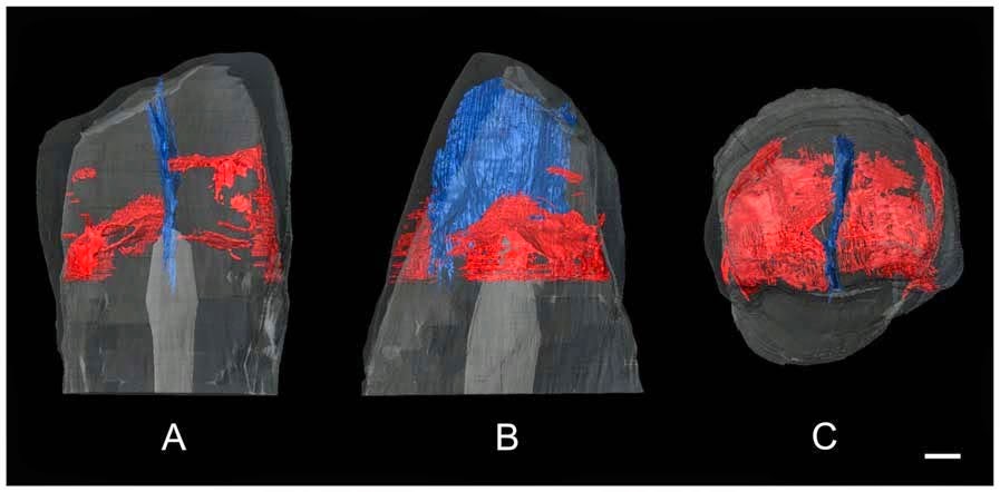
Right upper third premolar (PA832) (o: occlusal, B or b: buccal, M or m: mesial, L or l: lingual, d: distal). 3D reconstructions of the dentine surface (I–III) and the pulp cavity (IV) obtained from micro-CT scanning. The arrow points to the transverse crest. Solid lines indicate the bifurcation of the essential crest. Dotted lines indicate the mesial (I) and distal accessory ridges (II) and the well-demarcated central ridge onthe buccal surface (III). Sagittal sectional plane of PA832 (V). Cross-section of PA832 (VI) at the levelindicated by the red line. (I, II, III, IV, V, and VI) are not scaled. Xing et al. (2014).

The second tooth described is a left upper third premolar, with a complete crown but a broken root. The cusps of this tooth have been flattened exposing the dentine, and there are wear facets on the mesial and distal surfaces of the crown. The exposed dentine of the buccal cusp shows a pronounced mesial vertical furrow and a weaker distal furrow running towards the apex of the cusp. This tooth does not have a crest transversing the central groove.

Left upper third premolar (HXUP3) (o: occlusal, B or b: buccal, m: mesial, l: lingual, D or d: distal). The occlusal (I) and buccal (II) views of the dentine surface reconstructed from micro-CT scanning. Solid lines indicate the bifurcation of the essential crest. Dotted lines indicate mesial and distal accessory ridges. The arrow points to the vertical groove on the buccal surface. (I and II) are not scaled. Xing et al. (2014).

Transverse grooves crossing the central grooves of upper third premolars are common in Australopithecus and early African Homo, but become increasingly rare in later members of the genus Homo. However it is relatively more common in Early Pleistocene teeth from East Asia and Middle Pleistocene teeth from Europe. The strong medial groove seen in the dentine of the buccal cusp of the left premolar is typical of Early East Asian Hominins, and seen in some Middle Pleistocene specimens as well, including those from Zhoukoudian. This groove is completely absent from European Middle Pleistocene specimens, Neanderthals and all modern Humans. Such grooves are also seen in Homo ergaster specimens from the Late Pliocene and Early Pleistocene of East Africa, though it is less well developed in these. The asymmetrical oval outline of the crowns of these teeth is also seen in Australopithecus and early African Homo including Homo ergaster, as well as in the Early Pleistocene remains from the Sangiran Dome in Java, and the middle Pleistocene remains from Zhoukoudian and Chaoxian in China; this differs from the more symmetrical crowns seen in modern Homo sapiens, which is also seen in a Middle Pleistocene upper third premolar from in South China.

The triple root of the right tooth is more unusual; this is otherwise only known from Australopithecus and early Homo in Africa and in Early Pleistocene specimens from Java. The robust roots of the tooth are also shared with the Java specimens, with narrow, tapering roots typical of African and European Homo specimens.

From left to right, 3D reconstruction of the upper right premolar from Hexian from micro-CT scanning, Zhoukoudian PA67, Zhoukoudian 19, and Sangiran S7–35 (MB: mesiobuccal, B: buccal). Xing et al. (2014).

The upper right premolar is also exceptionally large, being wider than all specimens of Homo ergaster, Early Pleistocene Hominins from Europe or the specimens from Chaoxian or Panxian Dadong, and towards the upper end of the size range encountered in Early and Middle Pleistocene Hominins from East Asia, Middle Pleistocene Hominins from Europe and Neanderthals. It is longer than almost all known Hominin third premolars, except some Australopithecusand early Homo specimens from East Africa.

The third tooth described is an upper left first premolar. This is generally well preserved, although the buccal roots are broken off. The tooth is approximately square in outline. The upper surface is heavily worn, with large, oval-shaped wear facets, which have largely obliterated the upper surface of the tooth, though the presence of the four main cusps can still be detected by the grooves in the exposed dentine. This dentine surface is heavily crenulated, with crests and ridges on all cusps; the hypocone is large. The lingual root is very robust, divergent and flattened lengthways. It is 14.72 mm in length.

Left upper first molar (o: occlusal, B or b: buccal, m: mesial, l: lingual, D or d: distal). Sagittal sectional plane of PA836 (I). The occlusal (II) and lingual (III) views of dentine surface reconstructed from micro-CT scanning. Dotted lines point to the expression of mesial accessory ridges (II) and a Carabelli’s trait (III). (I, II, and III) are not scaled. Xing et al. (2014).

The hypocone of the first premolar has tended to decrease throughout the history of the Hominins, with large hypocones common in Australopithecus, and most Homo specimens from the Early and Middle Pleistocene, including those from East Asia, although the specimens from Zhoukoudian, which are close to the Longtan Cave Hominins both geographically and stratigraphically, have smaller hypocones. The outline of the tooth is similar to that of other East Asian Pleistocene specimens, and different from the more rhomboidal outlines seen in teeth from Pleistocene specimens from Europe and Africa, and from the more square teeth of modern Homo sapiens. The robust divergent root of the tooth is also typical of Early and Middle Pleistocene specimens from East Asia, and differs from those of Early Pleistocene Europeans, which diverge little if at all. However in modern Humans this trait is quite variable, with the Longtan tooth falling within the range of variation.

This tooth is also large, being wider than most known first molars of Middle Pleistocene Hominins from Europe or East Asia, Neanderthals or early modern Humans, though its width is typical for Homo ergaster and Early Pleistocene Hominins from East Asia. Its length is also typical for Early and Middle Pleistocene East Asian Hominins, Middle Pleistocene European Hominins, Neanderthals and early modern Humans, though it is longer than specimens from any known Homo ergaster and Early Pleistocene European Hominins.

The fourth tooth described is a left upper second molar. The roots of this tooth are completely missing and the tooth is broken along its cervical line. The cusps have been worn down and flattened, but not enough to expose the dentine. The outline of the tooth is trapezoidal, with the distal portion being narrower. There are four main cusps and a smaller fifth cusp visible, transversed by a series of grooves, the hypocone is medium-sized. Xing et al. calculate that the average enamel thickness of the tooth is 1.30 mm, but would have been 1.51 mm prior to wear.

Left upper second molar (o: occlusal, B or b: buccal, m: mesial, l: lingual, D or d: distal). The occlusal (I) and lingual (II) views of dentine surface reconstructed from micro-CT scanning. Dotted lines point to the mesial accessory ridges (I) of the occlusal surface and to the expression of a Carabelli’s cusp on the lingual face of the crown (II). (I and II) are not scaled. Xing et al. (2014).

The fifth tooth is a right upper second molar, also missing its roots, and partially broken along its cervical line. Again this tooth had been worn down and flattened, but not enough to expose the dentine. This tooth is also trapezoidal, with four main cusps, though there is no sign of a fifth cusp.

Right upper second molar (o: occlusal, B or b: buccal, M or m: mesial, l: lingual, d: distal. I and II: The occlusal (I) and lingual (II) views of dentine surface reconstructed from micro-CT scanning). Dotted lines point to the mesial accessory ridges of the occlusal surface and to the expression of a Carabelli’s cusp on the lingual surface of the crown. (I and II) are not scaled. Xing et al. (2014).

The hypercones of these teeth are typical for Early and Middle Pleistocene Hominins from East Asia, being smaller than those of Homo ergaster and other early Homo specimens from East Africa, but larger than those of Middle Pleistocene European Hominins, Neanderthals and Modern Humans. The Trapezoidal shape of the teeth is typical for Australopithecus, early Homo and Early and Middle Pleistocene Hominins from East Asia, but unlike the rhomboidal or triangular shape seen in Middle Pleistocene European Hominins, Neanderthals and Modern Humans. 

The teeth also have an average enamel thickness similar to those of East Asian specimens of Homo erectus and Middle Pleistocene Hominins from North Africa, and larger than that of most Middle Pleistocene European Hominins from Europe and Neanderthals, though within the range of modern Humans.

The mesial sectional plane of the Hexian upper left second molar (the red and grey areas indicate the reconstructed enamel and dentine, respectively, and green areas show how the occlusal wear was virtually restored). Xing et al. (2014).

Again these are large teeth, with the width exceeding all previously recorded Middle Pleistocen East Asian Hominins, and all but the largest Early and Middle European Hominins, Neanderthals or modern Humans, though the width does fall within the range of Homo ergaster and Early Pleistocene Hominins from East Asia. The length of the teeth is at the upper limit of, or outside the range of, the values seen in Middle Pleistocene East Asian Hominins, Early or Middle Pleistocene European Hominins, Neanderthals and modern Humans.

The sixth and seventh teeth described are a lower left second and third molar from the same individual, still attached by a fragment of alveolar bone (these were previously reported as the lower left first and second molars).

The second molar has some damage to the crown and root, and is severely worn down, preventing analysis of the morphology of the upper surface. The tooth has two roots, flattened slightly sideways and 15.07 and 16.84 mm long, respectively. The mesial root has two separate root canals, the distal root a single canal that splits in two close to the tip.

The third molar is much better prepared, with only minor damage to the enamel on the upper surface, although it is heavily worn, with a complex surface to the dentine surface that shows five main and one (or possibly two) minor cusps and numerous small ridges and crenulations. There are two roots which bifurcate (split in two) on their third portions and diverge (move apart) at their tips. These roots are 14.52 and 15.56 mm long. The pulp cavity has five horns, one for each of the main cusps, and three root canals, two mesial and one lingual.

Left lower second and third molars (PA834-1 and PA834-2) (o: occlusal, B or b: buccal, M: mesial, l: lingual, d: distal). The occlusal (I) and lingual (II) views of dentine surface reconstructed from micro-CT scanning of the third molar. The occlusal (III) and distobuccal (IV) views of the pulp cavity of the third molar reconstructed from micro-CT scanning. Sagittal sectional planes of the second (V) and third (VII) molars. Crosssection of the second (VI) and third (VIII) molars at the level indicated by the red lines. Dotted lines indicate theCusp 6 and 7 on the occlusal surfaces and the protostylid on the buccal surfaces of the crown. (I, II, III, IV, V,VI, VII, and VIII) are not scaled. Xing et al. (2014).

The eighth tooth described is also a lower left second molar. The root of this tooth is missing, but the crown is well preserved, if heavily worn, exposing patches of dentine. The crown of the tooth is an asymmetric oval in shape, and the tooth has five main cusps and two lesser cusps. The metaconis and hypoconid are in contact hyperconulid is large and displaces buccally, the sixth cusp is damaged, but appears medium sized, the seventh cusp is also medium sized.

Left lower second molar (o: occlusal, B or b: buccal, M or m: mesial, l: lingual, d: distal. I and II: The occlusal (I) and buccal (II) views of the dentine surface reconstructed from micro-CT scanning. Dotted lines indicate the Cusp 6 and 7 on the occlusal surfaces and the protostylid on the buccal surfaces of the crown. I and II are not scaled. Xing et al. (2014).

The ninth tooth described is another lower second left molar. This tooth retains its roots, and is also worn on its upper surface, leaving patches of exposed dentine. This tooth is also oval in outline and slightly asymmetric, the distal portion being slightly narrower than the mesial portion. There are five main cusps arranged in a ‘Y’ shape. There are two roots, both very robust, which bifurcate towards their tips. The mesial and distal roots are 15.96 mm and 16.00 mm long, respectively.

Left lower second molar (o: occlusal, B or b: buccal, M or m: mesial, l: lingual, d: distal). Xing et al. (2014).

The surfaces of the lower second molars from the Longtan Cave have an unusually complex surface, with seven cusps and numerous crenulations and ridges. This more typical of earlier Hominins, with complex arrangements being seen in Australopithecus and Early Pleistocene Hominins from Africa, Europe and Asia, but absent in Middle Pleistocene Hominins from Europe, as well as Neanderthals and Modern Humans. However this has been previously recorded in Middle Pleistocene East Asian Hominins. An oval shape to the upper surface of this tooth is otherwise only recorded in Early Pleistocene Hominins from Africa, and these have molars that are more symmetrical and elongate.  The presence of a seventh cusp has previously been recorded in Hominins from the Middle Pleistocene of Europe, and in Neanderthals. A ‘Y’ shaped arrangement of cusps seen in these teeth is the ancestral state in Hominins, but is highly unusual in Middle Pleistocene populations. The structure of the roots of these teeth is typical of Early Pleistocene Asian Hominins.

These teeth are also large, with a width falling within the range of Homo ergaster and Early Pleistocene Hominins from East Asia, but larger than otherwise seen in Middle Pleistocene East Asian Hominins or Early Pleistocene European Hominins, and towards the upper end of the range seen in Middle Pleistocene European Hominins and Neanderthals. Lengthwise these teeth are larger than any known Middle Pleistocene East Asian Hominin, Neanderthal or modern Human, and falls within the upper part of the range of Homo ergaster and Early Pleistocene Hominins from East Asia.

Buccal views of a sample of lower second molars from the East Asian mid-Middle Pleistoceneperiod (From left to right: Hexian PA834-1, Hexian PA838, Zhoukoudian PA70). Xing et al. (2014).

The lower third molar has a well-developed, buccally displaced hypoconulid, which is typical of Australopithecus and Hominins from the Early and Middle Pleistocen of Africa and Asia, differing from the reduced, centrally placed hypoconulids seen in Pleistocene European Hominins and modern Humans. The sixth cusp is also typical of Australopithecus and Hominins from the Early and Middle Pleistocen of Africa and Asia but not Pleistocene European Hominins and modern Humans. The root arrangement of this tooth also resembles Early Pleistocene East Asian specimens, with divergent roots, whereas those from the Middle Pleistocene Zhoukoudian Locality tend to converge.

Again this is a large tooth, with a width within the ranges of Homo ergaster and Early Pleistocene Hominins from East Asia but larger than any known Middle Pleistocene East Asian Hominins, Early or Middle Pleistocene European Hominins, Neandethals or modern Humans, and a length exceeding everything except Australopithecus and some early Homo specimens.

The final tooth described is a right central upper incisor. This has a small fracture on the root tip, but is otherwise in good condition. The tooth is shovel-shaped, with the front surface relatively flat, while the back is convex at the base. The root is robust, short and flattened.

High quality replica of the right upper central incisor (PA835) (o: occlusal, La or la: labial, M orm: mesial, li: lingual, d: distal). Xing et al. (2014).

The shovel shape seen in the incisor is seen in Australopithecus and some early Homo specimens, though the more pronounced shape seen in this tooth is more typical of Pleistocene Hominins from Eurasia, and in particular Neanderthals. This is another very large tooth, exceeding all but a few Australopithecus and early Homo specimens in size.

These teeth are generally ‘primitive’ compared to other middle Pleistocene East Asian ‘Homo erectus’, more closely resembling Early Pleistocene members of the group, but showing a mixture of features unlike any other Hominins. Xing et al. suggest this may be indicative of an unrealized level of diversity among East Asian Hominins, though they do not propose naming any new taxa on the fragmentary remains available at the current time.

See also…

http://sciencythoughts.blogspot.co.uk/2014/10/human-remains-from-middle-pleistocene.htmlHuman remains from the Middle Pleistocene of Normandy.                                              Early and Middle Pleistocene Human remains are extremely rare in northern Europe, having to date been...
Dentistry is known to have been practiced by the Ancient Egyptians, and several examples of putative...
http://sciencythoughts.blogspot.co.uk/2012/03/trying-to-find-peking-man.htmlTrying to find Peking Man.                                   Peking Man is a collective name given to a group of hominid fossils found at Zhoukoudian, near Beijing, between 1929 and 1937. This comprised remains from at least 15 individuals, assigned to the species Homo erectus, with an estimated age between 300 000 and 780 000 years old. In 1937 the invading Japanese army was drawing close to Zhoukoudian and the excavation...

Follow Sciency Thoughts on Facebook.