Showing posts with label Actinobacteria. Show all posts

Sunday, 17 September 2023

Ongoing Diphtheria outbreak in Nigeria appears to be worsening.

Since the week ending 2 July 2023, Nigeria has recorded an unusual increase in cases of Diphtheria across several states, according to a press release issued by the World Health Organization on 13 September 2023. From 30 June to 31 August 2023, a total of 5898 suspected cases were reported from 59 Local Government Areas in 11 states. In week 34 (ending 27 August 2023), 234 suspected cases have been reported from 20 Local Government Areas in five states, with one laboratory confirmed case from the 22 samples collected. Eighteen of these cases were epidemiologically linked and 141 were classified as clinically compatible.

Diphtheria is a highly contagious vaccine-preventable disease caused mainly by the Bacterium Corynebacterium diphtheriae, a form of Mycobacterium, which can be fatal in 5-10% of cases, with a higher mortality rate in young children.

The World Health Organization's most recent risk assessment of the Diphtheria outbreak in Nigeria has maintained the risk as high at the national level, and low at the regional and global levels. Public health measures such as vaccination response, enhanced surveillance for early case detection, case management and risk communication coordinated by the Nigeria Centre for Disease Control, in collaboration with the World Health Organization and other partners, are being implemented in response to the outbreak.

Since 27 April 2023, Nigeria has reported suspected cases of Diphtheria weekly to the World Health Organization. However, between 30 June and 31 August 2023, the country recorded an unusual increase in the number of confirmed Diphtheria cases. From 30 June to 31 August 2023, a total of 5898 suspected cases were reported from 59 Local Government Areas in 11 states across the country. The majority (99.4%) of suspected cases were reported from Kano (1816), Katsina (234), Yobe (158), Bauchi (79), Kaduna (45) and Borno (33). 

Diphtheria cases by year/epidemic-week in Nigeria, 1 May 2022 – 27 August 2023. World Health Organization.

Of the cumulative 8353 suspected cases reported since the outbreak was first reported in 2022, 4717 (56.5%) cases were confirmed (lab confirmed (169; 3.6%), epidemiologically linked (117; 2.5%) and clinical compatibility (4431; 93.9%)). While 1857 (22.2%) were discarded as not compatible with Diphtheria, 1048 (12.5%) cases are pending classification and 731 (8.8%) cases had unknown diagnosis. The case fatality ratio dropped slightly from 6.7% before April 2023 to 6.1%. Of the 4717 confirmed cases, 3466 (73.5%) were aged 1 – 14 years, of these 699 were aged 0-4 years, 1505 aged 5-9 years, 1262 (aged 10 – 14 years. More than half of the cases (2656; 56.3%) were females. Only 1074 (22.8%) of the confirmed cases were fully vaccinated against diphtheria, 299 (6.3%) were partially vaccinated. More than half of the cases (2801; 59.4%) were unvaccinated.      

Definitive diagnosis through laboratory molecular testing identified Corynebacterium diphtheriae and Corynebacterium ulcerans isolates as the species driving this outbreak, particularly Corynebacterium diphtheriae as the major etiologic pathogen. Antibiotic susceptibility tests for 62 isolates of Corynebacterium diphtheriae have been carried out and the findings revealed that all isolates were resistant to penicillin, and most were resistant to trimethoprim-sulfathiazole and ciprofloxacin, while being susceptible to erythromycin. Thus, erythromycin became the drug of choice in the management of this outbreak.

Drug sensitivity results of toxigenic Corynebacterium diphtheriae isolated in Nigeria, May 2022 – July 2023. Nigeria Centre for Disease Control and Prevention/World Health Organization.

Diphtheria is a highly contagious vaccine-preventable disease caused mainly by Corynebacterium diphtheria but also by Corynebacterium ulcerans. It spreads between people mainly by direct contact or through the air via respiratory droplets. The disease can affect all age groups; however, unimmunized children are most at risk.

Symptoms often come on gradually, beginning with a sore throat and fever. In severe cases, the Bacteria produce a poison (toxin) that causes a thick grey or white patch at the back of throat. This can block the airways, making it hard to breathe or swallow, and also creates a barking cough. The neck may swell in part due to enlarged lymph nodes.

Treatment involves administering Diphtheria antitoxin as well as antibiotics. Vaccination against Diphtheria has been effective in reducing the mortality and morbidity from Diphtheria dramatically. Diphtheria is fatal in 5-10% of cases, with a higher mortality rate in young children. However, in settings with poor access to Diphtheria antitoxin, the case fatality ratio can be as high as 40%.

Nigeria has recorded Diphtheria outbreaks in the past, notably in 2011 and 2022. In 2023, a previous outbreak of Diphtheria was recorded between January and April 2023 affecting 21 of the 36 states and the Federal Capitol Territory.

Nigeria is currently facing a second wave of a Diphtheria outbreak after a first wave of the outbreak was recorded between epidemiological week 52, 2022 (1 January 2023) and week 20, 2023 (22 May 2023). There is an increase in the affected population with a rise in the number of confirmed cases and related deaths reported in epidemiological weeks 31-33. There is an increased risk of transmission, with clusters and outbreaks reported in newly affected Local Government Areas, with currently 27 Local Government Areas reporting one clinically compatible case in the last three reporting weeks relative to 15 Local Government Areas that had active case in the preceding three weeks.

The low national coverage (57%) of the Pentavalent vaccine administered in routine immunization, and the suboptimal vaccination coverage in the paediatric population, with 43% of the target population unvaccinated, underscores the risk of further spread and the accumulation of a critical mass of susceptible population in the country with sub-optimal herd or population immunity. Vaccine coverage of 80–85% must be maintained to ensure community protection.

This emphasizes the urgent need to strengthen Diphtheria vaccination coverage nationwide, especially in the most affected states, such as Kano. Additionally, particular attention is necessary for regions experiencing insecurity challenges, like the Northwest, as it hampers vaccine accessibility. Due to insecurity, especially in Northeast Nigeria, vaccination coverage remains suboptimal.

Diphtheria antitoxin supply is currently very constrained and insufficient to respond to current demands, as there is only a limited number of manufacturers and large outbreaks are being reported in different regions of the world.  The Nigeria Centre for Disease Control and Prevention, with support from World Health Organization and other partners have procured 10 050 Diphtheria antitoxin vials for case management in response to the outbreak.

Diphtheria outbreaks are underreported in Nigeria. According to the 2021 Nigeria Multiple Indicator Cluster Survey and National Immunization Coverage Survey, the third dose of pentavalent vaccine coverage was 57% in 2021.

The control of Diphtheria is based on primary prevention of disease by ensuring high population immunity through vaccination, and secondary prevention of spread by the rapid investigation of close contacts to ensure prompt treatment of those infected.

Epidemiological surveillance ensuring early detection of Diphtheria outbreaks should be in place in all countries, and all countries should have access to laboratory facilities for reliable identification of toxigenic Corynebacterium diphtheria. Adequate quantities of Diphtheria antitoxin should be available nationally or regionally for the medical management of cases.

Vaccination is key to preventing cases and outbreaks, and adequate clinical management involves administering Diphtheria anti-toxin to neutralize the toxin and antibiotics reducing complications and mortality.

The World Health Organization recommends early reporting and case management of suspected diphtheria cases to initiate the timely treatment of cases, and follow-up of contacts, and ensuring a supply of Dihphtheria antitoxin.

The World Health Organization also advises that healthcare settings where Diptheria cases are likely to be encountered apply standard precautions, with focus on hand hygiene, personal protective equipment and equipment and environmental cleaning and disinfection droplet and contact precautions (at all times). That during screening/triage, medical personnel immediately place patients with symptoms of Upper Respiratory Tract Infection in a separate area until examined, and, if multiple cases are suspected, these should be cohorted with patients with the same diagnosis. Isolation areas should be kept segregated from other patient-care areas. Hospitals and medical centres should one meter between patients, and keep patient care areas well ventilated. Where possible, medical personnel should avoid patient movement or transport out of isolation area. If movement is necessary out of isolation area, have patient use a medical mask and cover any wounds/lesions on patient’s body.

Case management should be carried out following the World Heath Organization guidelines. In addition, high-risk populations such as young children under five years of age, school children, the elderly, close contact with diphtheria cases, and healthcare workers should be vaccinated on a priority basis. A coordinated response and community engagement can support further transmission and control of the ongoing outbreak.

Prophylactic antibiotics (penicillin or erythromycin, dependent on drug sensitivity) are indicated for close contacts of confirmed cases for seven days. If the culture is positive for toxigenic Corynebacterium spp., then the contact should be treated as a case with an antibiotic course for two weeks (Diphtheria antitoxin is not needed for asymptomatic cases or cases without a pseudomembrane).

Although travellers do not have a special risk of Diphtheria infection, it is recommended that national authorities remind travellers going to areas with Diphtheria outbreaks to be appropriately vaccinated in accordance with the national vaccination scheme established in each country prior to travel. A booster dose is recommended if more than five years have passed since their last dose.

See also...

Follow Sciency Thoughts on Facebook.

Follow Sciency Thoughts on Twitter


Saturday, 29 April 2023

Outbreak of Diphtheria in Nigeria claims 73 lives.

Since the beginning of 2023, 557 confirmed cases of Diphtheria have been detected in Nigeria, affecting 21 of the 36 states and the Federal Capital Territory. In December 2022, the Nigeria Centre for Disease Control and Prevention was notified of suspected Diphtheria outbreaks in Kano and Lagos States. From 14 May 2022 to 9 April 2023, 1439 suspected cases have been reported, of which 557 (39%) have been confirmed, including 73 deaths among the confirmed cases (a case fatality ratio of 13%), according to a press release issued by the World Health Organization on 27 April 2023. Nigeria has previously reported Diphtheria outbreaks, with the most significant reported in 2011 affecting the rural areas of Borno State, in the northeast of the country. Diphtheria is a highly contagious vaccine-preventable disease which spreads between people mainly by direct contact or through the air via respiratory droplets. The disease can affect all age groups, however unimmunized children are particular at risk. It is potentially fatal. The disease can be treated by administering Diphtheria antitoxin as well as antibiotics. Vaccination against Diphtheria has reduced the mortality and morbidity of Diphtheria dramatically.

The Nigeria Centre for Disease Control and Prevention was notified of suspected Diphtheria outbreaks in Kano and Lagos States on 1 December 2022. In January 2023, the number of confirmed cases increased, peaking at over 150 cases in epidemiological week 4 of 2023 (ending 28 January - an epidemiological week is a standardized method of counting weeks to allow for the comparison of data year after year); since then, a weekly decreasing trend has been observed. From 14 May 2022 to 9 April 2023, 1439, suspected Diphtheria cases were reported from 21 states in Nigeria, with the majority (83%) of cases reported from Kano (1188), Yobe (97), Katsina (61), Lagos (25), Sokoto (14) and Zamfara (13). Of the 1439 suspected cases, 557 (39%) were confirmed (51 laboratory-confirmed, 504 clinically compatible and two epidemiologically linked), 483 (34%) were discarded, and 399 (28%) are pending classification. Laboratory-confirmed cases were reported from Kano (45), Lagos (3), Kaduna (1), Katsina (1), and Osun (1) states. Among the 557 confirmed cases, 73 deaths were recorded, for a case fatality ratio of 13%. The case fatality ratio has dropped significantly since the beginning of the outbreak due to, among other factors, increased access to Diphtheria antitoxin.

Distribution of Diphtheria cases by state in Nigeria from epidemiological week 19, 2022 to epidemiological week 14, 2023. World Health Organization.

Nigeria had recorded diphtheria outbreaks in the past, but not on this scale. The most significant outbreak reported was between February and November 2011 in the rural areas of Borno State, north-eastern Nigeria, where 98 cases were reported. 

Diphtheria is a highly contagious vaccine-preventable disease caused by exotoxin-producing Mycobacterium Corynebacterium diphtheriae It spreads between people mainly by direct contact or through the air via respiratory droplets. The disease can affect all age groups, however unimmunized children are particular at risk. It is potentially fatal.  Symptoms often come on gradually, beginning with a sore throat and fever. In severe cases, the Bacteria produce a poison (toxin) that causes a thick grey or white patch at the back of throat. This can block the airways, making it hard to breathe or swallow, and also creates a barking cough. The neck may swell in part due to enlarged lymph nodes. Treatment involves administering Diphtheria antitoxin as well as antibiotics. Vaccination against Diphtheria has reduced the mortality and morbidity of diphtheria dramatically. Diphtheria is fatal in 5-10% of cases, with a higher mortality rate in young children. However, in settings with poor access to Diphtheria antitoxin, the case fatality ratio can be as high as 40%. 

Under the leadership of the Nigeria Centre for Disease Control and Prevention, coordination and monitoring of Diphtheria surveillance and response activities in the country are ongoing through the weekly Diphtheria National Technical Working Group meetings. Rapid Response Teams have been deployed to Katsina, Osun and Yobe States and re-deployed to Kano and Lagos States to support response activities. Harmonization of surveillance and laboratory data across states and laboratories is ongoing. Sensitization/training of clinical and surveillance officers has taken place in states where Rapid Response Teams have been deployed, on the presentation, prevention, and surveillance of Diphtheria. Cascaded training has been conducted in the effected states by some of the laboratory scientists/physicians trained at the Nigeria Centre for Disease Control and Prevention National Reference Laboratory in Abuja. Procurement for reagents and sample collection and transportation materials/media processes has been initiated. Drug sensitivity tests are ongoing at Nigeria Centre for Disease Control and Prevention National Reference Laboratory on isolates sent in from states. Distribution of Diphtheria antitoxin to the affected states has been ongoing since December 2022. Strengthening of routine immunization activities across the country continues. 

Diphtheria cases are under-reported in Nigeria, with few reports of outbreaks in the past. The last outbreak was reported between February and November 2011 in the village of Kimba and its surrounding settlements in Borno State, north-eastern Nigeria, where 98 cases were reported. The Diphtheria toxoid-containing vaccine third dose coverage in Nigeria is suboptimal. According to the 2021 Nigeria Multiple Indicator Cluster Survey and National Immunization Coverage Survey, the third dose of pentavalent vaccine coverage was 57% in 2021.

Distribution of Diphtheria cases by state in Nigeria from epidemiological week 19, 2022 to epidemiological week 14, 2023. World Health Organization.

The country is currently faced with several public health emergencies such as Lassa Fever, Cholera, Monkeypox, Meningitis and a humanitarian emergency in the northeast of the country. Due to insecurity, especially in north-eastern Nigeria, vaccination coverage remains suboptimal, especially in the areas controlled by non-state armed groups. Therefore, the outbreak of Diphtheria further complicates and strains the already overstretched resources. The global supply of Diphtheria antitoxin is limited, and this may affect the availability of the required doses in a timely manner.

The overall risk of Diphtheria in Nigeria was assessed as high at the national level, low at the regional level, and low at the global level.

The World Health Organization recommends that epidemiological surveillance ensuring early detection of Diphtheria outbreaks should be in place in all countries, and all countries should have access to laboratory facilities that allow for the reliable identification of toxigenic Corynebacterium diphtheriae. For the adequate medical management of cases, sufficient quantities of Diphtheria antitoxin should be available nationally or regionally.

The World Health Organization also recommends early reporting and management of suspected Diphtheria cases to initiate timely treatment of cases and follow-up of contacts and ensure the supply of diphtheria antitoxin. Case management should be carried out following the World Health Organization guideline and involve administering antitoxin to neutralize the toxin and antibiotics to kill the Bacteria, reducing complication and mortality.  

As vaccination is key to preventing cases and outbreaks, high-risk populations such as children under five years of age, schoolchildren, close contact of Diphtheria cases, and healthcare workers, should be vaccinated with Diphtheria-containing vaccines on a priority basis. A coordinated response and community engagement can support control of the ongoing outbreak.

Although travelers do not have a special risk of Diphtheria infection, it is recommended that national authorities remind travelers going to areas with Diphtheria outbreaks to be appropriately vaccinated in accordance with their national vaccination scheme. A booster dose is recommended if more than five years have passed since the last dose. The World Health Organization does not recommend any travel and/or trade restrictions to Nigeria based on the information available for this event.

See also...

Follow Sciency Thoughts on Facebook.

Follow Sciency Thoughts on Twitter.


Monday, 6 February 2023

Early Microbial colonisers of a short-lived volcanic island.

Micro-organisms are, unsurprisingly, typically the first organisms to colonise newly formed or exposed land surfaces, such as those exposed by a rockfall or glacial retreat, or the deposition of volcanic ash. These first colonisers typically comprise oligotrophic (able to survive on very limited nutrition) and autotrophic (able to generate their own energy through chemical reactions or photosynthesis) groups, which are able to survive in environments with very limited environments, as well as groups able to fix nitrogen and carbon obtained from the atmosphere. However, the precise nature of the earliest colonisers is not consistent, and varies from environment to environment. Thus, sediments exposed after the retreat of a glacier are typically first colonised by photosynthetic Cyanobacteria as are newly formed sand dunes, while newly laid lava-flow deposits are typically first colonised by chemolithic (able to obtain nutrients from rock) Micro-organisms.

Volcanic eruptions often create totally new land surfaces very rapidly, either by covering the existing landscape in a new layer of lava, ash, and tephra, or, less commonly, by creating totally new land-masses in the form of volcanic islands. These new volcanic islands are known as 'Surtseyan islands', in reference to the island of Surtsey, which was formed by a volcanic eruption on 14 November 1963. However, unlike Surtsey (which still exists), most such islands are very short lived, formed principally of soft volcanic ash, which is washed away by the sea within a few months, or at most one or two years. Surtseyan islands provide a completely new 'blank state' for micro-organisms to colonise, with little-or-no organic material within the new sediments, an unusually high level of heavy metals, and, frequently, intermittent exposure to toxic volcanic gasses.

Such environments are likely to be challenging to new microbial colonisers, as they are also for microbiologists hoping to study them. The extremely short-lived nature, and general instability, of most Surtseyan islands, makes it close to impossible to carry out field work upon them before they disappear. Persisting Surtseyan islands, however, present a much more interesting scenario, with the potential for biologists to study their long-term succession from a bare-volcanic environment to a developed terrestrial ecosystem. The two most recent persisting Surtseyan islands were Surtsey itself, which emerged in 1963, and Vulcão dos Capelinhos in the Azores, which emerged in 1957. Both of these were extensively studied by the biologists of the time, but in a period when the importance of Micro-organisms as environment shapers was poorly understood, an consequently little attention was paid to these organisms. The first survey of Micro-organisms on the island of Surysey did not take place until the year 2000, 37 years after the island formed. Based upon studies of other volcanic terrains and borehole evidence from the island itself (which expose evidence from early island surfaces covered up by subsequent volcanic activity) it has been suggested that the first Micro-organisms to settle on Surtsey were probably species capable of photosynthesis and lithotrophic species capable of oxidizing trace gasses, sulphur, and/or iron, and possibly some oligotrophic species capable of surviving on the very low levels of carbon and nitrogen found in volcanic sediments.

On 19 December 2014, the Hunga underwater volcano within the Kingdom of Tonga began a series of eruptions, which by 15 January had produced a new island, named Hunga Tonga Hunga Ha'apai, which connected the two older, and much smaller, islands of Hunga Tonga and Hunga Ha'apai into a single landmass. This new landmass included a cone of tephra and ash which rose to about 120 m above sealevel. Initially it was predicted that this new island would erode away within a few months, but this was not in fact the case, making the island the third such persistent Surtseyan island to have formed in the past 150 years. The new island in fact persisted until 2022, when it was destroyed by an explosive eruption of the Hunga volcano. However, the island was visited by scientists in October 2018 and again in October 2019, who collected samples of tephra from across its surface.

In a paper published in the journal mBio on 11 January 2023, Nicholas Dragone of the Department of Ecology and Evolutionary Biology at the University of Colorado Boulder, and the Cooperative Institute for Research in Environmental Science, Kerry Whittaker of the Corning School of Ocean Sciences at the Maine Maritime Academy, Olivia Lord of the Sea Education Association, Emily Burke of the School of Marine Science and Ocean Engineering at the University of New HampshireHelen Dufel of the Scripps Institute of Oceanography at the University of California San Diego, Emily Hite of the School of Earth Sciences and Environmental Sustainability at Northern Arizona University, Farley Miller and Gabrielle Page of the Université de Bretagne OccidentaleDan Slayback of Science Systems & Applications, Inc., and the Biospheric Sciences Lab at NASA's Goddard Spaceflight Center, and Noah Fierer, also of the Department of Ecology and Evolutionary Biology at the University of Colorado Boulder, and the Cooperative Institute for Research in Environmental Science, present the results of a study of the Micro-organisms from these samples, which aimed to answer the questions:  'What taxa were the earliest microbial colonizers of sediments on Hunga Tonga Hunga Ha'apai?' 'From where did these microbial colonizers originate?' and 'What metabolic strategies were used by these microbes to persist in the challenging environmental conditions found on the recently formed landmass?

The island of Hunga Tonga Hunga Ha’apai, Kingdom of Tonga (latitude, 20.536°S; longitude,175.382°W). The locations of the 32 surfaces where samples were collected are shown. The background image is from 19 August 2018 and is orthorectified. The inset image displays the islands of Hunga Ha’apai (west) and Hunga Tonga (east) on 11 September 2010, prior to the 2014–2015 eruption. Dragone et al. (2023).

Dragone et al. collected 32 samples from across the new landmass, at altitudes ranging from sealevel to the summit of the cone, at roughly 120 m higher. At the time of the collecting, some Plants and Animals had begun to settle on the island, although the majority of the samples were collected at sites away from such incursions.

The samples collected from unvegetated areas of the Hunga Tonga Hunga Ha'apai tuff cone show very low levels of nutrients, and organic carbon, but high levels of heavy metals and sulphur. The concentration of organic carbon in these tuff samples ranged from 0.19 to 0.50 mg per gram (with an average of 0.32 mg per gram), which was about ten times lower than the level from vegetated samples (i.e. samples collected from around plants, at the edge of the original landmass of Hunga Tonga). The tuff samples lacked any detectable nitrogen, while sulphur levels ranged from 0.1 to 19.8 mg per gram, with an average of 2.1 mg per gram, and iron levels ranged from 74 to 86 mg per gram, with an average of 80.1. Copper, vanadium, cobalt, and other metals were also present at levels far higher than typical of natural soils, but comparable to those often seen in contaminated soils from former industrial sites. 

All of the samples yielded both Bacterial and Archaean DNA, although the levels of DNA from tuff samples was typically two orders of magnitude lower than that from vegetated samples. The tuff samples were also apparently much less biodiverse, with an average of 108 variants on the 16SrRNA gene sequence (a highly conserved, but still variable, sequence found in all known Bacteria and Archaeans, as well as in the nuclei, mitochondria, and chloroplasts of Eukaryotes, which is considered extremely useful as a marker by Prokaryote taxonomists), compared to 473 in the vegetated samples. 

The Microbial community represented in the tuff samples was also distinct from the community found in the vegetated samples, dominated by members of the Bacterial phylum Chloroflexi making up 24.6% of the genetic reads, followed by Actinobacteria, 18.1% of genetic reads, Firmicutes, 15.7, and Proteobacteria, 15.5%. Present at lower concentrations were members of the Bacteroidetes, 6.2% of genetic reads, Planctomycetes, 5.4%, Acidobacteria, 3.3%, Cyanobacteria, 2.8%, Gemmatimonadetes, 1.5%, and candidate phylum WPS-2 'Eremiobacteria', 1.5%. The Archaean phylum Thaumarchaeota was also present in all samples, but in all cases at levels of less than 2% of the total number of genetic reads. 

The most commonly found Bacterial families were Acidiferrobacteraceae (Proteobacteria), Ktedonobacteraceae (Chloroflexi), and Sulfuricellaceae (Proteobacteria), all of which contain autotrophic chemolithotrophs capable of gaining energy by oxidizing sulphur or iron. However, many of the samples came from little known groups, so that 40% of the total genetic reads could not be classified to family level. This means that the majority of Bacteria which were colonising the new land surface belonged to taxa for which their ecological role is at best poorly understood, with only 52% belonging to families from which any member has ever been cultivated in the lab.

This Microbial community is quite distinct from that found in the initial stages of colonisation of new land surfaces in other terrestrial settings. Cyanobacteria, typically among the earliest settlers on new land surfaces in other environments, and thus widely considered to be indicative of such communities, were absent on Hunga Tonga Hunga Ha'apai. Dragone et al. suggest that this absence of Cyanobacteria may be linked to the presence of high concentrations of hydrogen sulphide, which is produced by most volcanic systems and known to be an inhibiting agent for Cyanobacteria. Members of the Phylum Chloroflexi dominated the Microbial community on Hunga Tonga Hunga Ha'apai. This dominance has not been seen elsewhere, but these Bacteria are known to be hydrogen sulphide-tollerant and are often found in volcanic settings with high hydrogen sulphide levels.

The Microbial community from Hunga Tonga Hunga Ha'apai does show some similarities to Microbial communities observed on older volcanic deposits at other sites. For example, on basaltic deposits in Iceland, the Bacterial orders Planctomycetales (Planctomycetota), Rhizobiales (Proteobacteria), Rhodospirillales (Proteobacteria), and Sphingomonadales (Proteobacteria) were apparently ubiquitous, being found in all samples, as were members of the Phylum Chloroflexi.

Theoretically, the most likely source of Microbial colonisers on a new island landmass are is the surrounding ocean, followed by gut Bacteria from Birds deposited in their feces. Neither of these seems to be a particularly likely source for the Microbes colonising Hunga Tonga Hunga Ha'apai. Another possibility is that Microbes could have migrated to the new landmass from the two original landmasses, Hunga Tonga and Hunga Ha'apai. However, while Dragone et al. did find some similarities between the Microbial community of Hunga Tonga Hunga Ha'apai and that of Hunga Tonga, this did not appear to be the main source of the new landmass's Microbiota. 

As an alternative, Dragone et al. suggest that the Bacteria may have originated from nearby volcanic and/or hydrothermal vent systems. Unfortunately, not information was available on the Microbial communities at submarine or subaerial geothermal systems in Tonga prior to the 2014-15 eruption, but the Microbes observed on Hunga Tonga Hunga Ha'apai does reflect that often found in such environments. Gene sequences associated with the Planctomycetales, Rhizobiales, Rhodospirillales, and Sphingomonadales have all been recovered from volcanic environments in Alaska, Hawai'i, and New Zealand. Dragone et al. also note that many of the most abundant gene sequences from the Hunga Tonga Hunga Ha'apai deposits, including the uncultivated Chloroflexi sequences, are common in marine water samples from the deep euphotic zone, and in particular around hydrothermal vents, on organic-poor seafloor surfaces, and in organic-poor deep marine sediments. The most abundant 16S rRNA gene sequence in the Hunga Tonga Hunga Ha'apai material, Chloroflexi AD3, is identical to a sequence recovered from sediments from the Brothers Volcano Complex in the Tonga-Kermadec Arc. Other gene sequences from Hunga Tonga Hunga Ha'apai are similar to sequences recovered from hot springs in Yellowstone National Park, and hydrothermal vent fields in the Atlantic and Pacific.

The island of Hunga Tonga Hunga Ha'apai (and the original islands of Hunga Tonga and Hunga Ha'apai) was a subaerial projection of the much larger submarine Hunga Volcanic Caldara, which covers a today area of about 16 km². This is an extremely active volcanic complex, with submarine venting recorded since 1912, and significant eruptions recorded in 2009, 2014-25, and 2022. It is possible that Microbes living in sediments on submarine parts of the volcano were transported to the new land surface during the 2014-15 eruption. Something similar has been observed on Surtsey, where Micro-organisms from subsurface sediment pore waters have been shown to be transported to the surface via fumerole systems. An alternative is that the Microbes could have been blown to the island from exposed volcanic surfaces on nearby islands. Hunga is one of about 20 active volcanic systems within the Kingdom of Tonga, the closest of which is the submarine Fonuafo’ou Volcano, only 25 km from Hunga Tonga Hunga Ha'apai. The nearest volcano which reaches the surface is Tofua, about 100 km away, while Late’iki, another submarine volcano about 200 km to the north, underwent an explosive eruption in 2019, which could have aerosolised Micro-organisms. Previous work in New Zealand has suggested that Microbes could be dispersed over 850 km following a volcanic eruption. Given the presence of gene sequences associated with deep marine sediments in the Hunga Tonga Hunga Ha'apai tuff samples, Dragone et al. suggest that many of the Microbes on the island are likely to have arrived from a subsurface environment.

The taxonomic identities of the Hunga Tonga Hunga Ha'apai Microbes, combined with our current knowledge of the ecological roles of these groups, suggests that the earliest stages of settlement on the island were dominated by hemolithotrophic Bacteria and anoxygenic phototrophs. Dragone et al. found gene sequences associated with the metabolism of sulphur, the oxidation of carbon monoxide and hydrogen, and bacteriochlorophyll-mediated anoxygenic photosynthesis in tuff samples at levels two-to-five times as high as in the vegetated samples, while genes associated with other Bacteria functions were present at roughly equal levels in both sets of samples. 

The high concentration of genes for sulphur metabolism in the Hunga Tonga Hunga Ha'apai tuff samples accords well with the high concentration of sulphur found in the same samples. Notably, genes associated with the metabolism of thiosulphate where far more abundant in the tuff samples than in the vegetated samples, notably thiosulphate reductase, thiosulphate sulphurtransferase, sulphur oxidation pathway, and thiosulphate dehydrogenase, were found in all samples. Thiosulphates serve as an intermediary phase in many sulphur metabolising pathways, with the effect that almost all sulphur-processing chemolithotrophic Microbes, including those from terrestrial volcanic systems and deep sea hydrothermal systems. Since the surface sediments on Hunga Tonga Hunga Ha'apai were well aerated and not likely to be lacking in oxygen, it is unlikely that the Bacteria here were engaged in the anaerobic reduction of sulphur, leading Dragone et al. to conclude that sulphur oxidation is likely to be the most important ecological strategy for these Micro-organisms.

No photosynthetic Cyanobacteria were found in any of the samples, nor were genes for oxygenic photosynthesis found. However, a number of genes associated with anoxygenic photosynthesis were found. These are known to be present in a wide range of Bacteria, including the Actinobacteria, which were a major component of the Hunga Tonga Hunga Ha'apai Microbiota, as well as more specific anoxygenic photosynthesis genes associated with the families Beijerinckiaceae (Proteobacteria) and Acetobaceraceae (Actinobacteria). This suggests that photosynthesis was occurring on Hunga Tonga Hunga Ha'apai, but that this was anoxygenic photosynthesis, in which sulphur is used as an electron doner.

Also widely present were genes associated with the oxidation of trace gasses, notably carbon monoxide and hydrogen. Many groups found within the samples, including the Ktedonobacteraceae and other members of the Chloroflexi are known to have this capability. The oxygenation of trace gasses is generally associated with Bacteria surviving in extremely resource-poor environments, such as Antarctic soils, and is also known to play an important role in more mature volcanic soils, 10-20 years after an eruption. Dragone et al. hypothesize that trace gas oxygenation may play the same role in volcanic soils as photosynthesis does in other newly colonised environments, enabling the very first Microbes to gain a foothold from which more developed communities can then develop.

Dragone et al.'s study indicates that Micro-organisms begin to settle on new volcanic islands very soon after their formation. However, unlike other new environments, the first colonisers on volcanic islands are not photosynthetic Cyanobacteria, but rather Bacteria capable of utilising the abundant sulphur resources of these environments, and oxidizing trace gasses. Furthermore, these Bacteria do not appear to have come from the neighbouring surface-marine or vegetated island environments, but rather to have originated from geothermal systems, potentially those deep beneath the sea or land surfaces. These Micro-organisms may well have reached the island as a result of volcanic activity elsewhere, which has the potential to disperse Micro-organisms over wide areas. It is quite possible that the Microbiota of the island went through a number of changes in the three years between the formation of the island, and the collection of the first samples there, and almost certain that, had the island survived, the Microbial community would have continued to evolve as the island developed from a bare rocky surface into a vegetated island. However, the complete destruction of the island by a new eruption in January 2022 made any further work impossible on that island, and scientists will have to wait for the formation of new volcanic islands elsewhere to take this field of study further foreward.

See also...

Follow Sciency Thoughts on Facebook.

Follow Sciency Thoughts on Twitter.


Wednesday, 15 April 2020

Glycomyces albidus: A new species of Actinobacteria from the rhizosphere soil of Wheat from Hebei Province, China.
Actinobacteria are Gram-positive, typically filamentous, aerobic Bacteria found in soils and aquatic ecosystems, where they play a significant role in the decomposition of Plant material and other organic matter, making them highly important in the formation and maintenance of soils. Some species of Actinobacteria are capable of 'fixing' nitrogen from the atmosphere (i.e. taking atmospheric nitrogen and using it to form nitrogen compounds that can be utilised by Plants). Members of the genus Glycomyces form an extensively branched vegetative mycelium with aerial hyphae.

In a paper published in the International Journal of Systematic and Evolutionary Microbiology on 3 April 2020, Lulu Qian, Liping Duan, Jiaying Lin, Yanming Yang, Jia Song, Xiangjing Wang, and Junwei Zhao of the Key Laboratory of Agricultural Microbiology of Heilongjiang Province at the Northeast Agricultural University, and Wensheng Xiang, also of the Key Laboratory of Agricultural Microbiology of Heilongjiang Province at the Northeast Agricultural University, and of the State Key Laboratory for Biology of Plant Diseases and Insect Pests at the Institute of Plant Protection of the Chinese Academy of Agricultural Sciences, describe a new species of Glycomyces isolated from the rhizosphere soil of Wheat plants from Hebei Province, China.

The new species is named Glycomyces albidus, where 'albidus' means ''whitish'. It is an aerobic, Gram-stain-positive Actinomycete that forms branched substrate hyphae and aerial mycelium that differentiates into straight or flexuous spore chains consisting of cylindrical spores. It grows well on a variety of media, tolerates up to 3.0 % NaCl (salt) and grows at temperatures between 10 and 45 °C, with an optimum temperature of 28 °C, and at pH 6.0–10.0, the optimum being pH 7.0.

Scanning electron micrograph of Glycomyces albidus grown on ISP 4 agar (International Streptomyces Project medium 4 or Inorganic Salt Starch Agar) at 28 °C for 4 weeks. Scale bar is 1 μm. Qian et al. (2020).

See also...

https://sciencythoughts.blogspot.com/2020/04/microbial-life-in-post-impact-chicxulub.htmlhttps://sciencythoughts.blogspot.com/2020/04/microfossils-from-palaeoproterozoic.html
https://sciencythoughts.blogspot.com/2020/03/rubinisphaera-italica-new-species-of.htmlhttps://sciencythoughts.blogspot.com/2019/07/sinomonas-gamaensis-new-species-of.html
https://sciencythoughts.blogspot.com/2019/06/acidaminococcus-provencensis-new.htmlhttps://sciencythoughts.blogspot.com/2019/06/thermoactinomyces-spp-thermophilic.html
Follow Sciency Thoughts on Facebook.

Wednesday, 17 July 2019

Sinomonas gamaensis: A new species of Actinobacteria from Chad.

Actinobacteria are Gram-positive, typically filamentous, aerobic Bacteria found in soils and aquatic ecosystems, where they play a significant role in the decomposition of Plant material and other organic matter, making them highly important in the formation and maintenance of soils. Some species of Actinobacteria are capable of 'fixing' nitrogen from the atmosphere (i.e. taking atmospheric nitrogen and using it to form nitrogen compounds that can be utilised by Plants). The genus Sinomonas are soil- or rock-dwelling Bacteria noted for a life-cycle in which they alternate between rod-shaped forms that form long filaments and coccoid (spherical) forms which form clusters. They are noted for the production of anti-fungal compounds, with some species being capable of fixing silver to form nanoparticles (which also have antimicrobial properties).

In a paper published in the journal Microorganisms on 8 June 2019, Yansong Fu, Rui Yan, Dongli Liu, Junwei Zhao, Jia Song, Xiangjing Wang, Lin Cui, and Ji Zhang of the Heilongjiang Provincial Key Laboratory of Agricultural Microbiology at the Northeast Agricultural University, and Wensheng Xiang, also of the Heilongjiang Provincial Key Laboratory of Agricultural Microbiology at the Northeast Agricultural University, and of the State Key Laboratory for Biology of Plant Diseases and Insect Pests at the Institute of Plant Protection of the  Chinese Academy of Agricultural Sciences, describe a new species of Sinomonas  from the Hadjer Lamis Region of Chad.

The new species is named Sinomonas gamaensis, meaning 'from Gama', in reference to the district of Gama; the new species was cultured from a soil sample collected from a cotton field in this area. The species was found to have formed rods after 12 hours of cultivation and cocci after 24 hours. The species was able to grow at temperatures of between 10°C and 45°C, with optimum growth at about 30°C, and at pH values of between 5 and 10, with an optimum of about 8.

Transmission electron micrograph of negatively staining cells of Sinomonas gamaensis after incubation for 12 hours (C) and 24 hours (D). Fu et al. (2019).

Sinomonas gamaensis was found to produce antifungal compounds which inhibited the growth of Exserohilum turcicum, the cause of Northern Corn Leaf Blight, a serious agricultural pest in cold temperate and highland regions.

The antagonistic activity of Sinomonas gamaensis (labelled as NEAU-HV1) against Exserohilum turcicum (a) and the antifungal activity of the supernatant and cell pellet of Sinomonas gamaensis against Exserohilum turcicum (b) Fu et al. (2019).

See also...

https://sciencythoughts.blogspot.com/2019/06/thermoactinomyces-spp-thermophilic.htmlhttps://sciencythoughts.blogspot.com/2019/02/petalonema-alatum-distinctive-northern.html
https://sciencythoughts.blogspot.com/2018/03/microbial-biodiversity-around-garga-hot.htmlhttps://sciencythoughts.blogspot.com/2017/11/streptomyces-asenjonii-new-species-of.html
https://sciencythoughts.blogspot.com/2015/06/microbial-sediments-from-early-to.htmlhttps://sciencythoughts.blogspot.com/2015/04/seeking-earths-earliest-fossils.html
Follow Sciency Thoughts on Facebook.

Sunday, 18 March 2018

Microbial biodiversity around the Garga Hot Spring in southern Siberia.

Microbial mats are thought to be the oldest biological communities on Earth, with a fossil record that dates back at least 3.5 billion years. Today these communities rend to be found in extreme environments where other organisms cannot thrive, such as hot springs or hypersaline lakes. The Baikal Rift Zone in southern Siberia is home to a number of hot spring systems with unusually alkaline waters, which are likely to be host to unique communities of mat-forming microbes, but which are relatively understudied.

In a paper published in the journal BMC Evolutionary Biology on 28 December 2017, Alexey Sergeevich Rozanov and Alla Victorovna Bryanskaya of the Institute of Cytology and Genetics of the Siberian Branch of the Russian Academy of Sciences, Timofey Vladimirovich Ivanisenko, also of the Institute of Cytology and Genetics of the Siberian Branch of the Russian Academy of Sciences, and of the Novosibirsk State University, and Tatyana Konstantinovna Malup and Sergey Evgenievich Peltek, again of the Institute of Cytology and Genetics of the Siberian Branch of the Russian Academy of Sciences, present the results of a study of the biodiversity of microbial mats around the Garga Hot Spring in the Barguzin Valley of the Republic of Buryatia in southern Siberia, which has the most alkaline known waters of any hot spring in the Baikal Rift Zone, with pH values of between 8.0 and 9.0.

 
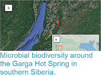
 (a) Eastern Siberia; (b, Eastern Baikal, Barguzin Valley, satellite photo. Red cross marks the location of the Garga hot spring (54°19′3.72″N, 110°59′38.4″E). Rozanov et al. (2017).

Rozanov et al sampled waters originating from the spring at four points. The first of there was close to the spring itself, with waters at temperatures in excess of 74°C, where there was a simple biofilm of white material was present. The second was located in a stream running from the spring towards the Barguzin River, where the waters were about 70°C, and a thicker biofilm with three distinct layers had developed. The upper two layers were yellowish, with the upper being dense and about 2 mm thick and the lower being thinner and about 2 cm. The bottom layer was about 1 cm thick, white, gelatinous and adhered to the substrate. The third point was further downstream, where the temperature had fallen to 55°C. Here the mat still had three layers, with the top layer being yellowish green and about 2 mm thick, the middle layer being membranous, gelatinous and whitish-green and about 2 cm thick, while the bottom layer was again 1 cm thick, gelatinous and white. The final point was further downstream, where the temperature had fallen to 45°C. Here there were again three layers, with the upper two layers being apparently identical to those at the third site, while the bottom layer as thinner, about 3-5 mm, and 'skin coloured'.

Microbial mats of the Garga hot spring. (a) Microbial mat of the upper reaches of the spring; red circle, second sampling point. (b) Microbial mat of the middle reaches; red circle, third sampling point. (c) Layers of the third sample. (d) Sampling scheme. Rozanov et al. (2017).

The sample taken from the first location contained a significant proportion of Archaeans (Prokaryotic micro-organisms resembling Bacteria, but only distantly related to them, and more closely related to Eukaryotes, about 20% of the sample, the highest proportion found at any site in the Baikal Rift Zone to date. Many of these belonged to the Crenarchaeota, a group widely associated with hot springs, with about 7.9% of the sample closely related to Thermopro teusuzonensis, a species isolated from acid hot springs on the Kamchatka Peninsula, and Vulcanis aetasouniana, a species from acidic springs in Japan. However, the sample also contained other, non-Crenarchaeote, Archaeans, with no apparent close relationships to any other described species.

The remaining three sites had essentially similar structures, with an upper layer dominated by Cyanobacteria (filament-forming photosynthetic Bacteria) of the genera Leptolyngbya, Synechococcus and Nostoc, all associated with hot springs in a variety of other locations around the world. The second layer contained lower numbers of Cyanobacteria, though they were still present, along with heterotrophic Bacteria (Bacteria that gain nutrition by consuming other Bacteria), principally Proteobacteria and Actinobacteria. The lowermost layer was anearobic and dominated by saprotrphic Bacteria such as Clostridia, obtain nutrition by breaking down organic material externally.

See also...

http://sciencythoughts.blogspot.co.uk/2017/12/understanding-role-of-biofilms-in.htmlhttp://sciencythoughts.blogspot.co.uk/2017/11/streptomyces-asenjonii-new-species-of.html
http://sciencythoughts.blogspot.co.uk/2017/10/algal-bloom-covers-much-of-western-lake.htmlhttp://sciencythoughts.blogspot.co.uk/2016/05/cuniculiplasma-divulgatum-new-species.html
http://sciencythoughts.blogspot.co.uk/2015/08/nisada-stipitata-new-species-of.htmlhttp://sciencythoughts.blogspot.co.uk/2015/06/microbial-sediments-from-early-to.html
Follow Sciency Thoughts on Facebook.

Sunday, 26 November 2017

Streptomyces asenjonii: A new species of Actinobacteria from the Atacama Desert.

Actinobacteria are Gram-positive, filamentous, aerobic Bacteria found in soils and aquatic ecosystems, where they play a significant role in the decomposition of Plant material and other organic matter, making them highly important in the formation and maintenance of soils. Some species of Actinobacteria are capable of 'fixing' nitrogen from the atmosphere (i.e. taking atmospheric nitrogen and using it to form nitrogen compounds that can be utilised by Plants). Members of the genus Streptomyces have long interested scientists for their ability to produce antibiotics and other potentially useful compounds, and recent discoveries of members of this genus living in extreme environments has opened up the possibility of further interesting discoveries.

In a paper published in the journal Antonie van Leeuwenhoek on 6 June 2017, Michael Goodfellow, Kanungnid Busarakam, and Hamidah Idris, of the School of Biology at Newcastle University, David Labeda of the National Centre for Agricultural Utilization Research, Imen Nouioui, and Roselyn Brown, also of the School of Biology at Newcastle University, Byung-Yong Kim of Seoul National University, Maria del Carmen Montero-Calasanz, again of the School of Biology at Newcastle University, Barbara Andrews of the Centre for Biotechnology and Bioengineering at the University of Chile, and Alan Bull of the School of Biosciences at the University of Kent at Canterbury, describe a new species of Streptomyces from the hyperarid Atacama Desert of Chile.

The species is named Streptomyces asenjonii, in honour of Juan Asenjo of the University of Chile, for his work on the Actinobacteria of the Atacama Desert. The species forms extensive colonies of branching filaments, with spiralling aerial hyphae that produce spores. It was able to grow at temperatures of between 10 and 50 °C and pH levels of between 5 and 11, though it grows best at a temperature of 37 °C and a pH of 7.5. 

Scanning electron micrograph of Streptomyces asenjonii showing hairy ornamented spores in open spirals following growth on oatmeal agar at 28 °C for 14 days. Scale bar is 1 μm. Goodfellow et al. (2017).

See also...

http://sciencythoughts.blogspot.co.uk/2017/05/kuphus-polythalamia-can-giant-free.htmlhttp://sciencythoughts.blogspot.co.uk/2016/12/understanding-worlds-highest-vascular.html
http://sciencythoughts.blogspot.co.uk/2015/06/microbial-sediments-from-early-to.htmlhttp://sciencythoughts.blogspot.co.uk/2015/04/seeking-earths-earliest-fossils.html
http://sciencythoughts.blogspot.co.uk/2015/01/the-fate-of-soil-microbes-during-end.htmlhttp://sciencythoughts.blogspot.co.uk/2014/11/calcifying-endosymbiotic-bacteria-in.html


Follow Sciency Thoughts on Facebook.