Showing posts with label Eukaryotes. Show all posts

Wednesday, 13 August 2025

Human Sleeping Sickness eliminated in Kenya.

The World Health Organization has officially confirmed that Kenya can be declared free of Human Sleeping Sickness (otherwise known as Human African Trypanosomiasis) in a press release issued on 8 August 2025. This is the second Neglected Tropical Disease to have been eliminated in Kenya, following the country being declared free of Guinea Worm, Dracunculus medinensis, in 2018, and makes Kenya the tenth country to be declared free of Human Sleeping Sickness. To date, 57 countries have been confirmed as having eliminated at least one of the seventeen recognised Neglected Tropical Diseases.

Human Sleeping Sickness is caused by a Protozoan, Trypanosoma brucei, which is an extracellular parasite infecting the blood plasma and other bodily fluids of its victims (unlike other parasitic Protozoans, such as the Malaria parasite Plasmodium spp., which infect the victim's cells). Trypanosoma brucei is a zoonotic infection, which is to say infection that affects Animals as well as Humans, and is typically carried by an Animal vector. This can create a reservoir of potential infectious agents in an Animal population, making such diseases difficult to eliminate. 

There are two subspecies of Trypanosoma brucei which infect humans, Trypanosoma brucei gambiense, which is found in West Africa, and Trypanosoma brucei rhodesiense, which is found in East and Southern Africa (and which was the form formerly found in Kenya). A third form, Trypanosoma brucei brucei, does not infect Humans, but can infect domestic Animals. All three known forms of Trypanosoma brucei infect a variety of Mammals (it is possible that other subspecies exist, but infect neither Humans nor domestic Animals, leading to their being overlooked), and are transferred from one host to another by the bite of the Tsetse Fly, Glossina spp.. Because of this, Humans involved in professions where they work closely with Animals, such as Animal husbandry or hunting, are particularly at risk of infection.

A smear of blood from a patient with Human Sleeping Sickness, stained with Giemsa (a histological stain which binds to areas of DNA with high levels of adenine-thymine bonding, making it useful for identifying parasitic organisms in blood), revealing two Trypanosoma brucei ssp. parasites. Centers for Disease Control and Prevention.

Because Trypanosoma brucei infections are not restricted to cells, the parasite is able to cross the blood-brain barrier with greater ease than most parasitic infections. The parasite breeds by binary fission, enabling its population within a host to increase exponentially. Once the population within the bloodstream become to high, the parasites begin to migrate within the body, frequently entering the cerebrospinal fluid and then the brain, where it caused Human Sleeping Sickness. As an Eukaryotic infection, Trypanosoma brucei is not vulnerable to antibiotics, and is typically treated with a form of chemotherapy which is also hazardous for the patient. As such, prevention of the disease is greatly preferable to treatment.

Human Sleeping Sickness was first recorded in Kenya in the early twentieth century, and has been the subject of strenuous control efforts ever since. A declaration of elimination for a disease is made at least ten years after the least recorded transmission of that disease within a country. In Kenya, the last reported case where the patient is believed to have contracted the disease within the country occurred in 2009, while the most recent reports of patients who are believed to have acquired the infection while out of the country (two patients) occurred in 2012. Despite this apparent success, Kenya has recently strengthened monitoring for Human Sleeping Sickness in counties where the disease was formerly endemic.

See also...

Monday, 6 May 2024

Multicellular Eukaryotic fossils from the Mesoproterozoic Chuanlinggou Formation of North China.

All life found on Earth today is made up of cells, with the vast majority of organisms still being unicellular; it is generally presumed that the earliest forms of life would have been single-celled Prokaryotes (organisms with cells which lack internal divisions and organelles).  Multicellularity has arisen numerous times within both Prokaryotic and Eukaryotic groups, although complex multicellularity, with cells differentiated into specialist forms and organised communication between cells, has only arisen six or seven times, and only in Eukaryotes.

The earliest widely accepted multicellular Eukaryotic fossils, filaments and spherical groups of cells, appear around the Mesoproterozoic-Neoproterozoic boundary, while filamentous Prokaryotes are known from the Archaean. Early multicellular Eukaryotes include Bangiomorpha pubescens, a putative Red Alga from 1050 million-year-old deposits in the Canadian Arctic, Eosolena loculosa, a Eukaryote of uncertain affinities from 1030 million-year-old deposits in Siberia, Arctacellularia tetragonala, another species of uncertain affinities from 1000 million-year-old deposits in the Democratic Republic of Congo, Proterocladus antiquus, a possible Green Alga from 950 million-year-old deposits in North China, Archaeochaeta guncho, another species of uncertain affinities from 950 million-year-old deposits in northwestern Canada, and Ourasphaira giraldae, a possible Fungi from 890 million year old deposits in the Canadian Arctic. 

Some putative multicellular Eukaryotes have also been recorded from earlier in the Mesoproterozoic, including Eosolena minuta, from 1500 million-year-old deposits in northern Siberia, or the carbonacious impressions of the Gaoyuzhuang Formation in North China, which can reach tens of centimetres across, or the possible Eukaryotic microfossils from the 1600 million-year-old Tirohan Dolomite of central India. The oldest examples of the coilled microfossil Grypania are currently dated to about 2100 million years before the present (i.e. Late Palaeoproterozoic) although it is debated whether this is a Eukaryote or a Cyanobacterium. Of similar age are the pyritic macrostructures of the Francevillian Biota of Gabon, though there is some debate as to whether there are of biological origins at all.

In 1989, micropalaeontologist Yan Yuzhong published a description of a filamentous Eukaryotic fossil from the 1630 million-year-old Chuanlinggou Formation of North China in the Bulletin of the Tianjin Institute of Geology and Mineral Resources. At this time journals were only available in print, and the Bulletin, which was printed in Chinese, had almost no circulation outside of China. Furthermore, the quality of the images in Yan's paper were rather poor, leading to the publication being largely overlooked.

In a paper published in the journal Science Advances on 24 January 2024, Lanyun Miao and Zongjun Yin of the State Key Laboratory of Palaeobiology and Stratigraphy at the Nanjing Institute of Geology and PalaeontologyAndrew Knoll of the Department of Organismic and Evolutionary Biology at Harvard University, Yuangao Qu of the Institute of Deep-sea Science and Engineering of the Chinese Academy of Sciences, and Maoyan Zhu, again of the State Key Laboratory of Palaeobiology and Stratigraphy at the Nanjing Institute of Geology and Palaeontology, and of the College of Earth and Planetary Sciences of the University of the Chinese Academy of Sciences, re-examine the Chuanlinggou Formation fossils, and discuss the implications of these for the origin of multicellular Eukaryotic life.

Samples of grey shale were collected from the Wengjiazhuang Section of the Chuanlinggou Formation in Kuancheng County of Hebei Province, which has been dated to 1634.8 million years before the present (±6.9 million years) using uranium/lead ratios in zircons. Zircons are minerals formed by the crystallisation of cooling igneous (or in this case, impact) melts. When they form, they often contain trace amounts of uranium, which decays into (amongst other things) lead at a known rate. Since lead will not have been present in the original crystal, it is possible to calculate the age of a zircon crystal from the ratio between these elements. Microfossils were then extracted from these shales by acid maceration.

This technique recovered flattened greyish or pale brown filamentous fossils, which Miao et al. describe as Qingshania magnifica, the name used by Yan Yuzhong for his material. These are not the only filamentous fossils derived from the Chuanlinggou Formation shales, but are significantly larger than other forms, supporting the idea that they are Eukaryotic in origin, while the other forms are Prokaryotic, probably Cyanobacteria. The 278 individual specimens Miao et al. identified ranged from 20 to 194 μm in diameter, with a maximum length of 860 μm. The filaments were straight or curved, and made up of smooth-walled cells, more than 20 of which were present in the longest specimens. These cells are generally cylindrical in shape, with a cell length of 15 to 190 μm. Terminal cells, where preserved, are hemispherical. None of the specimens had any form of external sheath or holdfast.

Transmitted-light photomicrographs of Qingshania magnifica from the Chuanlinggou Formation. (A) to (D) and (K) Filaments with cells of varying length and width. (E) Four-celled filament with hemispherical terminal cell. (F) and (G) Filament with notably decreasing cell width toward one end. Note that (F) and (G) represent the same specimen; (F) lost the narrowest part of the filament as shown in (G). (H to J) Filaments displaying more uniformity of cell dimensions. (L) Two-celled filament with ovoid terminal cell. All specimens were handpicked from organic residues of acid maceration and photographed in wet mounts, except for (K), which was photographed from a permanent strew mount. Solid and empty gray triangles in (A), (C), and (K) indicate the longest and the shortest cells, respectively, within single filaments. tb, transverse band (interpreted as cross wall); tr, transverse ring (interpreted as partially preserved cross wall). Scale bars 50 μm (A) to (E), (I), (J), and (L) and 100 μm (F) to (H) and (K). Miao et al. (2024).

The specimens show considerable variation, with the largest being ten times as wide as the smallest, individual cells being cylindrical, barrel-shaped, or cup-shaped, and filaments being of even width or tapering towards one end. Despite this variation, Miao et al. treat them all as a single species, suggesting that the variations reflect s different growth or developmental stages within the population.

Micrographs of Qingshania magnifica from the Chuanlinggou Formation. (A) Transmitted-light photomicrograph of a five-celled filament with constant width and dark narrow transverse bands. (B) Scanning electron microscope image of (A) showing surface features and the preservation as a complete compression. Note the obliquely compressed cross wall of the right terminal cell showing smooth surface and no other particular features. (C) to (E) Magnifications of (B), showing smooth wall surface and the well-defined contact between adjoining cells manifested by a very shallow groove (marked by cyan arrowheads) along transverse bands. (C) and (E) represent dashed boxes in (A) and (B); (D) corresponds to the dashed box in (C). Scale bars, 50 μm (A) and (B), 10 μm (C), and 2 μm (D) and (E). Miao et al. (2024).

Some of the filaments have small, round-to-ovoid structures within some of their cells. These structures are faint, but always contained entirely within the cell, making it unlikely that they are separate structures superimposed upon the filaments. Inclusions within cells, from both Proterozoic and Phanerozoic settings, have variously been interpreted as endocysts, collapsed cytoplasm, or organelles. The fossils are interpreted as being compressed cell walls, which makes it likely that structures withing them would be endocysts. A variety of Eukaryotic and Prokaryotic groups produce endospores in response to worsening conditions (such as the end of a growing season), but these tend to have protective envelopes thicker than the outer cell wall, which is not the case with these structures. However, the structures are found only in larger cells, and are only slightly smaller than the smallest cells, which suggests that they may be some form of asexual reproductive spore; similar spores are produced by some extant filamentous Algae, such as Urospora wormskioldii.

Transmitted-light photomicrographs of Qingshania magnifica with a small round or ovoid inclusion from the Chuanlinggou Formation. (A), (C), and (D) Filaments with constant width. (B) and (E) Magnifications of dashed boxes in (A) and (C), respectively, showing details of round inclusions. (F) Filament of notably varying width. Note that the middle cell of the filament is cyathiform in shape. (G and H) Magnifications of dashed box in (F) and (D), respectively. All specimens were handpicked from organic residues of acid maceration and photographed in wet mounts. Scale bar, 50 μm (A), (C), (D), and (F). Miao et al. (2024).

Microscale Raman and Fourier transform infrared spectroscopic investigations of the composition of the filaments suggested that the cell walls of Qingshania magnifica were composed largely of aromatic compounds, with a lower proportion of aliphatic compounds, with the aliphatic compounds forming long chains with little branching. This is not sufficient to make any  assessment of the taxonomic status of Qingshania magnifica on its own, but is quite distinct from the composition of Cyanobacterial cells found in the same deposits.

The original specimens of Qingshania magnifica described by Yan in 1989 were identified from thin sections of yellowish-green shales, and had a maximum width of about 250 μm and were up to 6000 μm in length. Yan identified these as Green Algae, placing them in the modern family Ulotrichaceae. Miao et al.'s specimens are slightly smaller, but preserve more detail, allowing for a more detailed reconstruction.  They interpret Qingshania magnifica as a simple multicellular organism with large cells and a degree of morphological variation, with a life cycle that involved spores produced within cells, which then produced thin filaments, which grew into thicker filaments, which were capable of producing more spores.

A wide range of both Prokaryotic and Eukaryotic organisms produce filaments of cells today. Among Prokaryotes, these include at least eleven phyla of Bacteria and one of Archaeans. The most sophisticated filamentous Prokaryotes are Cyanobacteria, which produce a range of forms including straight, tapering, and branching filaments. However, no known Cyanobacterium, or other Prokaryote, living or fossil, closely resembles Qingshania magnifica. Filamentous Eukaryotes include Algae such as Archaeplastids (the group that includes both Red and Green Algae) and Ochrophytes (the group that includes Brown Algae, Golden Algae, and Diatoms), as well as filamentous Fungi and Oomycetes (Water Molds). The cells of Qingshania magnifica are completely surrounded by cell walls, which suggests that each cell acquires its own nutrition, by either photosynthesis or osmotrophy (absorbing nutrients from the environment). This is also quite different from the hyphal structure seen in Fungi and Oomycotes, even the septate forms. making it unlikely that Qingshania magnifica could be assigned to either of these groups. Furthermore, molecular clock estimates suggest that Fungi did not appear till about 1000 million years ago, and Oomycotes probably around the dawn of the Cambrian.

Based upon this analysis, Miao et al. conclude that Qingshania magnifica is most likely to have been a Eukaryotic Algae. This is consistent with molecular clock analyses, which suggest plastids (chloroplasts) were first acquired by unicellular Algae during the Palaeoproterozoic. The morphology of Qingshania magnifica is also consistent with younger fossils interpretted as Green Algae, as well as several modern members of that group. However, Miao et al. do no conclude there is sufficient evidence to confidently place Qingshania magnifica within the Green Algae, as originally proposed by Yan, instead concluding that it could be a Green Algae, a Red Algae, a stem group Archaeplastid, or even a member of an entirely extinct Eukaryotic group. Whichever of these is true, Qingshania magnifica provides strong support for a Late Palaeoproterozoic appearance of the crown group Eukaryotes, rather than a Late Mesoproterozoic one, which has sometimes been proposed. 

Overview of early evolution of the Eukarya along with fossil records. (A) Simplified Eukaryotic tree with divergence time estimates of major branches by molecular clock study. LECA, Last Eukaryotic Common Ancestor. Dashed grey lines represent hypothetical stem-group Eukaryotes, which are extinct. Abbreviation: Pha., Phanerozoic. (B) Representative fossil records of early Eukaryotes. The oldest unambiguous Eukaryotic fossils are unicellular forms, e.g., Tappania plana and Shuiyousphaeridium macroreticulatum from the approximately 1650 million-year-old Ruyang Group; Dictyosphaera macroreticulata, Germinosphaera alveolata, and Valeria lophostriata from the Changzhougou Formation and lowermost Chuanlinggou Formation of North China. The Qingshania magnifica represents the current oldest convincing multicellular Eukaryote from the approximately 1635 million-year-old upper Chuanlinggou Formation of North China. The oldest Red Alga is Bangiomorpha pubescens from the approximately 1050 million-year-old Hunting Formation, Canada. The oldest Green Alga is Proterocladus antiquus from the approximately 950 million-year-old Nanfen Formation of North China. The oldest putative Fungus is Ourasphaira giraldae from the approximately 890 million-year-old Grassy Bay Formation of Canada. The oldest Amoebozoans are vase-shaped microfossils, e.g., Cycliocyrillium torquata from the approximately 750 to 730 million-year-old Kwagunt Formation, part of the Chuar Group of Arizona. Scale bars, 500 μm for the oldest Green Alga and 50 μm for all other specimens. Miao et al. (2024).

See also...

Friday, 11 November 2022

Halamphora minima: A new species of Diatom from the mudflats of Hampyeong Bay, Korea.

Diatoms are single celled algae related to Kelp and Water Moulds. They are encased in silica shells with two valves. During reproduction the cells divide in two, each of which retains one valve of the shell, growing a new opposing valve, which is slightly smaller and fits flush within the older valve. This means that the Diatoms grow smaller with each new generation, until they reach a minimum size, when they undergo a phase of sexual reproduction, giving rise to a new generation of full-sized cells. The genus Halamphora was first described in 1895 as a subgenus of Amphora, but was elevated to a genus in its own right in 2009. It currently contains 154 species, with a global distribution, predominantly in marine and brackish water environments. Fifteen of these species have been recorded from Korea to date.

In a paper published in the journal Phytotaxa on 8 November 2022, Sung Min An of the Department of Microbial Resources at the National Marine Biodiversity Institute of Korea, Jihoon Kim of the Department of Biodiversity, also at National Marine Biodiversity Institute of Korea, and Nam Seon Kang, Kichul Cho, Jung Lee, and Fun Song Kim, also of the Department of Microbial Resources at the National Marine Biodiversity Institute of Korea, describe a new species of Halamphora from Hampyeong Bay, on the west coast of South Korea.

Samples were collected from an intertidal mudflat in Hampyeong Bay in July 2018, and cultured at the National Marine Biodiversity Institute of Korea. The recovered cells were then examined under light and scanning electron microscopes, with DNA being extracted to determine whether these Diatoms were a new species, and how they were related to other species within the group. The new species is named Halamphora minima, in reference to its small size in comparison to other members of the genus.

Map of sampling localities in Hampyeong Bay, the west coast of Korea. Type locality of Halamphora minima (35º03’41.94’’ N, 126º24’40.06’’ E). An et al. (2022).

The valves are only 5.9-7.4 μm in length and 2.4-3-3 μm in width, whereas most members of the genus exceed 15 μm in length, and the largest can exceed 80 μm. The tests of Halamphora minima have a deeply convex dorsal margin and a nearly straight ventral margin. The ventral side is expanded, and the valves on this size sealed by a series of striae. The raphe (a slit in the ventral edge of the shell) is straight on the and well developed. There is no axial longitudinal line on the dorsal edge. The surfaces of the shell are covered by a series of striae (lines) single on the ventral surface and double on the dorsal surface.

SEM micrographs of Halamphora minima. (11) External whole valve view, with central area (asterisk), and dorsal raphe ledge (arrow). (12) Internal whole valve view. (13) Detail of external valve apex,with dorsal raphe ledge (arrow). (14) Detail of internal valve apex showing tongue-like proximal helictoglossae (double asterisk), poorly developed distal helictoglossae (arrowhead), and internal longitudinal rib (arrow). (15) Detail of internal dorsal areolae occluded by hymens (arrows). (16) Dorsal girdle bands with two rows of poroids. (17) frustules showing the ventral girdle bands with dorsal raphe ledge (arrow). Scale bars: (11)-(14), (16), (17) 1 μm, (15) 0.5 μm. An et al. (2022).

See also...


Follow Sciency Thoughts on Facebook.

Follow Sciency Thoughts on Twitter.


Thursday, 24 December 2020

Looking for the origin of the Eukaryotes.

The Eukaryotic cell is strikingly distinct from its much simpler Prokaryote relatives, possessing not only a nucleus, but also a complex cytoskeleton, a sophisticated endomembrane system, and mitochondria, the last of these the result of an ancient endosymbiosis with a Proteobacterium. How this complex cell evolved has long been a puzzle, in part because of the lack of living intermediate taxa that would help determine the sequence in which these distinctive Eukaryotic characters appeared. The recent discovery of the Asgard Archaea, a group closely related to eukaryotes that possesses homologues of several Eukaryote genes, has heightened interest in this problem, as they represent, in a sense, the transitional forms that have long been sought. Nonetheless, there will always be evolutionary transitions that cannot be inferred using modern taxa, as is well illustrated by the Birds, whose evolutionary history would be a mystery without the rich fossil record of the Dinosaurs. The fossil record of early Eukaryotes traditionally has been dismissed as useless for reconstructing Eukaryogenesis, in part because the important transitions involve characters which are typically not preserved (e.g. the nucleus, mitochondria, cytoskeleton), and in part because the prevailing view of early Eukaryote evolution assumes these transitions took place long before the first Eukaryotes show up in the fossil record. 

In a paper published in the journal Interface Focus on 30 March 2020, Susannah Porter of the Department of Earth Science at the University of California at Santa Barbara argues that there are fossil proxies that could be used for inferring the relative order in which some Eukaryotic characters evolved, and focus on four in particular: (i) excystment structures (medial splits or pylomes, i.e. circular openings in the cyst wall) which imply the capacity to form cysts; (ii) spines and pylomes, which require the cell to be able to change its shape and thus imply a complex cytoskeleton; (iii) sterane biomarkers which imply sterol synthesis; and (iv) evidence of Eukaryotes living in oxic habitats, which implies aerobic respiration, and thus the possession of mitochondria. Contrary to widespread assumptions, current evidence allows the possibility that some of these characters evolved significantly later than the first recognisable Eukaryotic fossils appeared, potentially enabling us to identify their time of appearance in the fossil record.

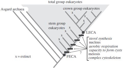
Before discussing the early Eukaryote fossil record, it is useful to review some common terms. Crown Group Eukaryotes are defined as the last common ancestor of all living Eukaryotes plus all of its descendants, both extinct and extant. This last common ancestor is commonly referred to as the Last Eukaryotic Common Ancestor. Stem Group Eukaryotes are those lineages that diverged prior to the appearance of the Last Eukaryotic Common Ancestor, but after the split with the group’s closest living relatives, a clade of Asgard Archaea in the case of Eukaryotes. Stem Groups are, by definition, extinct, and possess some, but not all, of the characters that define the Crown Group. Stem and crown groups together make up the Total Group. The term First Eukaryotic Common Ancestor, is often used to refer to the initial lineage of Total Group Eukaryotes, just after its split from its closest living relative. Eukaryogenesis refers to the interval between the First Eukaryotic Common Ancestor and the Last Eukaryotic Common Ancestor, when the characters that define the Crown Group evolved.

 
Diagram illustrating important terms and the evolution of selected characters that define modern Eukaryotes. Characters in italics are the focus of this paper. Note that the evolutionary sequence of characters that evolved after the First Eukaryotic Common Ancestor and before the Last Eukaryotic Common Ancestor cannot be resolved without the fossil record. Porter (2020).

The oldest widely accepted evidence of Eukaryotes is large (greater than 100 μm), spiny, ornamented, organic-walled microfossils found in latest Palaeoproterozoic rocks (about 1650 million years old). Throughout the Mesoproterozoic, organic-walled microfossil assemblages include a variety of Eukaryotic fossils, but it is not until the latest Mesoproterozoic and early Neoproterozoic that there is evidence for modern (i.e. crown group) Eukaryotes. This includes a handful of fossils plausibly assigned to various modern Eukaryotic supergroups, the first appearance of Eukaryotic steranes, and evidence of other Crown Group innovations like tests and biomineralised scales.

The most common explanation for these patterns is that while Crown Group Eukaryotes appeared more than 1.65 nillion years ago, they remained minor components of Mesoproterozoic ecosystems until their dramatic diversification in the early Neoproterozoic. Evidence offered in support of this view includes the absence of steranes from Mesoproterozoic rocks, which indicates that Eukaryotes must have been severely limited in abundance, and widespread ocean anoxia during this time, which means that early Eukaryotes, which require oxygen for aerobic respiration, must have been spatially restricted to the few habitats where sufficient oxygen was present. Porter evaluates the evidence for early crown group Eukaryotes and suggest instead that the data allow another end-member possibility: crown group Eukaryotes did not appear until the late Mesoproterozoic, and early Mesoproterozoic Eukaryotes had not yet acquired the capacity for aerobic respiration or sterol synthesis.

There are two end-member models of early Eukaryote evolution, which differ with respect to the age of the Last Eukaryotic Common Ancestor. If the Last Eukaryotic Common Ancestor appeared early (e.g. 1600–1800 million years ago), then Crown Group Eukaryotes must have inhabited Mesoproterozoic seas and are probably well represented among Mesoproterozoic Eukaryote fossil assemblages. Crown Group Eukaryotes would have been capable of aerobic respiration, and therefore would have lived in oxygenated environments (at least some of the time), and would have had the capacity to synthesize sterols. If, however, the Last Eukaryotic Common Ancestor appeared near the end of the Mesoproterozoic, then, depending on when in stem group evolution these characters evolved, it is possible that all early Mesoproterozoic Eukaryotes were anaerobic, unable produce sterols, or both. These different scenarios have different implications for our ability to trace Eukaryogenesis in the fossil record. If the Last Eukaryotic Common Ancestor appeared early, before the first recognisably Eukaryotic fossils, then those characters must have appeared in taxa that either did not leave a fossil record or are not recognisably Eukaryotic, making it difficult if not impossible to know the order in which they evolved. If, however, the Last Eukaryotic Common Ancestor appeared much later than the oldest recognisably Eukaryotic fossils, then it might be possible to track the appearance of different Crown Group characters through the Mesoproterozoic.

 
Models of early Eukaryote evolution. If the Last Eukaryotic Common Ancestor appears early, then Eukaryotes with cysts, a complex cytoskeleton, sterols and the capacity for aerobic respiration must have lived during Mesoproterozoic time. In this model, the lack of detectable steranes during this time would reflect limited abundance of Eukaryotes, particularly Crown Eukaryotes. Eukaryogenesis (orange box) would have preceded the record of recognisably Eukaryotic fossils (indicated by grey box). If the Last Eukaryotic Common Ancestor appeared late, then Mesoproterozoic rocks dominantly preserve Stem Group Eukaryotes, many of which may be anaerobic or lacked the ability to synthesise sterols, or both. Stages in Eukaryogenesis may be observable, given overlap with the fossil record. Porter (2020).

So, when did the Last Eukaryotic Common Ancestor appear? Unfortunately, molecular clock estimates for the age of the Last Eukaryotic Common Ancestor span a range of over 1 billion years, making it difficult to have much confidence in any particular estimate. Several arguments, however, favour a younger age for the Last Eukaryotic Common Ancestor. First, older ages for the Last Eukaryotic Common Ancestor seem to be driven primarily by the inclusion of Bangiomorpha pubescens, a presumed Bangiophyte Red Algal fossil. Many of these analyses used a now-outdated age for Bangiomorpha pubescens (1198 million years versus the recent report of 1.047 billion year), and this presumably accounts for some of the discrepancy. In addition, it is also possible that Bangiomorpha pubescens is neither a Bangiophyte nor even a Crown Group Red Alga. Other more deeply diverging Red Algal species exhibit similar filamentous forms, similar multicellular holdfasts and packets of spores that form in a similar way to the wedge-shaped cells of Bangiomorpha pubescens, suggesting these characters could have evolved multiple times early in Rhodophyte evolution. Bangiomorpha pubescens is therefore perhaps better interpreted as a part of Total Group Red Algae, as it could represent part of the Stem Group, rather than the Crown Group, of this clade. Notably, the analysis by Diana Chernikova, Sam Motamedi, Miklós Csürös, Eugene Koonin, and Igor Rogozin, assigned Bangiomorpha pubescens to Total Group Red Algae (using it to calibrate the split between Red and Green Algae), and yielded age estimates for the Last Eukaryotic Common Ancestor that were much less offset from those using Phanerozoic calibrations only (e.g. 1200–1400 million years with Bangiomorpha pubescens versus 1100–1300 million years without Bangiomorpha pubescens). This analysis used the now-outdated calibration age for Bangiomorpha pubescens (1100–1200 million years), suggesting that both a corrected age and more conservative taxonomic assignment for Bangiomorpha pubescens might remove the discrepancy altogether, with Phanerozoic and Proterozoic calibrations aligning in support of a younger (1100–1300 million years ago) age for the Last Eukaryotic Common Ancestor.

 
Selected molecular clock constraints on the initial divergence within crown group eukaryotes (i.e. the age of the Last Eukaryotic Common Ancestor). Estimates that do not use the fossil Bangiomorpha pubescens as a calibration point are 200–300 million years younger than those that do. Porter (2020).

Second, though estimates for the Last Eukaryotic Common Ancestor vary widely, there is consistent agreement among molecular clock analyses that the Eukaryotic supergroups diverged within 300 million years of the Last Eukaryotic Common Ancestor. Thus, if the Last Eukaryotic Common Ancestor appeared early (1600–1800 million years ago), then there should be evidence for Eukaryotic supergroups roughly 1300–1500 million years ago. Though the Mesoproterozoic is not as well sampled as the Neoproterozoic, thus far the picture seems to be that Eukaryotic supergroups diverged in the latest Mesoproterozoic and early Neoproterozoic, with fossils plausibly assigned to the Archaeplastida, Amoebozoa and Opisthokonta, appearing 1050–750 million years ago, along with other innovations widespread among living Eukaryotes (such as biomineralized scale microfossils; 811 million years ago). Thus, if Eukaryotic supergroups diverged 1050–750 million years ago, then these molecular clock analyses are telling us that the Last Eukaryotic Common Ancestor appeared around 1350–1050 million years ago.

Finally, models of phylogenetic diversification suggest it is highly unlikely that a Crown Group would survive at low diversity for approximately half its life (e.g. 800 million years) before it radiated. Rather, clades that survive to the present day (by definition any crown group) tend to start off with higher net rates of diversification relative to other (nonsurviving) clades. This is because clades that happened to have diversified rapidly early in their history are simply more likely to survive to become Crown Groups. It is not easy to persist at low diversity for hundreds of millions of years; there is just too much risk of going extinct. These phylogenetic models also indicate that after the Crown Group emerges, the Stem Group rapidly declines. This again reflects survivorship bias; if they did not decline rapidly, it is unlikely they would have gone extinct, which by definition, they must have, given that they are Stem Groups. Thus, the interval of overlap between Stem and Crown Group Eukaryotes should be short; if the Last Eukaryotic Common Ancestor did appear around 1.6–1.8 billion years ago, there should be few Stem Group Eukaryotes present by late Mesoproterozoic time. These results do not hold in special cases, e.g. when the Crown diversification follows a mass extinction, and, therefore, it is not easy to know how to apply them to the particular case of Eukaryotes. However, there is no obvious evidence for mass extinction in the Mesoproterozoic or early Tonian. Regardless, the null model suggests that once the Last Eukaryotic Common Ancestor had appeared, Crown Eukaryotes should have diversified rapidly. Thus, if Crown Group Eukaryotes diversified in the early Neoproterozoic, then the null model favours a younger (i.e. late Mesoproterozoic) age for the Last Eukaryotic Common Ancestor. Given reasons to favour a younger Last Eukaryotic Common Ancestor, Porter now turns to the fossil record and review the evidence for when important Eukaryogenic characters evolved.

Many Eukaryotes enter a resting stage during times of environmental stress, forming resistant-walled structures known as cysts. Encystation is so widespread, found among all the Eukaryotic supergroups, that it seems likely that the Last Eukaryotic Common Ancestor was also able to encyst, or least possessed a capacity to readily evolve cysts such that cysts evolved again and again within Crown Group Eukaryotes. Cysts are most easily recognised in the fossil record by the presence of excystment structures, pre-programmed openings by which the cells escape. Two types are common in Proterozoic fossils: medial splits, in which the cyst opens along an equatorial line often dividing the cyst into two separate hemispheres; and well-defined (typically circular) openings, known as pylomes. Fossils exhibiting medial splits are common in assemblages throughout the Proterozoic, including those with the oldest recognized Eukaryotes. Possible pylomes are reported in Dictyosphaera macroreticulata from 1744 to 1411 million years ago Ruyang Group, North China, with definite occurrence by 1100 million years ago (Leiosphaeridia kulgunica in the Atar/El Mreïti Group, Mauritania).

 
Examples of fossils and characters. (a), (b) Bangiomorpha pubescens from the late Mesoproterozoic Hunting Formation, Somerset Island, arctic Canada. Note cross-section in (b) showing distinctive wedge-shaped cells. (c), (f ) Excystment structures in Eukaryotic microfossils. (c) Circular pylome with partially detached lid, in Kaibabia gemmulella. (f) Medial split, in Leiosphaeridia crassa. Both (c) and (f) are from the late Tonian Chuar Group, Grand Canyon, USA. (d), (e) Indirect evidence for a complex cystoskeleton in Eukaryotic microfossils. (d) Irregularly distributed hollow processes in Tappania plana. (e) Spiny ornamentation in Dictyosphaera macroreticulata. Both (d) and (e) are from the early Mesoproterozoic Ruyang Group, North China. Porter (2020).

In Eukaryotes, the cytoskeleton is involved in controlling cell shape, cell movement, phagocytosis and intracellular trafficking. Though traditionally considered a diagnostic character of Eukaryotes, the discovery of actin and tubulin homologues in Bacteria and Archaea indicated cytoskeletal building blocks were already present in the First Eukaryotic Common Ancestor, and, in fact, recent discoveries of numerous homologues of cytoskeletal genes in Asgard Archaea suggest that the First Eukaryotic Common Ancestor had a dynamic actin cytoskeleton, perhaps even capable of phagocytosis. Nonetheless, many Eukaryotic cytoskeletal genes do not have homologues in the Archaea, suggesting that Eukaryotes have a much more complex cytoskeleton than their closest Archaeal relatives. It is difficult to know how to translate these genotypic differences into phenotypic differences we might observe in the fossil record, however. In the past, the presence of irregularly distributed, branching processes in Eukaryotic fossils has been used as evidence for a Eukaryotic cytoskeleton (in Tappania plana), but the presence of similar, irregularly distributed, long, sometimes branching protrusions extending from Asgard Archaeal cells recently described from culture suggests this might not be a uniquely Eukaryotic feature. Similarly, it is difficult to know whether complex ornamentation or the presence of spines in a fossil implies a complex (Eukaryotic) cytoskeleton or whether the First Eukaryotic Common Ancestor could have produced these with its more primitive cytoskeleton.

 
Proxy records for four characters of crown group Eukaryotes (coloured circles), and inferred age constraints on their appearances (coloured arrows): (a) cysts, represented by microfossils with excystment structures (medial splits or pylomes); and (b) the presence of a complex cytoskeleton, represented by microfossils with spines or pylomes, which indicate an ability of the cell to change its shape; (c) sterol synthesis, represented by sterane biomarkers; (d ) aerobic respiration, represented by the occurrence of Eukaryotes in oxic habitats (grey circles indicate evidence is ambiguous). Note that, with the exception of steranes, the absence of a proxy record is not taken to imply the absence of the character, age constraints in (a), (b), (d), show hard minimums but no maximums. Note also that age constraints in (a)–(d) do not take into account data from other records, e.g. fossil evidence for crown group Eukaryotes at 1.05 billion years. Porter (2020).

Thus, rather than focusing on proxies for a complex cytoskeleton, it might make sense to focus on features that the Eukaryotic cytoskeleton confers, recognising that some of these might have been present in the First Eukaryotic Common Ancestor. In particular, the ability for a cell to change shape, a key feature of the Eukaryotic cytoskeleton that is linked to the ability to phagocytose,  might be inferred through several fossil proxies. Two have already been mentioned: evidence for an actively growing (and remodelling) cell wall (as in Tappania plana) and the presence of spines in cell walls or cysts, which probably formed via the extension of long, narrow protrusions of the cell membrane. Another proxy might be the presence of pylomes, as it might imply that the cell changed shape, squeezing through the hole to escape the cyst. 

The distribution of sterols across the tree of Eukaryotes suggests that they were present in the Last Eukaryotic Common Ancestor, although it is possible that lateral gene transfer is at least in part responsible for this distribution. Archaea do not appear to have sterol genes, suggesting that the capacity for sterol synthesis appeared during the First Eukaryotic Common Ancestor–Last Eukaryotic Common Ancestor transition. (Note that while some bacteria produce sterols, their sterol synthesis genes are generally thought to have been acquired from Eukaryotes via lateral gene transfer). Thus far, biomarker studies of Mesoproterozoic rocks have failed to turn up convincing evidence of indigenous Eukaryotic steranes; this is the case even in units with diverse Eukaryotic fossils, in facies that would be favourable to sterol-producing Eukaryotes (i.e. oxic, nutrient-rich). Though this has generally been interpreted to indicate either an extremely low abundance of Eukaryotes or preservational bias, it may instead be that the absence of Eukaryotic steranes reflects the fact that sterol synthesis had not yet evolved. This would be consistent with a late appearance of Last Eukaryotic Common Ancestor (i.e. less than 1100 million years ago), but would also be consistent with an earlier appearance if the genes for sterol biosynthesis were transferred via lateral gene transfer among early Crown Group Eukaryotes. (Note that a recent molecular clock analysis concluded that some sterol biosynthesis genes were present in early Eukaryotes about 2.3 billion years ago (1.75–3.05 billion years ago). However, this does not imply that they were forming Eukaryotic sterols but rather might have been forming ‘protosterols’).

Today, mitochondria are essential for Eukaryotic respiration in aerobic habitats. Thus, it is reasonable to assume that evidence for aerobic respiration in early eukaryotes implies the presence of mitochondria in these organisms. (Note, however, that the converse is not true: evidence for an anaerobic lifestyle does not imply that these Eukaryotes lacked mitochondria.) Despite frequent acknowledgement in the literature that late Palaeoproterozoic and Mesoproterozoic Eukaryotic fossils are probably best interpreted as Stem Group Eukaryotes, there is a widespread assumption that nonetheless they must have been aerobic (and therefore possessed mitochondria). In part, this might reflect assumptions about the age of the Last Eukaryotic Common Ancestor, but it might also be based on the idea that aerobic respiration is required for the relatively large size and complexity of late Palaeoproterozoic and Mesoproterozoic Eukaryotic microfossils. Discoveries of anaerobic Eukaryotes over the last decade, however, have shown that this is not the case. Some anaerobic Protists (including free-living forms) can reach hundreds of micrometres in size and possess complex structures, including elaborate attachment sites, complicated cytoskeletons, large nuclei and thousands of flagella. In some cases, there is evidence that large size and complexity evolved within ancestrally anaerobic lineages, indicating that aerobic respiration is neither necessary to sustain large size and complexity, nor required to evolve it. Thus, the fairly large, ornamented Eukaryotic cells found in late Palaeoproterozoic and Mesoproterozoic rocks cannot be assumed to have been aerobic. (Whether their large size and complexity implies the presence of mitochondria per se, however, is more difficult to know).

A better proxy for aerobic respiration in early Eukaryote fossils is evidence that those Eukaryotes lived in oxygenated environments. Currently, the most widely used tool for reconstructing local redox conditions in an ancient water column is iron speciation, which uses the ratio of highly reactive iron versus total iron in a sample as a proxy for water column anoxia. Ratios of 0.22 or less are taken to indicate fully oxygenated water column, whereas highly reactive iron versus total iron ratios of 0.38 or higher suggest water column anoxia (ratios in between are ambiguous). Unless there is reason to think the organisms were infaunal, the presence of Eukaryotic microfossils in oxic samples should generally indicate that those organisms were aerobic. (While Eukaryotic fossils recovered from anoxic samples have traditionally been interpreted to be planktonic Eukaryotes that lived in the oxic surface layer of a stratified water column, it is also possible they were anaerobic and lived lower in the water column or on the seafloor). The link is not clear-cut, however, because both the iron speciation proxy and microfossil assemblages are integrated records, providing information about redox conditions and communities over some interval of time: samples that indicate an oxic water column might have witnessed short episodes of anoxia, and therefore might host fossils of anaerobic Eukaryotes. Conversely, the absence of Eukaryotes from oxic samples does not imply aerobic Eukaryotes did not exist, as they may be missing for other reasons, such as a lack of food, preservational bias or dilution due to high Cyanobacterial fossil abundance. Thus, no single sample should be taken as definitive evidence for (or against) aerobic Eukaryotes. But a consistent pattern across many samples and many units through time could provide compelling evidence for the absence of aerobic respiration.

Unfortunately, there are only a handful of studies in which iron speciation and fossil occurrence data come from the same samples, and the data are too sparse to come to any definitive conclusions about when aerobic Eukaryotes evolved. Nonetheless, unpublished work on the roughly 780–730 million year old Chuar Group (USA) suggests that the proxy may be useful: numerous oxic shale samples from throughout the Galeros Formation contain well-preserved Eukaryotic fossil assemblages that show the same species richness as those from anoxic samples providing good evidence for aerobic Eukaryotes at this time (not surprisingly) and suggesting there is no inherent preservational bias against organisms that lived in fully oxygenated water columns. Further back in time, the data become more difficult to interpret, and the evidence for aerobic Eukaryotes becomes less certain. In a palaeoecological study of the roughly 1100 Ma Atar/El Mreïti Formation of northwest Africa, there are only two fossiliferous oxic samples, only one of which preserves definitive Eukaryote species as minor components (less than 3% of the assemblage). However, the anoxic samples, which are much more numerous, are not dissimilar: although these can record abundances of Eukaryotes up to greater than 25% of the assemblage, the median Eukaryote abundance is nonetheless 0%. An analysis of drill core from the 1400 million year old Kaltasy Formation (Russia) revealed a suite of oxic samples, but definitive eukaryotes are rare or absent. This stands in contrast with more diverse assemblages from unknown redox habitats preserved in roughly coeval units from Australia and the USA. In the roughly 1450 million year old Roper Group, diverse Eukaryotes occur in environments interpreted to have been oxygenated, but iron speciation data are based on an outdated extraction method, and more recent research suggests that marine anoxia characterised the depositional basin of the Roper Group.

We know that First Eukaryotic Common Ancestor was an anaerobic organism, and that the oldest fossilised Eukaryotes lived in oceans that were dominantly anoxic. Thus, the default assumption should be that the earliest Eukaryotes to show up in the fossil record were anaerobic, with the onus on palaeontologists to show this is not true. The very limited data for Mesoproterozoic Eukaryotes discussed by Porter do not provide strong evidence either way: though a few Eukaryotic specimens are reported from oxic samples, the possibility that they reflect brief appearances of anaerobic organisms in anoxic conditions cannot be ruled out. A critical reading of the available data, therefore, would suggest that while aerobic Eukaryotes were present by roughly 800 million years ago, there is no definitive evidence that Eukaryotes occupied oxic habitats during the Mesoproterozoic. Contrary to widespread assumptions about early Eukaryotes, aerobic respiration, and therefore possibly mitochondria,  might have been acquired late in Mesoproterozoic time, and late in stem group evolution.

The four characters can be mapped onto a phylogeny of early Eukaryotes, where the appearance of the Last Eukaryotic Common Ancestor is placed at 1100 million years. The limits of this approach are well illustrated: while the fossil record can provide definitive minimum age constraints on the appearance of these characters, cysts and a complex cytoskeleton by about 1650 million years ago, aerobic respiration (and thus mitochondria) by about 800 million years ago and sterol synthesis by 820 million years ago, it is more difficult to place maximum age constraints on the appearance of these characters. This relies on evidence of absence, which is particularly a problem with the fossil record, where preservational controls are always a concern. However, hypotheses regarding the timing of the origin of these characters remain testable using the rock record. For example, if continued sampling of well-preserved Mesoproterozoic and Palaeoproterozoic rocks consistently yields biomarkers, but never steranes, it may suggest that the strata in question predate sterol biosynthesis. Similarly, the timing of the evolution of aerobic respiration may be constrained by strata that consistently yield well preserved and abundant Eukaryotic body fossils in anoxic habitats but negligible occurrences in oxic habitats. While the absence of evidence in these scenarios does not necessarily provide definitive evidence that Eukaryotes lacked these distinctive characters, the simplest explanation is that these characters had not yet evolved.

The prevailing view of the early eukaryote fossil record is that the Last Eukaryotic Common Ancestor appeared by 1.6–1.8 billion years ago, and that the first Eukaryotes we see had the capacity for sterol synthesis and aerobic respiration. If this is true, then, given what we know about the Archean and Palaeoproterozoic fossil record, we have little hope of reconstructing Eukaryogenesis. But there is good reason to think this is not true. Molecular clock analyses allow the possibility that Last Eukaryotic Common Ancestor arose much later, a scenario favoured by current views that crown group diversification occurred in the latest Mesoproterozoic and Tonian. The consistent lack of detectable steranes in Mesoproterozoic rocks, especially in units that should host them, points to the possibility that Eukaryotic sterol synthesis had not yet evolved. Finally, there is no clear-cut evidence that Eukaryotes lived in oxic habitats during the Mesoproterozoic, permitting the possibility that aerobic respiration, and perhaps mitochondria, which are essential for aerobic respiration,  was acquired late in Eukaryote evolution. Continued studies of body fossil and biomarker assemblages placed within a palaeoenvironmental and redox context may allow us to track Eukaryogenesis in the fossil record, and identify the environmental conditions that allowed the appearance of such complex life.

See also...


Online courses in Palaeontology. 

Follow Sciency Thoughts on Facebook.

Follow Sciency Thoughts on Twitter.


Friday, 10 April 2020

Microfossils from the Palaeoproterozoic Hutuo Group of Shanxi Province, China.

Geological and geochemical evidence has revealed that the Neoarchean–Paleoproterozoic period was vitally important for Earth’s evolution. The earliest ‘snowball event’ and major glaciation occurred during this period. This was followed by a great oxidation event, which caused an abnormal positive shift in global carbon isotopes and is referred to as the Lomagundi Event. The emergence of oxygen-producing photosynthetic organisms that led to the sudden increase in atmospheric oxygen has been the focus of several studies. However, until now, convincing fossil records from this key geological time interval (i.e. latest Neoarchean to Palaeoproterozoic) are scarce. During this period, the biosphere experienced multiple geological events, but little is known about it and what is known is dependent on molecule clock dating analyses and estimates. To better understand the biosphere during this time, the metamorphosed Palaeoproterozoic deposits of the Hutuo Group at Wutai Mountain in Shanxi Province, China provide an excellent stratigraphic sequence in which to study well-preserved fossil records of this key period in Earth’s evolution.

In a paper published in the journal Precambrian Research on 5 February 2020, Leiming Yin and Fanwei Meng of the Nanjing Institute of Geology and Palaeontology, Fanfan Kong of the School of Resource and Earth Science at the China University of Mining and Technology, and Changtai Niu, also of the Nanjing Institute of Geology and Palaeontology, describe the results of a study of the microfossils of the Hutuo Group, and describe a number of these.

The Wutai Mountains are located in the Xinzhou area of Shanxi Province, China, between 38°50′ North and 39°05′ North, and between 113°29′ East and 113°44′ East. The Wutai Mountains of the central Trans-North China Orogen are a typical region for the investigation of Precambriansequences. The Precambrian strata in the Wutai Mountains can be divided into the Neoarchean Wutai Group, and the overlying Palaeoproterozoic Hutuo Group, separated by an unconformity.

Geology of Wutai Mountains showing the sample localities. Yin et al. (2020).

The Hutuo Group is distributed in an area of about 1500 km², from northernmost Taihuai-Sijizhuang on the south slope of Wutai Mountain to southernmost Shizui-Dingxiang, and from the upper Taishan River in the east, to Yuanpingqi village in the west. Although the Hutuo Group underwent a major tectonic movement (the ‘Lulianng Movement’) to show strong fold, which has still completely reserved many primary deposited structures, such as cyclothems (alternating stratigraphic sequences of marine and non-marine sediments), wavemarks, cross-bedding, etc. The Hutuo Group is characterised by thick carbonate and silicified rocks and has been divided into three subgroups. At the base is the Doucun Subgroup, which is dominated by terrigenous clastic sediments. This is overlain by the Dongye Subgroup, which is characterised by claret sandstone or slate in the lower part, and transitions upward into interbedded sandstone and carbonate with Stromatolites. It is dominated by dolomitic carbonate in its upper part. This is unconformably overlain by the Goujiazhai Subgroup, which consists of sandstone and local conglomerates. 

The Hutuo Group was previously determined to be more than 2366 million years old, based zircon Uranium-Lead ages (when zircon forms it often contains trace amounts of uranium, which decays into, amongst other things, lead at a known rate; since lead  will not have been present in the original crystal matrix, it is possible to calculate the age of a zircon from the ratio between these elements) from metamorphosed basic volcanic lava. More recent zircon age estimates made with more sensitie techniques are significanty younger, typically between 2150 and 1950 million years old.

Yin et al. investigated the Wenshan, Hebiancun, and Tianpengnao Formations of the Dongye Subgroup of the Hutuo Group for microfossils. The Wenshan Section is located 17.6 km southwest of Wutai County (38°36′ North and 113°7′ East). It comprises the lowermost Wenshan Formation, which is about 500 m thick and composed of metamorphic slate, and in consideration lithology of the single; 7 rock samples of slate were collected from the upper part of this section. The overlying Hebiancun Formation, which is about 1400 m thick and contains well-developed silicified carbonates containing Stromatolites and chert-like concretions. In order to obtain more possible preservation of fossil material from this deposited sequence, especially those of silicified Stromatolites and chert-like concretions, 59 rock samples were collected from middle-upper part of the Hebiancun Formation. In addition, and 76 phyllite rock samples from the uppermost Tianpengnao Formation of the Dongye Subgroup were collected from the Geziling Section, which is about 4 km east of Wutai County (38°44′ North, 113°17′ East).

Stratigraphic column of the Palaeoproterozoic Hutuo Group and sampling horizons of the Dongye Subgroup. Yin et al. (2020).

Standard palynological maceration was mainly used to obtain organic-walled microfossils from 12 slate samples of the Wenshan Formation and 76 phyllite samples from the Tianpengnao Formation. Samples of 50 g were cleaned and macerated with hydrocholric acid (37%) and hydrofluric acid (40%). Organic residues were either concentrated by heavy liquid with a specific gravity of 2.1–2.2, or poured through a 10 μm nylon mesh, and fixed slices were prepared with Canada balsam for the mounting medium of slices and sealed by paraffin. To obtain more fossil material and checking out possible contamination, which was mainly aimed at the phyllite of the Tianpengnao Formation, repeated macerations were performed. In result, total 14 samples including 2 slate samples of the Wenshan Formation and 12 phyllite samples of the Tianpengnao Formations produced organic-walled microfossils.

Additionally, 5 rock thin sections of phyllite of the Tianpengnao Formation (at least 6 mm in thickness) were cleared in distilled water were etched in dilute 8% hydrofluoric acid for 2–3 min, then cleared with distilled water, which were repeated processing in six times. Such etched rock thin sections were observed under scanning electron microscope to show preserved specimens in situ.

Outcrop photographs of the Tianpengnao Formation in the Geziling section and the Hebiancun Formation in the Wenshan section. (A), (B) Black phyllite or greyish-green phyllite interbedded with carbonates in the lower (A) and upper (B) parts of the Tianpengnao Formation. (C) Siliceous concretions in dolostone of the Hebiancun Formation. Yin et al. (2020).

The Tianpengnao Formation (at least 1.95 billion years old), which is the the uppermost formation of the Dongye Subgroup contains a higher diversity of organic-walled microfossils than the underlying formations. Organic-walled microfossils from the Tianpengnao Formation were mostly obtained through palynological preparation of phyllite. They were strongly carbonized and appeared as opaque vesicles. Individual specimen showed wall folds with a few fine spines under the scanning electron microscope. Similar opaque specimens in situ also found in thin sections. of fuchsia phyllite from the lower part of the Tianpengnao Formation. However, greyish-green phyllite from the upper part of the Tianpengnao Formation yielded much better preserved organic-walled microfossils. 

The microfossils found by Yin et al are all considered to be Acritarchs or Cyanobacteria.

Acritarchs are unicellular Eukaryotic organisms (organisms with cells with a discrete nucleus) that appear in the fossil record from about 3200 million years ago until the end of the Permian, and possibly later (depending on what is classified as an Acritarch). They're affinities are unclear, and the group is probably paraphyletic (not all members sharing a common ancestry), though the majority are thought to have been unicellular planktonic Algae or the resting cysts of other unicellular organisms.

Cyanobacteria are filament-forming photosynthetic Bacteria found across the globe and with a fossil record dating back over 3.5 billion years. They are thought to have been the first organisms on Earth to obtain carbon through photosynthesis, and it is also thought that the chloroplasts (photosynthetic organelles) of eukaryotic plants and algae are descended from Cyanobacteria that lived symbiotically within the cells of ancient eukaryotes. Cyanobacteria are often known as Blue-Green Algae, but this is somewhat misleading, as the term Algae is otherwise restricted to photosynthetic eukaryotes (no other group of photosynthetic Bacteria are referred to as Algae), and because not all Cyanobacteria are blue-green in colour; many are dark green or even black.

The first Acritarch described by Yin et al. is assigned to the genus Dictyosphaera, but not to species level. A single specimen was obtained from a siliceous lens in a dolostone containing Stromatolite from the Hebiancun Formation, at the Wenshan Section. This is a spheroidal vesicle, thin-walled, with very fine net-like ornamentation on its surface, forming a polygonal or subrounded mesh with 3–6 μm in diameter; the vesicle diameter is about 52 μm; no excystment structure was observed.

The Achritarch Dictyosphaera sp., in a in thin sections of siliceous material obtained from the Wenshan Section of the Hebiancun Formation. Scale bar is 10 μm. Yin et al. (2020).

The second Acritarch described is placed on a new species and genus, and given the name Dongyesphaera tenuispina, where 'Dongyesphaera' means 'sphere from Dongye' and 'tenuispina' means 'fine-spined'. Six specimens of this Acritarch were obtained by palynological maceration of material from the upper part of the Tianpengnao Formation. They are spheroidal to sub-spheroidal vesicles, the walls of which are psilate (lacking in ornamentation) and prominently wrinkled; with fairly short, conical processes or protrusions of varying length (0.5–4.2 μm, typically 0.8–1.5 μm), their termination showing as round; vesicles are 30–35 μm in diameter; no excystment opening was observed.

Dongyesphaera tenuispina, obtained from the upper greyish-green phyllite of the Tianpengnao Formation in the Geziling Section by palynological maceration. Scale Bars are 10 μm. Yin et al. (2020).

Acritarchs of the genus Leiosphaeridia were found in both palynological maceration and thin section from phyllite of the Tianpengnao Formation, siliceous lenses in dolostone containing Stromatolites of the Hebiancun Formation, and slate of the Wenshan Formation. spp. were obtained by palynological maceration and thin section. Most are strongly carbonized and even opaque. Some specimens obtained from phyllite of the upper part of the Tianpengnao Formation are less carbonized. None of these are assigned to species level by Yin et al. The specimens are spheroid vesicles, with a circular outline in compressed specimens; the wall surface typically is psilate or with inconspicuous ornament; some specimens show irregular folds; the vesicle diameter is 33–65 μm; no excystment structure was observed.

Leiosphaeridia spp., (A) obtained by palynological maceration from the upper greyish-green phyllite; (C) and (F) obtained by palynological maceration from the lower amaranth phyllite; (J) found in a thin section of the lower amaranth phyllite. Yin et al. (2020).

A single specimen is assigned to the genus Satka. This is a spherical colony-like specimen aggregated with many cell-like spheroids that has been compressed and appears to be enveloped by a thin outer membrane. The included spheroids are deformed and show different shapes and sizes. Single spheroids are 3–7 μm in diameter, and the whole vesicle about 40 μm in diameter. This specimen comes from a greyish-green phyllite of upper part of the Tianpengnao Formation, from the Geziling location.

Satka sp., from the upper greyish-green phyllite. Yin et al. (2020).

The first Cyanobacteria described by Yin et al. are assigned to the species Eoentophysalis belcherensis. These are irregular clusters that contain small and large spheroidal to sub-spheroidal cell-like units singly or in pairs; they are in a crowded arrangement, with a common thin envelope. Cell-like units are typically 1.8–2.5 μm in diameter; irregular clusters are 15–25 μm across and are frequently stuck together. These were foiund in thin sections of siliceous lenses in dolostone containing Stromatolites from the Hebiancun Formation.

Eoentophysalis belcherensis, from the Hebiancun Formation. Scale bar is 10 μm. Yin et al. (2020),

Seven specimens found in thin sections of two samples from siliceous concretions in dolostone of the Hebiancun Formation are assigend to a new species of Eoentophysalis; which is given the specific name hutuoensis, meaning 'from Hutuo', in reference to the Hutuo River in the Wutaishan area of Shanxi Province, China. Eoentophysalis hutuoensis comprises cell-like units spheroidal, ellipsoidal or deformed by compression, and 2.5–12 μm in diameter; mostly single, a few in possible pairs and irregular clusters; characteristically crowded in colonies which are enveloped by a thick sheath-like material. Therse are several hundred cell-like units arranged in clusters or extensive colonies, that are enveloped by an opaque sheath-like material (about 5–8 μm thick) and characterized by suborbicular holes (at least 1 μm in diameter); nearly all cell-like units are single, and more distinct in laser scanning confocal microscope images. Some of show ‘lining structure’ (or remains of plasmolysis). Clusters or colonies 78–126 μm across.

Eoentophysalis hutuoensis under laser scanning confocal microscope. Yin et al. (2020).

The third Cyanobacterium described by Yin et al. is assigned to the species Sphaerophycus medium. A single specimen from a siliceous lens in a dolostone containing Stromatolite of the Hebiancun Formation comprises an irregular clump of cells about 135 μm long and 86.5 μm wide; the cells generally not mutually deformed. Individual cells are spheroidal and ellipsoidal and 4.5–12.5 μm in diameter; cell walls are about 0.5 μm thick.

Sphaerophycus medium, from a siliceous lens in a dolostone containing Stromatolite of the Hebiancun Formation. Yin et al. (2020).

The fourth Cyanobacterium described is placed in the genus Pseudodendron, but not assigned to species level. This comprises a single compressed organic carbon specimen that has two short branches connected with a single, main ‘tube-like’ filament along two sides; the surface is nearly psilate and no preserved cellularity or outer enveloped material is observed. The ends of both branches and the main filament are truncated. The main filament 140 μm long, and 5–8 μm wide; the branches 20–48 μm long and 4–4.5 μm wide. Since only one incomplete specimen was found, its placement in Pseudodendron is tentative. The specimen was observed in a thin sections of siliceous lens from a dolostone containing Stromatolite of the Hebiancun Formation from the Wenshan Section.

Pseudodendron sp., from a thin sections of siliceous lens from a dolostone containing Stromatolite of the Hebiancun Formation from the Wenshan Section. Yin et al. (2020).

The final Cyanobacteria described is Siphonophycus kestron. Many specimens of this were found in thin sections of dolostone containing Stromatolites of the Hebiancun Formation. They are single filamentous microfossils, unbranched, nonseptate, surface smooth; only preserved as carbon membrane-like remains due to degradation; and 5–8 μm across.

Specimen of Siphonophycus kestron, from a thin section of dolostone containing Stromatolite of the Hebiancun Formation. Yin et al. (2020).

The Paleoproterozoic (2.5-1.6 billion years ago) was a critical period in  Earth’s evolution. During it, important global events, such as glaciation, atmospheric oxygenation over the early period, and the following Lomagundi-Jatuli isotopic event occurred. 

The Hutuo Group, as a typical Palaeoproterozoic sequence in the North China Craton, is distributed through the Wutai and Luliang mountains in Shanxi Province, China. The lithological character of the group is characterized by metamorphic deposits and volcanic rocks. Based on recent uranum-lead isotopic age dating, the Hutuo Group is constrained to the period between 2.14 and 1.95 billion years ago. Carbon isotope excursions in the Hutuo Group have been documented as a response to the Palaeoproterozoic global glaciation. Glaciogenic diamictite (a type of lithified sedimentary rock that consists of nonsorted to poorly sorted terrigenous sediment) has also been discovered in the Shijiazhuang Formation of the Hutuo Group at Wutai Mountain, North China which suggests a locally protracted glacial event could have extended to the Wutaishan area. Based on the uranium-lead isotopic ages and chronological framework of the Hutuo Group, a three-stage evolution in the Carbon¹³ isotope curve has been recognized in carbonates of the Hutuo Formation. The lower to middle part of the Dongye Subgroup shows oscillating positive and negative Carbon¹³ values that range between −5.2 and +2.7 parts per thousand relative to the Pee Dee Belemnite Standard (an increase in the proportion of Carbon¹³ relative to Carbon¹² is often indicative of an increase in photosynthesis, as photosynthetic organisms preferentially extract Carbon¹² from the atmosphere, whereas carbonate forming ones incorporate both in in a proportion reflecting atmosphere composition). 

The microfossils from the Wenshan and Hebiancun Formations would be the fossil records of the geological period manifested by the aftermath of a positive excursion of Carbon¹³ (the Lomagundi-Jatuli isotopic event). The transition from an abnormally high organic carbon burial rate to massive oxidation of organic matter. The microfossils from the uppermost part of the Dongye Subgroup, (i.e. the Tianpengnao Formation), would represent the remains of Microphytoplankton during the geological period characterized by fluctuating Carbon¹³ levels.

The Palaeoproterozoic Hutuo Group was deposited in supra-tidal to sub-tidal environments. Furthermore, the Dongye Subgroup was followed by a remarkable transition of geochemical environments. In such palaeoenvironments, phosphates deposited on the continental margin of North China during late early Palaeoproterozoic. In the middle of the Hebiancun Formation, phosphatic deposits developed in association with dolomitic carbonates and a few terrigenous clasts in the studied Wenshan section. Several phosphatized specimens of spheroidal and filamentous Cyanobacteria and Leiosphaeridia-like forms were found in the phosphatic horizon. Typically, these specimens are poorly preserved, possibly due to late oxidation. Some specimens of Eoentophysalis hutuoensis were preserved as compressed carbon membranes. Such well-preserved Palaeoproterozoic microfossils, especially Eoentophysalis as multicellular colonies, have rarely been reported before. To understand their detailed morphological structure and elemental composition, scanning electron microscope associated with energy spectrum test was used for individual specimen of Eoentophysalis hutuoensis. Many sub-spherical individual cells were embedded within or enveloped by a Carbon membrane. The main elements detected were Carbon, Silicon, Calcium and Magnesium. This could suggest that Cyanobacteria colonies were primitively buried in the dolomitic carbonate and silicification during diagenesis resulted in their preservation. Common early diagenetic silicification was observed in the carbonates as distinct chert layers or concretions intercalated within dolostones of the Hebiancun Formation.

Eoentophysalis belcherensis, from the Hebiancun Formation. Scale bar is 10 μm. Yin et al. (2020),

A few degraded coccoid and filamentous cyanobacteria microfossils have previously been reported from the Hebiancun Formation. Some poorly preserved organic-walled microfossils obtained by palynological maceration have been described from the Doucun Subgroup. Some of those specimens that showed triangular, polygonal and boat-shaped forms that were plausibly interpreted as being like Eukaryotic Protists. In morphological feature discrimination, those specimens probably resulted from taphonomic alteration or were contaminants. Eukaryotic microfossils, except multicellular forms, are normally characterized by a Eukaryotic cytoskeleton and endomembrane system, morphogenetic characters like a multilayered wall, distinct surface ornamentation, and excystment by partial rupture or a circular opening. The oldest fossil evidence for Eukaryotic Protists (e.g. Tappania and other ornamented forms) have been documented from about 1.41 billion years ago elsewhere in the world. The new genus Dongyesphaera described by Yin et al. from the Tianpengnao Formation has a distinct fine spinous ornament on the vesicle wall, which would be recognized as eukaryotic protist. Additionally, a specimen identified as Dictyosphaera sp. was found in the Hebiancun Formation. The morphological genus Dictyosphaera has mostly been described from Late Palaeoproterozoic to Mesoproterozoic sediments in China, Australia and America. It is characterised by a multilayered vesicle wall, polygonal network ornamentation and possible excystment structure and is interpreted as a Eukaryotic Protest. At present, just one specimen identified as Dictyosphaera sp., by displaying network ornamentation on its vesicle surface, was found in the Hebiancun Formation, with an age of approximately 2010 million years. This suggests that the Eukaryotic Protist exercised metabolic activities rarely observed to have occurred in the early Palaeoproterozoic ocean. Up to the middle Palaeoproterozoic sequence, more specimens ornamented with fine conical spines, named as Dongyesphaera tenuispina, occurred in the greyish-green phyllite of the upper part of the Tianpengnao Formation (aged at about 1950 million years). Additionally, in siliceous lenses of dolostone containing stromatolite in the Hebiancun Formation, many Coccoid Cyanobacteria, such as Eoentophysalis and Eogloeocapsa were preserved, and some squashed Leiosphaerids, typically over 50 μm in diameter, have been preserved in situ. No obvious surface ornament was observed. However, the occurrence of the individual specimen of Dictyosphaera in the Hebiancun Formation implies that possible Eukaryotic organisms already existed around 2026 million years ago. Therefore, the microfossil evidence from Yin et al.'s study suggests that Eukaryotic organisms occurred earlier than, at least 2000 million years ago and quite lower morphological diversity of Eukaryotic organisms at the geological epoch. The recent discovery of early Precambrian microfossils, e.g. fungus-like mycelial fossils from 2.4 billion-year-old basalts of the Ongeluk Formation in South Africa, could suggest that Eukaryotic organisms may have occurred earlier than previously thought.

Photomicrographs of microfossils from the Hebiancun and Wenshan Formations in the Wenshan section. (A), (B) Leiosphaeridia sp., (A) broken specimen in thin section of silicified slate; (B) obtained by palynological maceration. (C) Degraded Coccoidal Cyanobacterium-like aggregation in a thin section of flint within crystalline dolostone, (D), (E) Eoentophysalis hutuoensis, in a thin section of a siliceous concretion in dolostone. Yin et al. (2020).

On the basis of microfossils found in samples from the Dongye Subgroup of the Palaeoproterozoic Hutuo Group in the Wutai Mountains of Shanxi Province, China, Yin et al. conclude the following: (1) Based on published geological data, there was an increased influence of oxygen on the carbon cycle during deposition of the Dongye Subgroup. For instance, phosphatised microfossils in phosphatic deposits of the Hebiancun Formation underwent stronger oxidation and show indistinct morphological aspects. (2) Owing to a remarkable increase in oxygen during the early Palaeoproterozoic, Eukaryotic Protists exercised metabolic activities rarely occurred in suitable environment, although Cyanobacteria were dominant in the early Palaeoproterozoic ocean. (3) The morphological diversity of Cyanobacteria appears to increase after about 2.0 billion years ago, especially the occurrence of many colonylike forms, such as the morphological genera Eoentophysalis and Sphaerophycus. Follow on research of microfossil records obtained from terrestrial clastic sediments of lower part of the Hutuo Group and greenschist of Neoarchean ‘Gaofan Subgroup’ is a potential project to reveal evolution of Earth’s life before and after global glaciation event.

See also...

https://sciencythoughts.blogspot.com/2020/01/understanding-influence-of-large-bolide.htmlhttps://sciencythoughts.blogspot.com/2019/07/acritarchs-from-ediacaran-of-camaqua.html
https://sciencythoughts.blogspot.com/2019/06/evaluating-possibility-that-iron-oxides.htmlhttps://sciencythoughts.blogspot.com/2019/02/petalonema-alatum-distinctive-northern.html
https://sciencythoughts.blogspot.com/2018/03/microbial-biodiversity-around-garga-hot.htmlhttps://sciencythoughts.blogspot.com/2017/10/algal-bloom-covers-much-of-western-lake.html
Follow Sciency Thoughts on Facebook.