Showing posts with label Sponge. Show all posts

Monday, 15 December 2014

A new species of Sponge from the Late Ordovician of Sweden, Germany and the Netherlands.


Reworked Late Ordovician Sponges have been collected from Miocene to Pleistocene across a wide area of northern Europe for over two centuries. These are associated with the course of the Baltic River System, which drained much of northern Europe for much of this time period. There are four main assemblages of such fossils recognised. The German-Dutch Sponge Assemblages are collected from the border area between Germany and the Netherlands, and were deposited on the Baltic River Delta in the Early Pleistocene. The material here is found in brown silicified limestones and chert (these are common materials in fossil assemblages rich in Sponges, as the silica spicules of the Sponge dissolve, then precipitate out with chemical changes in the buried sediments, replacing other materials), and is referred to as the Brown Sponge Assemblage. The deposits around Lausitz, to the southeast of Berlin, were deposited in the Middle Miocene, and comprise largely bluish-grey or black silicified material, known as the Lavendel Blaue Hornsteine. The Island of Sylt in northwestern Germany produces reworked material laid down in the Pliocene; this is dominated by material from the Lavendel Blaue Hornsteine, with a small amount of Brown Sponge Assemblage material. The Brown Sponge Assemblage is also found on the Swedish island of Gotland, in the Baltic, where a wide variety of Sponges have been recognised.

In a paper published in the journal Scripta Geologica in March 2014, Freek Rhebergen of Emmen in the Netherlands describes a new species of Anthaspidellid Demosponge from reworked Ordovician material from Gotland, the Dutch-German border region and the Island of Sylt.

The new species is named Brevaspidella dispersa, where ‘Brevaspidella’ means ‘little-short-shield’, in reference to the shape of the Sponge, short and cylindrical with a shield-like top, and ‘dispersa’ means ‘disperse’ a reference to the scattered osculi (exhalent openings) on its surface (Sponges are filter feeders that pump water in through tiny canal-openings all over their surface, and out through one or more larger openings known as ‘osculi’). The species is described from specimens from the Museum of Gotland, the Swedish Museum of Natural History, the Archiv für Geschiebekunde in the Geologisch-Paläontologisches Institut und Museum of the University of Hamburg and several private collections.

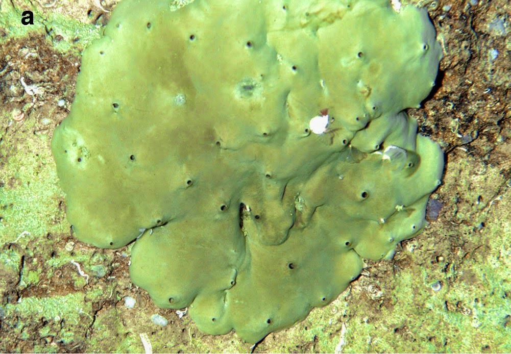
 Specimen of Brevaspidella dispersa from unknown locality on Gotland, Sweden. Flat to slightly concave upper surface with cluster of osculi and some converging lateral canals. Rhebergen (2014).

Brevaspidella dispersa is a small Sponge, typically less than 5 cm across, with the height and diameter about the same. The upper surface is slightly concave with scattered osculi, the base flat or slightly concave with a thickened dermal layer with concentric wrinkles.

Brevaspidella dispersa. (A, B) Specimen from beach near Västlanda, Gotland, Sweden. (A) View on upperside with dispersed osculi and radial surface canals. (B) Lateral view with concentrically wrinkled dermallayer in the lower part. (D, E) Specimen from unknown locality on Gotland, Sweden. (D) The top shows morethan 50 osculi, scattered over the flat surface. (E) Side view. Dermal layer poorly developed and restrictedto the base. Rhebergen (2014).

See also…


The shallow water reefs around Bonaire and Klein Curaçao in the Caribbean Netherlands are well studied and are considered a biodiversity hotspot, but the...
 
Sponges (Porifera) are generally considered to be the oldest extant animal group, with a fossil record that extends considerably into the Precambrian; phylogenomic analysis suggests they are the sister group to all other animals, which also suggests an early origin for the group.

Chalinid Dermosponges are among the hardest Sponges to classify taxonomically due to their simple anatomies and variable morphologies. They are encrusting Sponges with skeletons made up of...


Follow Sciency Thoughts on Facebook.

Thursday, 6 November 2014

Calcifying Endosymbiotic Bacteria in a Non-Sclerosponge.


Sponges (Porifera) are generally considered to be the oldest extant animal group, with a fossil record that extends considerably into the Precambrian; phylogenomic analysis suggests they are the sister group to all other animals, which also suggests an early origin for the group.

Calcified tissues appear suddenly in Sponge-like fossils in the late Ediacaran Period then in a variety of metazoan groups (multi-cellular Animals) at the start of the Cambrian, an event widely known as the Cambrian Explosion. This is generally considered a major step forward in animal evolution, as mineralized body parts are extremely useful both in defending against predation and penetrating the defences of potential prey, and serve as excellent support for muscles, although many modern sea creatures are still able to survive and thrive without any mineralized tissues.

Many, perhaps all, sponges contain colonies of symbiotic bacteria, and it has been suggested that this relationship may also date back to the Precambrian. This makes sponges good subjects for scientists studying the origins of symbiotic relationships between Animals and Bacteria.

Sponges can secrete skeletal elements (spicules) of either calcium carbonate (Class Calcarea) or silica (Classes Hexactinellida and Dermospongeiae),with one group in Class Dermospongeiae, the Sclerosponges, capable of producing both. It has previously been suggested that Coraline (calcium carbonate secreting) Dermosponges may have gained the ability to biomineralizecalcium carbonate by horizontal gene transfer from a Bacterial donor.

In a paper published in the journal Evolution in October 2012, Maria Uriz and Gemma Agell of the Centre d’Estudis Avançats de Blanes, Andrea Blanquer, also of the Agell of the Centre d’Estudis Avançats de Blanes, as well as the Observatoire Océanologique at Université Paris-Sorbonne, and Xavier Turon and Emilio Casamayor, again of the Centre d’Estudis Avançats de Blanes, discuss the discovery of endosymbiotic calcifying Bacteria in non-calcifying Dermosponges of the genus Hemimycale, and the implications of this for the origins of calcification in metazoans.

Uriz et al.collected specimens of Hemimycale from northeast Spain, the Mediterranean, the Red Sea and East Africa. These were dried in a stove at 80°C for 48 hours then weighed, then placed in a second oven at 600°C to remove the organic portion, leaving silica and calcium carbonate. Finally the samples were boiled in nitric acid to remove the calcium carbonate, before being washed and redried to give the weight of the silica spicules. Samples were also subjected to Scanning Electron and Transmission Electron Microscopy, Energy-Dispersive X-ray Analysis and in situ DNA hybridization with bacterial and archaeal probes.

The Calcibacteria harbouring Sponge, Hemimycale columella, from the Atlantic and Mediterranean. Uriz et al. (2014).

The sponges were found to contain 30-60% calcium carbonate (dry weight). They contained numerous Coccoid Calcibacteria within vacuoles in specialist cells (Calcibacteriocytes). These Calcibacteria secreted calcareous coatings as they matured, which built up within the calcibacteriocytes. As this happened the calcibacteriocytes migrated to the surface of the sponge, eventually lysing at the surface to contribute to a Calcibacterial coating.

During the sponges reproductive cycle maternal calcibacteriocytes surrounded the embryo, and were then phagocytised, releasing the Bacteria into the embryo’s mesohyl, where they were absorbed by embryonic proto-calcibacteriocytes.

The Calcibacteria harbouring Sponge, Hemimycale sp., from the Indo-Pacific region. Uriz et al. (2014).


Hemimycale hosts a low diversity of bacteria compared to many sponges, but these bacteria make up about 60% of the dry bodyweight, compared to about 38% by volume for most higher diversity sponges. This is not a direct comparison and highlights a need for further study in the field.

Work is underway to identify the Calcibacteria found; 16S rRNA gene tag-pyrosequencing suggests that 65% of the bacteria present within Hemimycale are Alphaproteobacteria, though it does not confirm that these are the Calcibacteria. Mitochondria are modified Alphaproteobacteria, and many symbiotic bacteria found in other invertebrates belong to this group, making them good candidates.

Dividing Calcibacteria entrapped within the calcareous coat (SEM); scale bar is
600 nm. Uriz et al. (2014).

Calcarious nodules have been reported in Sponges before, but this is the first time that endosymbiotic Calcifying Bacteria have been reported; Uriz et al. suggest that this trait might be quite widespread, but that it may be widely overlooked by the practice of treating glass sponges with nitric acid to release the silica spicules.

Uriz et al. consider that the Calcibacteria appear to be fully adapted to a symbiotic lifestyle; they lack normal bacterial cell walls, live within specialized cells within the host, and do not contain much cytoplasmic material of their own. The relationship appears to be a reasonably old one, since the bacteria are found in all members of the genus studied, in widely varying ecological conditions, and there is a well-developed mechanism for propagating the bacteria from one generation to the next, and the relationship appears to be beneficial to the sponge; very little eats Hemimycale, despite a poorly developed silica skeleton and no obvious chemical defences, and the sponges appear to grow faster than related species in similar environments.

High density of Calcibacteria released from broken calcibacteriocytes after squeezing the sponge (SEM); scale bar is 1 μm. Uriz et al. (2014).

Studies of the Coralline Demosponge Astrosclera willeyana suggest that the genes responsible for calcification in this sponge are fully integrated into the sponge genome, but that these genes appeared to be of prokaryotic (Bacterial) origin. Furthermore, since the same genetic pathway is used in the related Astrosclera queenslandica, which is thought to have shared a last common ancestor with Astrosclera willeyana in the Triassic, this horizontal gene transfer must have happened at least 265-220 million years ago.

Lynn Margulis’sendosymbiotic hypothesis suggests that eukaryotic cells evolved at least in part by the integration of symbiotic bacteria into the cells as organelles. This is widely accepted, though scientists vary in the degree to which they agree with the theory; most accept that mitochondria and chloroplasts have bacterial origins, but not all accept such an origin for other organelles. Uriz et al.’s findings suggest that the Calcibacteria within the cells of Hemimycale are currently evolving towards full integration as organelles.

It has previously been suggested that the development of mineralized tissues in animals was triggered not by a need to develop a skeleton to support the body, but by a need to remove (toxic) calcium ions from the cells. Under this scenario, biomineralization occurred initially as an excretory process, probably provoked by an abrupt rise in environmental calcium, then was co-opted for skeleton building later.

The presence of endosymbiotic calcifying bacteria in Hemimycale, combined with the probable prokaryotic origin of the calcifying genes in Astrosclera, suggest a possible scenario in which Calcifying Bacteria were co-opted by early metazoans, then eventually integrated completely into the animal genomes. Calcifying Bacteria are known to have developed long before the origin of multi-cellular life-forms, making it plausible that a variety of different animals may have co-opted Bacteria for this purpose, particularly if they all suffered a simultaneous rise in environmental calcium which needed to be overcome.

See also…

Chalinid Dermosponges are among the hardest Sponges to classify taxonomically due to their simple anatomies and variable morphologies. They are encrusting Sponges with skeletons made up of...


Theonellid Sponges are predominantly deepwater Sponges found across the globe, with a rigid skeleton made up of interlocking silica spicules. They are noted for the production of an array of unusual chemicals, the majority of which are thought to be produced by...


Unlike most Sponges (Porifera), which feed by filter feeding water pumped through their bodies, Carnivorous Sponges (Cladorhizidae) feed by capturing Crustaceans and other small animals on hooked spicules on filaments, then digesting them externally.  The group are predominantly found in deep water, where carnivory is presumed to be a better feeding strategy than filter...


Follow Sciency Thoughts on Facebook.

Tuesday, 29 July 2014

Two new species of Chalinid Dermosponge from Western Australia.

Chalinid Dermosponges are among the hardest Sponges to classify taxonomically due to their simple anatomies and variable morphologies. They are encrusting Sponges with skeletons made up of individual spicules which support their structure, but are not helpful taxonomically. Species are often distinctively coloured, but this is not visible in preserved specimens, making comparison to museum specimens extremely difficult. Chalinid Dermosponges are found globally and at all depths, but are most abundant and diverse in shallow subtidal waters (i.e. just below the low-tide level).

In a paper published in the journal Zootaxa on 9 July 2014, Jane Fromont of the Aquatic Zoology Department at the Western Australian Museum and David Abdo of the Marine Ecology and Monitoring Section of the Department of Fisheries of the Government of Western Australia describe two new species of Chalinid Dermosponge from temperate coastal waters off the coast of Western Australia, both of which are placed in the genus Haliclona.

The first new species is named Haliclona djeedara, which means 'brown' in the Nyoongar language, spoken by the indigenous people of south Western Australia. Haliclona djeedara is an erect, lobate, encrusting Sponge, light brown or beige in colour. It is firm, but compressible with a springy texture and large internal canals. It was found living on limestone reefs at depths of 3-30 m, in temperate waters south of 30˚ south, from Jurien Bay in the north to Bremer Bay in the south. It was observed giving birth to cylindrical parenchymellae larvae (planktonic Sponge larvae consisting of clumps of ciliated cells) in February, which is summer in Western Australia. The individual Sponges are gonochoric (sexed, can be male or female).

Underwater image of Haliclona djeedara. David Abdo in Fromont & Abdo (2014).

The second new species is named Haliclona durdong, meaning 'green' in the Nyoongar language. Haliclona durdong is an erect or massive Dermosponge with a smooth, velvety surface. It is compressible and easily damaged. It was found living on limestone reefs at depths of 3-30 m, in temperate waters south of 30˚ south, from Jurien Bay in the north to Bremer Bay in the south. It gives birth to to cylindrical parenchymellae larvae in the summer; individuals can apparently be gonochoric or hermaphroditic. 

Underwater image of Haliclona durdong. David Abdo in Fromont & Abdo (2014).

See also…


Theonellid Sponges are predominantly deepwater Sponges found across the globe, with a rigid skeleton made up of interlocking silica spicules. They are noted for the production of an array of unusual...


Unlike most Sponges (Porifera), which feed by filter feeding water pumped through their bodies, Carnivorous Sponges (Cladorhizidae) feed by capturing Crustaceans and other small animals on hooked spicules on filaments, then digesting them externally.  The group are predominantly found in deep water, where carnivory...


 A hypercalcified Chaetetid Demosponge from the Late Carboniferous of northwest Spain.

Sponges (Porifera) are considered to be the most primitive form of animals. They lack differentiated cells, and can reform if...


Follow Sciency Thoughts on Facebook.

Saturday, 28 June 2014

Two new species of Theonellid Sponge from the Great Barrier Reef.

Theonellid Sponges are predominantly deepwater Sponges found across the globe, with a rigid skeleton made up of interlocking silica spicules. They are noted for the production of an array of unusual chemicals, the majority of which are thought to be produced by symbiotic bacteria living within the Sponges. Many of these chemicals have anti-fungal properties.

In a paper published in the journal Zootaxa on 11 June 2014, Kathryn Hall and Merrick Ekins of  Marine Environments at the Natural Environments Program at the Queensland Museum and John Hooper of Marine Environments at the Natural Environments Program at the Queensland Museum and the Eskitis Institute for Drug Discovery at Griffith University, describe two new species of Theonellid Sponge from the Great Barrier Reef, Australia. Both new species have been placed in the genus Theonella, which are a morphologically conservative (tend to all be the same shape) and usually bright yellow-orange in colour, though some species are blue, and which are predominantly Indo-Pacific in distribution.

The first new species is named Theonella deliqua, meaning ‘lacking’ or ‘wanting’ in reference to the species’ lack of desmas (explain). This is a bright orange Sponge with a smooth surface, incorporating portions of green Algae cream Snail shells and other material into its body. It forms thin sheets (roughly 50 μm thick) over the shells of the Snail Tenagodus, with smaller shells and fragments of shell of the same species incorporated into its body-mass. Snails interior to the sponge are all deceased, but living Snails appeared to be being incorporated at the edge of the Sponge-mass. The species was found living on the seafloor on the inner Barrier Reef south of Wreck Island at depths of between 40 and 45 m.

Portion of Theonella deliqua fixed in alcohol. Scale bar is 2 cm. Hall et al. (2014).

Theonella deliqua, Micrographs (SEM). (A) Overview of sponge, showing empty shells of a species of Tenagodus; scale bar is 1 mm. (B) Detail of snail shell, showing slit, which is definitive for species of Tenagodus; scale bar is 1 mm. (C) Detail of snail shell, showing Theonella deliqua forming thin encrusting sheets over the shell; scale bar is 1 mm. (D) Detail of region lying between aggregated snail shells; note the accumulation of debris and foreign spicules, including broken calthrops; scale bar is 100 μm. Hall et al. (2014).

The second new species is named Theonella maricae, after Mary Kay Harper of the College of Pharmacy at the University of Utah in honour of her work on the chemistry of Theonellid Sponges. The species forms thin sheets (about 50 μm thick) over a variety of substrates, and incorporates a range of material into its body, including Gastropod shells, Diatoms, broken Coral debris, algal material and quartz sand. The species was found on the inner Barrier Reef sea floor southeast of Guthrie Shoal.

Portion of Theonella maricae fixed in alcohol. Scale bar is 2 cm. Hall et al. (2014).

Detail view of Theonella maricae, showing thin sheets over debris, including filamentous algae, gastropod shells and other carbonates; scale bar is 500 μm. Hall et al. (2014).

See also…


Unlike most Sponges (Porifera), which feed by filter feeding water pumped through their bodies, Carnivorous Sponges (Cladorhizidae) feed by capturing Crustaceans and other small animals on hooked spicules on filaments, then digesting them externally.  The group are predominantly found in deep water, where carnivory...


Sponges (Porifera) are considered to be the most primitive form of animals. They lack differentiated cells, and can reform if disassociated by (for example) shoving them through a sieve. On the other hand they cannot be considered colonies of single-celled organisms, as they have definite structures, bodies with more-or-less set shapes consisting of networks of pores and channels through which water is pumped; the...


Sponges (Porifera) are considered to be the most primitive form of animals. They lack differentiated cells, and can reform if disassociated by (for example) shoving them through a sieve. On the other hand they cannot be considered colonies of single-celled organisms, as they have definite structures, bodies with...


Follow Sciency Thoughts on Facebook.

Friday, 30 May 2014

Four new species of Carnivorous Sponges from the American West Coast.

Unlike most Sponges (Porifera), which feed by filter feeding water pumped through their bodies, Carnivorous Sponges (Cladorhizidae) feed by capturing Crustaceans and other small animals on hooked spicules on filaments, then digesting them externally.  The group are predominantly found in deep water, where carnivory is presumed to be a better feeding strategy than filter feeding, though a few shallow water forms are known. Spicules apparently from Carnivorous Sponges have been found from deposits as old as the Jurassic, implying that the group is at least this old, though it has been suggested that the group is paraphyletic, resulting from convergent evolution by different lineages within the Sponge group Poecilosclerida. Carnivorous Sponges have often been collected in the vicinity of chemosynthetic communities, and one species, Cladorhiza methanophila, has been demonstrated to host symbiotic methane oxidizing bacteria within its tissues.

In a paper published in the journal Zootaxa on 9 April 2014, Lonny Lundsten of the Monterey Bay Aquarium Research Institute, Henry Reiswig of the Department of Biology at the University of Victoria and the Natural History Section of the Royal British Columbia Museum, and William Austin of the Khoyatan Marine Laboratory, describe four new species of Carnivorous Sponges from the Pacific Coast of North America.

The first new species described is placed in the genus Asbestopluma, and given the specific name monticola, meaning ‘mountain dweller’, as it was discovered living on the Davidson Seamount off central California. Asbestopluma monticola is a branching Sponge with a ‘bottle-brush’ arrangement of filaments reaching 28 cm tall and 19 cm wide. As well as on the Davidson Seamount it was found living on a rocky outcrop within the Monteray Canyon, closr to the North Californian coast, as well as off the coast of Oregon, giving the species a known range of over 1000 km.

Colony of Asbestopluma monticola on the Davidson Seamount. Lundsten et al. (2014).

Asbestopluma monticola was found living at an average depth of 1236 m, in areas where oxygen concentration was low and the average temperature was 3.18˚C. Small Crustacean prey was observed adhered to the body of the Sponges, in various states of decomposition. The species occurred in mixed communities, alongside other Sponges, Corals, Crustaceans, Echinoderms and Fish. Small Pandalid Shrimps were observed climbing about on the branches of the Sponges.

(A) Filaments of Asbestopluma monticola. (B & C) Images of prey in various states of decomposition. Lundsten et al. (2014).

Asbestopluma monticola, spicules: (A) large styles 1, (B) large styles 2, (C) large styles 3, (D) microacanthotylostrongyle, (E) sigma, (F) palmate anisochela. Lundsten et al. (2014).

The second new species described is also placed in the genus Asbestopluma, and is given the specific name rickettsi, in honour of the marine biologist Edward F. Ricketts, who was immortalized as ‘Doc Ricketts’ in John Steinbeck’s Canary Row the first specimens of Asbestopluma rickettsi were collected by the Monterey Bay Aquarium Research Institute’s ROV Doc Ricketts.

Asbestopluma rickettsi is a branching Sponge with a ‘bottle-brush’ arrangement of filaments reaching 21.78 cm tall and 12.38 cm wide. The Sponge was found living in a community of chemosynthetic organisms in a low oxygen basin off the coast of California, north of La Jolla. The community also included Vesicomyid Clams, Tube Worms and Bacterial Mats. The average depth at the site was 1031 m and the average temperature 3.93˚C. Asbestopluma rickettsi was not observed capturing Crustacean prey, but rather was found to be feeding on methane-oxidizing Bacteria; it could not be determined whether these Bacteria were symbionts (living within the tissues of the Sponge) or were being ingested and consumed from the neighbouring Bacterial Mats.

Specimen of Asbestopluma rickettsi. Lundsten et al. (2014).

Asbestopluma rickettsi, spicules: (A) large styles 1, (B) large styles 2, (C) large styles 3, microacanthotylostrongyle (D), sigma (E), palmate anisochelae 1 (F), palmate anisochelae 2 (G). Lundsten et al. (2014).

The third new species is placed in the genus Cladorhiza, and given the specific name caillieti, in honour of Gregor Cailliet of Moss Landing Marine Laboratories, a noted ichthyologist and deep sea biologist. It was discovered on the Endeavor Segment, of the Juan de Fuca Ridge hydrothermal vent field, off the coast of British Columbia, where it was found hanging from the underside of overhanging basalt ledges at an average depth of 2149 m in temperatures averaging 1.87˚C. Cladorhiza caillieti is an unbranching Sponge with a bottle-brush arrangement of filaments reaching up to 9.17 cm long and 3.6 mm wide. It was part of a community that included other Sponges, as well as Gorgonian Corals, Crinoids and Serpulid Worms. Numerous Crustacean prey, in various states of decomposition were found adhered to the filaments of the Sponges.

Cladorhiza caillieti: in situ image of numerous specimens attached to the underside of overhanging ledges(A), collection of specimens (B). Lundsten et al. (2014).

Collected specimens of Cladorhiza caillieti in the lab. Lundsten et al. (2014).

Partially digested Crustacean adhered to a filament of Cladorhiza caillieti. Lundsten et al. (2014).

Cladorhiza caillieti spicules: large styles 1 (A), 2 (B) and 3 (C), sigma 1 (D) and 2 (E). Lundsten et al. (2014).

Cladorhiza caillieti spicules: sigma 3 (A), sigmancistra (B), unguiferate anisochelae 1 (C) and 2 (D). Lundsten et al. (2014).

The final new species described is also placed in the genus Cladorhiza and given the specific name evae, in honour of Eve Lundsten, the wife of Lonny Lundsten. The species was discovered on a hydrothermal chimney at a depth of 2373 m, in the Gulf of California east of Cabo Pulmo where the Lundstens honeymooned. Cladorhiza evae is an unbranching Sponge with a bottle-brush arrangement of filaments that reaches 18.7 cm long and 3.1 mm wide. The area had low oxygen concentrations an average temperature of 2.02˚C, but was home to a diverse community including numerous Crabs, Worms and Fish. A variety of small prey were found adhered to the Sponges in various states of decomposition.

Cladorhiza evae group of individuals in situ on the hydrothermal chimney where they were discovered. Lundsten et al. (2014).

Cladorhiza evae, Collected specimens in the lab. Lundsten et al. (2014).

Prey in various states of decomposition on the filaments of Cladorhiza evae. Lundsten et al. (2014).

Cladorhiza evae spicules: large styles 1 (A), 2 (B), and 3 (C), sigma 1 (D) and 2 (E). Lundsten et al. (2014).

Cladorhiza evae spicules: sigmancistra (A), unguiferate anisochelae (B). Lundsten et al. (2014).

See also…


Sponges (Porifera) are considered to be the most primitive form of animals. They lack differentiated cells, and can reform if disassociated by (for example) shoving them through a sieve. On the other hand they cannot be considered colonies of single-celled organisms, as they have definite structures, bodies with more-or-less set shapes consisting of networks of pores and channels through which water is pumped; the individual cells feeding separately by filtering food from the water in these channels...


Sponges (Porifera) are considered to be the most primitive form of animals. They lack differentiated cells, and can reform if disassociated by (for example) shoving them through a sieve. On the other hand they cannot be considered colonies of single-celled organisms, as they have definite structures, bodies with more-or-less...


Sponges (Porifera) are considered to be the most primitive form of animals. They lack differentiated cells, and can reform if disassociated by (for example) shoving them through a sieve. On the other hand they cannot be considered colonies of single-celled organisms, as they have definite structures, bodies with more-or-less set shapes consisting of networks of pores and channels...


Follow Sciency Thoughts on Facebook.

Friday, 17 January 2014

A hypercalcified Chaetetid Demosponge from the Late Carboniferous of northwest Spain.

Sponges (Porifera) are considered to be the most primitive form of animals. They lack differentiated cells, and can reform if disassociated by (for example) shoving them through a sieve. On the other hand they cannot be considered colonies of single-celled organisms, as they have definite structures, bodies with more-or-less set shapes consisting of networks of pores and channels through which water is pumped; the individual cells feeding separately by filtering food from the water in these channels. They are the only extant group of animals with a fossil record that extends significantly into the Precambrian.

In a paper published in the journal Palaeontologia Electronica in August 2013, Diego Corrochano of the Departamento de Geología at the Universidad de Salamanca and Ronald West of the Kansas State University, describe a new form of Chaetetid Demosponge (a group of Sponges that played a major role in reef-building in the Late Palaeozoic; they still exist but play a less prominent role in modern ecosystems) from the Late Carboniferous of León in northwest Spain.

The new species is named Loiscupula bachendensi, where 'Loiscupula' derives from 'Lois' a village close to the site where it was found, and 'cupula' meaning 'cup-shaped', and 'bachendensi' derives from the Bachende Formation, in which the specimens were found. Loiscupula bachendensi has a flattened cup-shape, reaching 5 cm high and 32 cm across. The underside shows a number of concentric rings, possibly reflecting a growth pattern. The upper side is dotted with cylindrical features, which can be simple or branches and reach up to 3.4 cm above the cup. Internally the Sponge has a structure of polygonal or cylindrical tubes.

Loiscupula bachendensi. (2) Side view showing the flat cup-shaped skeleton, with the central part of the specimen lower than the outer edges. (3) Top view, showing the wackestone matrix filling the cup. (4) Underside of the sponge, showing the plate-shape and the concentric rings around a central point; note the projected structures developed within each ring and usually preserved as eroded (truncated) protuberances (arrow). Corrochano & West (2013).


The preserved skeletons of Loiscupula bachendensi are composed of course calcite, with occasional preserved fascicular needles. Corrochano & West believe the original skeleton have been composed of aragonite.

Idealized reconstruction of Loiscupula bachendensi. Note the low conical growth form; (A) presumed attachment point; (P) projections on lower surface; (G) grooves; (R) ridges; (P) plate like skeleton; (C) cylindrical/branching features (extended mamelons/chimneys) on upper surface. Corrochano & West (2013).


Follow Sceincy Thoughts on Facebook.

Thursday, 16 January 2014

Two new species of calcareous Sponge from the Weddell Sea.

Sponges (Porifera) are considered to be the most primitive form of animals. They lack differentiated cells, and can reform if disassociated by (for example) shoving them through a sieve. On the other hand they cannot be considered colonies of single-celled organisms, as they have definite structures, bodies with more-or-less set shapes consisting of networks of pores and channels through which water is pumped; the individual cells feeding separately by filtering food from the water in these channels. They are the only extant group of animals with a fossil record that extends significantly into the Precambrian.

In a paper published in the journal Zootaxa on 25 July 2013, Hans Rapp of the Centre for Geobiology and Department of Biology at the University of Bergen, Christian Göcke of the Forschungsinstitut und Naturmuseum Senckenberg, Ole Tendal of the Zoological Museum of the University of Copenhagen and Dorte Janussen, also of the Forschungsinstitut und Naturmuseum Senckenberg, describe two new species of calcareous Sponge (Calcarea, sponges that have skeletons composed of calcium carbonate), from the Eckström Shelf of the Eastern Weddell Sea, Antarctica.

The first new species is placed in the genus Clathrina and given the specific name brandtae in honour of Angelika Brandt of the University of Hamburg, organizer of two expeditions to the Antarctic. Clathrina brandtae is a yellowish Sponge with a body made up of a loose arrangement of tubes, roughly 1 cm long and 1.5 cm wide. It has a skeleton made up of simple three pointed elements. The Spomge was found at depths of between 118 and 595 m.

Clathrina brandtae. (A) Preserved specimen from the Weddell Sea (os = osculum). (B) Spicules, from left: ste = subregular tetractine, rte = regular tetractine, rtri = regular triactine and aa = apical actine of a tetractine. (C) Wall of asconoid tube seen from the interior. Rapp et al. (2013).


The second new species is placed in the genus Leucetta and given the specific name delicata, meaning delicate. Leucetta delicata is a beige sponge with an ovular to cylindrical shape, reaching 2.2 by 1.2 cm. It was found at a single site at a depth of 595 m.

Leucetta delicata. (A) Preserved specimen. os = osculum. (B) Spicules. Bottom left: rte = regular tetractine with very short apical actine. Remaining spicules: rtri = regular triactines of variable size. (C) Cross section of the body wall. cx = cortex. ca = canal. atr = atrium. (D) Cross section of the cortical region. cx = cortex. cch and arrow = choanocyte chamber (scale bar as in E). (E) Cross section of the atrial region. atr = atrium. Rapp et al. (2013).


See also A Chambered Glass Sponge from the Early Devonian of northern SpainA mysterious Glass Sponge from the Early Cambrian of south-east China, New Glass Sponges from the North Atlantic and The oldest animals - Pre-Ediacaran Sponges from Namibia(?) 

Follow Sciency Thoughts on Facebook.

Friday, 20 December 2013

A new species of Sponge dwelling Pontoniine Shrimp from Sabah State, Malaysia.

The Pontoniine Shrimps are an unusual group of Decapod Crustaceans (the group that also includes Crabs, Lobsters and most 'Shrimps') noted for their tendency to form commensal relationships with other invertebrates. Species are known which have such relationships with Sponges, Corals, Jellyfish, Hydrozoans, Siphonophores, Soft Corals, Snails, Clams, Starfish, Brittlestars, Crinoids, Sea Urchins, Sea Cucumbers and Sea Squirts. They are found in all tropical and sub-tropical marine waters, but are at their most abundant and diverse in the waters of the tropical Indian and western Pacific Oceans.

In a paper published in the journal Zootaxa on 6 August 2013, Charles Fransen of the Department of Marine Zoology at the Naturalis Biodiversity Center in Leiden describes a new species of Sponge dwelling Pontoniine Shrimp from Sabah State, Malaysia.

The new species is placed in the genus Nippontonia, which previously contained only a single species, the Sponge-dwelling Nippontonia minirostris from the Japanese Ryukyu Islands and Taiwan. The new species is named Nippontonia christellae, in honour of the author's sister-in-law, Christel van Eijnatten, for her 'inspiring perseverance and positivism in conquering life again after it was almost taken from
her'. 

Nippontonia christellae is ~10 mm Shrimp, translucent and dotted with brown or green chromatophores (coloured cells). It was found living inside the Sponge Acanthostrongylophora ingens, and another unknown black Sponge at a depths of 10-15 m off the coast of Ligitan Island in the Celebes Sea off the east coast of Sabah State Malaysia (i.e. northern Borneo).

Nippontonia christellae, (top) female carrying eggs inside the Sponge Acanthostrongylophora ingens, and (bottom) line drawing of the same. Scale bar is 4 mm. Fransen (2013).

The approximate location of Ligitan Island, where Nippontonia christellae was discovered. Google Maps.


Follow Sciency Thoughts on Facebook.

Saturday, 2 November 2013

A Chambered Glass Sponge from the Early Devonian of northern Spain.

Sponges (Porifera) are considered to be the most primitive form of animals. They lack differentiated cells, and can reform if disassociated by (for example) shoving them through a sieve. On the other hand they cannot be considered colonies of single-celled organisms, as they have definite structures, bodies with more-or-less set shapes consisting of networks of pores and channels through which water is pumped; the individual cells feeding separately by filtering food from the water in these channels. They are the only extant group of animals with a fossil record that extends significantly into the Precambrian.

In a forthcoming paper in the journal Acta Palaeontologica Polonica available online from 17 June 2013, Martin Nose of the SNSB-Bavarian State Collection of Palaeontology and GeologyRadek Vodrážka of the Institute of Geology at the Academy of Sciences of the Czech Republic and Luis-Pedro Fernández and Isabel Méndez-Bedia of the Departamento de Geología at the Universidad de Oviedo, describe a species of Chambered Glass Sponge from the Early Devonian of northern Spain.

 The new species is placed in the genus Casearia, and given the specific name Casearia devonica. It is the oldest member of the genus by around 200 million years, as well as being the oldest known Chambered Glass Sponge and the first Chmabered Glass Sponge from the Palaeozoic. Chambered Glass Sponges are otherwise known from the Late Triassic to the Late Jurassic.

Casearia devonica is a straight or branching Chambered Sponge, comprising up to 18 chambers, each 0.5 to 5.0 mm in height. It was discovered in shales of the Valporquero Formation near the village of Colle in the Cantabrian Mountains in northern Spain. These shales are thought to have formed in mud mounds between Coral outcrops, in waters bellow the storm wave-base. Modern Glass Sponges no longer form similar chambered shapes, but they do typically live bellow the wave-base, being found across the globe in waters deeper than 100 m.

Chambered Hexactinellid Sponge Casearia devonica in thin section. Scale bar is 5 mm.


Follow Sciency Thoughts on Facebook.