
The Sterkfontein Caves site, part of the wider ‘Cradle of Humankind’ complex in Gauteng State, South Africa, has yielded the largest known collection of specimens assigned to the Plio-Pleistocene Hominin species Australopithecus africanus. The site comprises a series of karstic caves (i.e. caves created by the action of water percolating through soft limestone) that would have been encountered by the Hominins both as cave openings at the surface that could provide potential shelter and as potholes into which they could fall. Specimen StW 431 is a partial Australopithecus africanus skeleton discovered at Sterkfontein in 1987. This specimen's lumbar vertebrae showa number of deformaties, with both bone overgrowth and erosion. This has been diagnosed variously as brucellosis, spondylosis deformans, osteophytic formation,and osteoarthritis of the facet joints.
In a paper published in the South African Journal of Science on 30 January 2017, Edward Odes of the School of Anatomical Sciences at the University of the Witwatersrand, Alexander Parkinson of the Evolutionary Studies Institute, also at the University of the Witwatersrand, Patrick Randolph-Quinney, also of the School of Anatomical Sciences and Evolutionary Studies Institute at the University of the Witwatersrand, and of the School of Forensic and Applied Sciences at the University of Central Lancashire, Bernhard Zipfel, agian of the Evolutionary Studies Institute, and of the School of Geosciences at the University of the Witwatersrand, Kudakwashe Jakata, again of the Evolutionary Studies Institute at the University of the Witwatersrand, Heather Bonney of the Department of Earth Sciences at the Natural History Museum and Lee Berger, once again of the Evolutionary Studies Institute at the University of the Witwatersrand, re-examine skeleton StW 431, and draw new conclusions about its taphonamy and osteopathology.

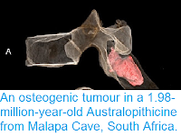
StW 431 – a partial skeleton of Australopithecus africanus discovered at Sterkfontein Caves in 1987. Stw 431 represented only the third partial skeleton attributed at the time to Australopithecus africanus, and represents the only probable male skeleton attributed to this taxon to date. Odes et al. (2017).
Odes et al. examined the fourth and fifth vertebrae of StW 431, using micro computed tomography to examine the internal structures of the bone. They were able to identify areas of both erosion and deposition of bone, though unlike previous studies, which had examined only the exterior of the bones, they conclude that, while the excess bone deposition cleary happened while the individual was alive, the bone erosion was almost certainly post-mortem, and is consistent with damage caused by Insects, such as Southern African Termite, Trinervitermes trinervoides, or Dermestid Beetle, Dermestes maculatus.

Micro-CT orthoslice views of the L4 vertebra of StW 431: (a) transverse superior, (b) sagittal midline, (c) transverse inferior and (d) anterior. Note the bilateral osteophytic formation and two zones of erosion evident on the inferior endplate surface of the L4 in (c). There is no evidence of sclerosis or new bone formation around the margins of the cavities (c). Also note areas of new bone formation ranging from open woven (c) to sclerotic (d). Odes et al. (2017).
Based upon this Odes et al. conclude that the individual was suffering from osseous proliferation (osteophytosis), cosistant with a degenerative spinal joint disease. Such osseous proliferation is a fairly common condition, typically caused by erosion of the cartilage, which can lead to bone rubbing against bone, leading to bone damage which the body tries to heal through new growth,

Micro-CT orthoslice views of the L5 vertebra of StW 431: (a,b) transverse to the midline (close to surface), (c) transverse to inferior, (d) coronal midline, (e) sagittal superior to bottom. Note new bone formation on the anterior wall (e) and major osteophytic formation at the antero-superior margin indicating remodelling of the cortex, revealing a sclerotic margin, and as crenulated buttresses of porous bone devoid of internal trabeculae (a,b). There is no evidence of sclerosis or reactive bone formation around the cavity margins (a). The inferior endplate (c) exhibits an osteophytic formation as a thin ordered sclerotic rim around inferior circumference with no presence of buttressing. Notice the channel interpreted as invertebrate damage in (e). Odes et al. (2017).
See also...


Follow Sciency Thoughts on Facebook.