Showing posts with label Palaoentology. Show all posts

Friday, 1 December 2023

Squirmarius testai: A new species of Cyclostome from the Carboniferous Mazon Creek Lagerstӓtte.

Vermiform (Worm-like) fossils assigned to the species Nemavermes mackeei have been described from the 308.6–308.4 million year old Mazon Creek Lagerstӓtte of Illinois, and the 330.3–323.4 million year old Bear Gulch Limestone in Montana. These are between 40 and 140 mm in length, and between 3 and 16 mm in width, and have been interpreted as free-living marine Nematodes. 

In a paper published in the Journal of Paleontology on 1 December 2023, Victoria McCoy of the Department of Geosciences at the University of Wisconsin-Milwaukee, and the School of Geography, Geology, and the Environment at the University of Leicester, Jack Wittry of the Field Museum of Natural History, Hamed Sadabadi of the Department of Materials Science and Engineering at the University of Wisconsin-Milwaukee, and Paul Mayer, also of the Field Museum of Natural History, re-examine specimens assigned to Nemavermes mackeei, and conclude that the taxon is not valid, and that the specimens assigned to this species belong to a variety of different Vermiform Animals, including a new species of Cyclostome (the group of Jawless Fish which includes modern Hagfish and Lampreys).

Examination of the holotype of Nemavermes mackeei, FMNH PE 21551, shows that it is a proboscis of the well-known (but hard to interpret) Mazon Creek fossil Tullimonstrum gregarium. In taxonomy, a specimen is deemed to belong to a species if it can be shown to belong to the same species as the holotype. Since the holotype of Nemavermes mackeei, FMNH PE 21551, clearly belongs to the same species as the holotype of Tullimonstrum gregarium, PE 10504, which was named first, then Nemavermes mackeei must be considered to be a junior synonym of Tullimonstrum gregarium, rather than a valid species.

Comparison of the holotype of Nemavermes mackeei to the proboscis and buccal apparatus of Tullimonstrum gregarium. (1) Holotype of Nemavermes mackeei, FMNH PE 21551; (2) line drawing of (1), showing in particular the projected shape of the bifurcate portion of the buccal apparatus; (3) Tullimonstrum gregarium proboscis with buccal apparatus, FMNH PE 39890, showing sharp bend in the proboscis, and the differential preservation between the proboscis and buccal apparatus; (4) Tullimonstrum gregarium proboscis with buccal apparatus, FMNH PE 39375, showing two sharp bends in the proboscis; (5, 6)Tullimonstrum gregarium buccal apparati, FMNH PE 28739 (5) and FMNH PE 31063 (6), showing the distinctive shape of the buccal apparatus and the preservational differences between the buccal apparatus and the rest of the proboscis. Scale bars are 10 mm. McCoy et al. (2023).

Many specimens assigned to Nemavermes mackeei, however, are clearly not specimens of Tullimonstrum gregarium, but of a vermiform Animal, which McCoy et al. interpret as a new species of Cyclostome, and name Squirmarius testai, where 'Squirmarius' means 'one who squirms' and 'testai' honours fossil hunter Tom Testa, who collected many of the specimens assigned to the new species, and who referred to these fossils as 'squirms'.

Specimens of Squirmarius testai: (1) holotype, FMNH PF 17809; (2) FMNH PF 17810; (3) FMNH PF 17812; (4) FMNH PF 17811; (5) LF 2101; (6) LF 5664, which was collected by David Douglass and donated by the David and Sandra Douglass collection to the Lauer Foundation for Paleontology, Science, and Education in Wheaton Illinois for this study. Scale bars are 10 mm. McCoy et al. (2023).

Squirmarius testai is a vermiform Animal, frequently showing soft tissue preservation, with a tapering posterior end (tail) and a blunter anterior end (head). None of the specimens preserve any fins, including specimens where the areas of a Cyclostome where fins would be expected are well preserved. The stem Hagfish, Gilpichthys greenei, also from the Mazon Creek Lagerstӓtte, is also interpretted as beign finless, so this is not an implausible state for a Cyclostome from this era. However, in some specimens of the Lamprey, Mayomyzon pieckoensis, and the Hagfish, Myxinikela siroka, fins are not preserved, though other specimens are known to have fins, suggesting that preservation can vary in Mazon Creek Cyclostomes.

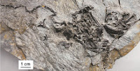
Detailed morphology of Squirmarius testai holotype, FMNH PF 17809: (1) whole body with boxes indicating regions that correspond to (2) box A, (3) box B, and (4) box C, and dots indicating the spots that correspond to (5) dot D and (6) dot E; ; (2) head, box A in (1), showing the blunt point at the anterior end of the body, with two eyes preserved as dark ovals with a white dot in the right eye that might represent a lens; (3) gut near the middle of the body, box B in (1), showing the flat kaolinite preservation; (4) tail, box C in (1), showing the narrow point at the posterior end of the body, the gut with dark preservation ending before the posterior point of the tail (white arrow), and the lack of a tail fin; (5) SEM image in SE mode of the leftmost eye, dot D in (1), showing ovoid melanosomes along with siderite crystals with moulds of ovoid melanosomes; (6) SEM image in BSE mode of the gut with dark preservation in the tail, dot E in (1), with siderite crystals without melanosome moulds, and abundant pyrite microcrystals and occasional pyrite framboids. Scale bars are 10 mm (1); 2 mm (2)–(4); 10 μm (6); 5 μm (5). McCoy et al. (2023).

Several specimens assigned to Squirmarius testai have dark segmented structures which McCoy et al. interpret as myomeres (blocks of skeletal muscle tissue arranged in sequence). Also present in many is a lighter structure running through the centre of the body, interpreted as a gut, and some unidentified darker regions, which may be internal organs.

Detailed morphology of Squirmarius testai, FMNH PF 17812: (1) whole body with boxes indicating the regions that correspond to (2) box A, (3) box B, (4) box C, and (5) box D;  (2) head or anteriormost preserved part of the body, box A in (1), showing the mottled dark coating and two irregular, differently sized dark patches that are unlikely to be eyes; (3) triangular or C-shaped organ, box B in (1), directly anterior to gut; note also the dark mottled covering; (4) body region, box C in (1), showing dark mottled covering and 3D gut; (5) tail, box D in (1), showing the dark mottled covering, the gut ending before the tip of the tail (white arrow), and nothing that clearly represents a tail fin. Scale bars are 10 mm (1); 2 mm (2)–(5). McCoy et al. (2023).

Many specimens have eyes preserved as dark brown-black ovals on both the part and counterpart, with some having central infills of kaolinite, which could represent lenses. Under the scanning electron microscope, it is possible to see ovoid microbodies and moulds of microbodies, which are the size and shape of melanosomes. Melanosomes have been documented in a range of fossil lagerstӓtte, including other Chordates at Mazon Creek, and therefore are not implausible structures to find preserved in Squirmarius testai. Importantly, very few groups of Animals have melanostomes within their eyes; outside of the Chordates, these structures are found only in the eyes of Box Jellyfish and some Flatworms, neither of which resemble Squirmarius testai in form, supporting the idea that this Animal is a Chordate.

None of these features is consistent with Squirmarius testai being a Nematode, including its size, which would be remarkably large in a free-living Nematode (some modern parasitic Nematodes get much larger, but free-living forms tend to be microscopic), supporting McCoy et al.'s belief that Squirmarius testai is a Cyclostome, and probably a stem Hagfish.

See also...

Online courses in Palaeontology

Follow Sciency Thoughts on Facebook.

Follow Sciency Thoughts on Twitter.


Thursday, 28 July 2016

Photogrammetry as a tool in Morphometric Analysis.

Morphometric analysis is a tool used by palaeontologists, archaeologists, anthropologists and forensic pathologists to analyse and compare specimens. It relies on taking numerous measurements of an object such as a bone or shell, and comparing both these measurements and ratios between measurements to those obtained from other specimens in order to establish relationships between them. Traditionally these measurements have been obtained using tape measures and callipers, but modern scientists often use more sophisticated tools such as structured light scanners, which are capable of building highly detailed three dimensional models of specimens. Photogrammmetry has been used as a tool in landscape analysis since the mid nineteenth century. It relies on taking overlapping images of a landscape, from a number of different vantages, in order to help cartographers and geomorphologists build up a three dimensional model of the landscape. The advent of digital photography has led to a far wider use of photogrammetry in the archaeology and other fields in the past two decades, with it becoming regularly used to build models of archaeological sites or even individual buildings. Recently some scientists have begun using photogrammetry to build models of smaller items, including archaeological artefacts and Human remains.

In a paper published in the Journal of Archaeological Science: Reports on 18 July 2016 a team of scientists led by Allowen Evin of the Institutdes Sciences de l'Evolution at Université de Montpellier, the Department of Archaeology at the University of Aberdeen, and the Department of Archaeology, Classics and Egyptology at the Universityof Liverpool, describe a process by which three dimensional computer models were constructed using digital photographs, and the results compared to the results of a morphometric model made to by structured light analysis.

Evin et al. examined a series of five modern Wolf skulls from the collection of the Muséum Nationald'Histoire Naturelle in Paris, as part of a study into morphometric changes to the cranium during the domestication of Dogs. In order to do this they created two models, one using a Breuckmann StereoScan structured light scanner and the other a model constructed from a series of photographs taken with a an 8 mega-pixel digital single-lens reflex (DSLR) Canon EOS 30D camera, mounted with a CanonEF 24–105mmf/4 L IS USM lens.

In order to take the photographs the specimens were placed on a rigid cardboard sheet with a calibrated referential pattern, and photograped from three different elevations (approximately 0°, 15° and 40°) using a tripod mounted camera, which was moved around the specimen taking images at 10° intervals (i.e. 36 sets of three images in a circle). This was then repeated with the skull the other way up, to give a model of the complete specimen. These images were then turned into a three dimensional model using a VisualSFM software package.

(A) Schematic representation of the set-up and camera positions used for the acquisition of the photographs. (B) Fixed dimensions reference pattern used to scale the models and enhance the performance of key-points detection/matching and camera calibration algorithms. Evin et al. (2016).

The photogrammatic model was then compared to the model made using the Breuckmann StereoScan structured light scanner (which rotates the specimens on its own automated turntable, creating a model using its own Optocat software package) using a computed mesh-to-mesh deviation map to compare the topology of the two models. This found that the two models matched to an average of 0.088 mm, with the only significant differences occurring within the nasal cavity and occipital foramen, areas which are generally considered extremely difficult to measure accurately, and which are not usually included in morphometric analyses.

Models obtained with photogrammetry (top) and the Breuckmann structured light scanner (bottom) with the cloud-mesh distances visualisation (middle). Differences are expressed using the colour scale on the left. Evin et al. (2016).

Evin et al. note the total tine spent with each specimen while capturing the images used to create the photogrammetric model was about 15 minutes, while the Breuckmann scanner required about forty five minutes to scan each specimen, although having scanned the specimens the Breuckmann scanner was able to produce a model almost instantaneously, while the camera method required considerable further input from the users, so that the Breuckmann scanner method took less time overall.

However since the Breuckmann scanner is not portable and is reliant on specimens being brought to it, Evin et al. felt that on the whole the advantages of the camera outweighed those of the scanner, as it could potentially be used in situations where specimens could not be moved, was considerably cheaper and captured additional information about the colour and surface texture of the specimen that were not recovered with the scanner.


See also...

http://sciencythoughts.blogspot.co.uk/2014/11/x-ray-computed-tomography-studies-of.htmlX-ray Computed Tomography studies of two Woolly Mammoth calves from Russia.            The Woolly Mammoth, Mammuthus primigenius, is thought to have diverged from the earlier Steppe Mammoth Mammuthus trogontherii in northeast...
http://sciencythoughts.blogspot.co.uk/2014/11/using-morphometric-analysis-to.htmlUsing morphometric analysis to understand the nature of Canid remains from Plio-Pleistocene Hominid sites from East Africa.                                                                        Morphometric analysis is a method used...
http://sciencythoughts.blogspot.co.uk/2014/04/reconstructing-paluxy-river-dinosaur.htmlReconstructing the Paluxy River Dinosaur Chase Sequence.                                            In 1940 palaeontologist Roland Bird of the American Museum of Natural History in New York described and partially excavated a sequence of Dinosaur footprints along the Paluxy River at Glen Rose in...

Follow Sciency Thoughts on Facebook.

Monday, 16 November 2015

Turtle remains from the Late Miocene to Early Pliocene Black Rock Sandstone of Victoria, Australia.

Australia has a diverse assemblage of Sea Turtles today, with six of the seven living Sea Turtle species found in Australian waters and one found nowhere else. The continent also has an extensive fossil record of Cretaceous Turtles, but no post-Cretaceous fossil Turtles have been found in Australia to date. This is odd, as Turtles have excellent preservational potential, having dense bony shells and spending much of their lives in shallow marine environments, and because Cainozoic fossil Turtles are known from New Guinea and New Zealand, making it highly unlikely that Sea Turtles were absent from Australia during this time.

In a paper published in the journal Alcheringa on 16 October 2014, Erich Fitzgerald and Lesley Kool of Geosciences at Museum Victoria describe the discovery of fragmentary Turtle remains from the Late Miocene to Early Pliocene Black Rock Sandstone at Beaumaris on Port Philip Bay in Victoria State, Australia.

The Black Rock formation is noted for a diverse vertebrate assemblage, including Sharks and Rays, Bony Fish, Penguins, other Seabirds, Sireneans, Seals, Baleen and Toothed Whales, and some terrestrial Birds and Marsupials, as well as invertebrates such as Molluscs and Echinoderms and microfossils such as Foraminiferans.

Only two Turtle bones were recovered from the site, an almost complete dentary (lower jawbone) and the proximal tip of a costal bone (plate on the dorsal portion of the shell derived from a modified rib). This is not enough to assign the remains to any particular Turtle, though they do resemble other specimens assigned to the genus Pacifichelys, which has previously been described from California and Peru.

Isolated proximal end of a costal of an unidentified Turtle in: (A, dorsal; (B) visceral; and (C) proximal views. Specimen whitened with ammonium chloride. Fitzgerald & Kool (2014).

See also...

http://sciencythoughts.blogspot.co.uk/2015/10/turtle-eggs-from-late-cretaceous-of.htmlTurtle eggs from the Late Cretaceous of Madagascar.                                               Turtles are unique among living Amniotes (Vertebrates that can lay...
http://sciencythoughts.blogspot.co.uk/2015/10/pappochelys-rosinae-proto-turtle-from.htmlPappochelys rosinae: A Proto-Turtle from the Middle Triassic of Badem Württemberg, Germany.                                                             Turtles have a fairly good fossil record, as would...
http://sciencythoughts.blogspot.co.uk/2015/10/desmatochelys-padillai-protostegid.htmlDesmatochelys padillai: A Protostegid Turtle from the Early Cretaceous of Colombia.        The Protostegid Turtles were a group of Turtles known from the Cretaceous that are thought to have been members of the Chelonioidea, the group that includes the two living Marine Turtle groups, the Chelonidoidea (Sea Turtles) and Dermochelyidae (Leatherback...
Follow Sciency Thoughts on Facebook.

Saturday, 19 September 2015

Isthminia panamensis: A South American ‘River Dolphin’ from the Late Miocene of Panama.


There are four species of River Dolphins known today; Inia geoffrensis and Pontoporia blainvillei from South America, Platanista gangetica from South Asia and the probably extinct Lipotes vexillifer from China (Lipotes vexillifer, the Yangtze River Dolphin, is considered Critically Endangered under the terms of the International Union for the Conservation of Nature's Red List of Threatened Species, as there were less than 50 adult individuals surviving when the species was last assessed in 2003, all living within a few hundred kilometers of one-another on a single river). These were formerly thought to comprise a single group, but are now thought to represent three distinct lineages that separately invaded freshwater environments from the sea. The fossil record of the Asian River Dolphins is poor, but the South American River Dolphins, Inioidea, have a fairly good fossil record, with freshwater fossils known from a number of sites in South America, and older marine specimens from South America, North America and Europe.

In a paper published in the journal Peer J on 1 September 2015, Nicholas Pyenson of the Department of Paleobiology at the National Museum of Natural History and the Departments of Mammalogy and Paleontology at the Burke Museum of Natural History and Culture, Jorge Vélez-Juarbe of the Department of Mammalogy at the Natural History Museum of Los Angeles County and the Florida Museum of Natural History at the University of Florida, Carolina Gutstein of the Department of Paleobiology at the National Museum of Natural History and the Comisión de Patrimonio Natural at the Consejo de Monumentos Nacionales, Holly Little, also of the Department of Paleobiology at the National Museum of Natural History, and Dioselina Vigil and Aaron O’Dea of the Smithsonian Tropical Research Institute, describe a new species of South American ‘River Dolphin’ from the Late Miocene Chagres Formation on the Caribbean coast of Panama.

The new species is named Isthminia panamensis, where ‘Isthminia’ means ‘isthmus-Inia’ a reference to the similarity between the fossil, which was found on the Isthmus of Panama, and the living Inia geoffrensis and ‘panamensis’ means ‘from Panama’. It is described from an almost complete skull, a complete set of mandibles (lower jaws) and an incomplete right scapula and two carpals. The specimen is estimated to be between 5.8 and 6.1 million years old.

Skull in dorsal, anterior, and posterior views. Dorsal views of the skull of Isthminia panamensis from (A) photographs and (B) orthogonal digital three-dimensional polygon model prepared from CT data, with lighting and colour modifications using the Smithsonian X 3D browser. (C) Anterior and (D) posterior views of the skull of Isthminia panamensis from orthogonal digital three-dimensional polygon model prepared from CT data. Anatomical Abbreviations: adif, anterior dorsal infraorbital foramen; alis, alisphenoid; fr, frontal; gf, glenoid fossa of squamosal; ju, jugal; la, lacrimal; max, maxilla; mc, maxillary crest; me, mesethmoid; na, nasal; nar, bony narial opening or naris; nuc, nuchal crest; pa, parietal; pdif, posterior dorsal infraorbital foramen; pmax, premaxilla; popf, postorbital process of the frontal; socc, supraoccipital; sopf, supraorbital process of frontal; smf, suprameatal fossa; sq, squamosal; zpsq, zygomatic process of squamosal. Pyenson et al. (2015).

Isthminia panamensis is estimated to have been about 285 cm long in life, making it large for a Inioid, though not the largest species known. It has an extremely long snout compared to the skull, though this is typical for an Inioid, being one of the more obvious distinguishing features of the group. It had 15 teeth on each side of its upper jaw and 18 teeth on each side of its lower jaw; most of these teeth are straight and lack wear facets, though the canines and premolars curve backwards slightly, and some of these do show wear.

Mandibles in ventral and lateral views. Ventral views of the mandibles of Isthminia panamensis from (A) photographs and (B) orthogonal digital three-dimensional polygon model prepared from CT data, with lighting and colour modifications using the Smithsonian X 3D browser. (C) Left lateral and (D) right lateral views of the mandibles of Isthminia panamensis from orthogonal digital three-dimensional polygon model prepared from CT data. Anatomical Abbreviations: ap, angular process of mandible; cp, coronoid process of mandible; mef, mental foramen or foramina; ms, mandibular symphysis. Pyenson et al. (2015).

Isthminia panamensis shows a number of morphological traits more consistent with a fully marine lifestyle than a freshwater one, and the Chagres Formation is interpreted as having been an open water marine environment not close to any estuary or riverine environment. This is interesting as, while marine Inioids are well known in the fossil record, the majority of these are thought to have lived before the group invaded freshwater environments, or at to have descended from members of this group that diverged from the true ‘River Dolphins’ before this invasion. Isthminia panamensis appears to have been more closely related to the living River Dolphin Inia geoffrensis than the other South American River Dolphin species, Pontoporia blainvillei. This has implications for the history of the Inioids as a group, as it implies that either the ancestors of Inia geoffrensis and Pontoporia blainvillei invaded freshwater environments separately, or that the ancestors of Isthminia panamensis moved from a marine environment to a freshwater environment and back again. Either of which scenarios undermines the simple view of a single transition from one habitat to another by a group which gave rise to all later River Dolphins in South America, and suggests a more complex history for the group.

Reconstruction of Isthminia. Life reconstruction of Isthminia panamensis, feeding on a Flatfish, which would have been abundant in the neritic zone of the late Miocene equatorial seas of Panama. Julia Molnar in Pyenson et al. (2015).

See also…

A fossil Porpoise from the Early Pliocene of Antwerp Harbour.
Porpoises, Phocoenidae, are small Dolphins, Delphinida, found today across most of the world’s oceans, but with a fossil record restricted almost entirely to the North Pacific. Only a single fossil species from outside the Pacific Basin has been described,Septemtriocetus bosselaersi from Pliocene sediments at Verrebroek Dock in Antwerp...


A fossil Dolphin from the Late Miocene of Hokkaido, Japan, which may be the oldest True Dolphin.
Dolphins, Delphinidae, are the most diverse group of Whales (or indeed any form of Marine Mammals) alive today, with 36 described species including charismatic animals such as Bottlenose Dolphins and Killer Whales. Historically a large number of fossils have also been...


A new species of Dolphin from the Early Miocene of Peru.
The Squalodelphinidae are a small group of small to medium-sized Toothed Whales known from the Miocene of Europe and North...


Follow Sciency Thoughts on Facebook.

Sunday, 2 August 2015

Calcified Lizard eggs with preserved embryos from the Early Cretaceous of Thailand.


Among living Vertebrate groups, Lizards show the most diverse range of reproductive strategies, with species known that reproduce sexually and parthanogenically  (check spelling – a form of asexual reproduction in which the female fertilizes her own eggs, rather than producing clones as in some Insects and Plants), as well as egg laying and viviparous (live-birthing) Lizards being known. The majority of Lizards produce soft-shelled eggs, with casings with a leathery or parchment-like casing, similar to the eggs of Turtles and Crocodiles, however some members of one group, the Geckoes, produce calcified eggs, similar to those of Birds. Calcified Lizard eggs are known in the fossil record from the Early Cretaceous onwards, and have generally been referred to the Gekkota, however there is no strong basis for this assumption, as these eggs have not been found in close association with adult Lizards nor produced examinable embryos, so the possibility that members of other Lizard groups produced calcified eggs in the past cannot be ruled out.

In a paper published in the journal PLoS One on 15 July 2015, Vincent Fernandez of the European Synchrotron Radiation Facility and the Evolutionary Studies Institute at the University of the Witwatersrand, Eric Buffetaut of the Laboratoire de Géologie de l’Ecole Normale Supérieure at the Centre National de la Recherche Scientifique, Varavudh Suteethorn of the Palaeontological Research and Education Centre at Mahasarakham University, Jean-Claude Rage of the Sorbonne Universités and the Muséum national d’Histoire Naturelle, Paul Tafforeau, also of the European Synchrotron Radiation Facility, and Martin Kundrát of the Subdepartment of Development and Evolution at Uppsala University and the Institute of Physiology of the Academy of Sciences of the Czech Republic, describe a series of calcified Lizard eggs with preserved embryos from the Early Cretaceous Sao Khua Formation of Sakhon Nakhorn Province in northeast Thailand.

Material and geological settings. (A) Map of Thailand showing outcrops of the Sao Khua Formation (in green) and (B) close-up on north-eastern-Thailand with location of Phu Phok; (C) and photograph of 4 of the eggs from Phu Phok (SK1-1, SK1-2, SK1-3 and SK1-4). Scale bar is 1 cm. Fernandez et al. (2015).

The eggs were collected during official field campaigns of the Royal Thai Department of Mineral Resources and examined at the European Synchrotron Radiation Facility in Grenoble, and have subsequently been placed in the collection of the Sirindhorn Museum in Phu Kum Khao. When collected the eggs were thought to have been produced by a Theropod Dinosaur, but examination with phase contrast synchrotron microtomography (multiple X-rays used to build up a three dimensional computer model of the internal structure of an object) revealed the presence of  disarticulated embryonic Lizard skeletons in the eggs.

Three-dimensional rendering of two fossil eggs and their enclosed embryonic bones from Phu Phok. (A) SK1-2. (B) SK1-1. Colours: red, skull and mandible; yellow, vertebrae; grey, ribs; green, pectoral and pelvic girdle; blue, limbs. Scale bar is 5 mm. Fernandez et al. (2015).

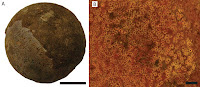
The eggs were all crushed, but are interpreted to have been about 18 mm in height and 11 mm in maximum diameter, giving them a volume of about 1.15 cm3. The shells of the eggs appear to comprise a single layer of calcitic material overlaying an inner layer of fibrous material. The surface ornamentation of the eggs is nodular, with two distinct sizes of nodules, one tall and one smaller. Beneath these nodes are funnel-shaped canals, with their tips opening at the tips of the nodes and with wider depressions on the inner surface; these are interpreted as pore-canals (which would have allowed the living egg to breath and regulate moisture). The calcite layer is comprised of large crystals arranged in a columnar manner, arranged in a fan-shaped pattern around the pores/nodes.

Eggshell morphology and microstructure of the eggs from Phu Phok. (A) 3D rendering of a portion of the surface of the eggshell of SK1-2 showing the distribution of nodes. (B) Tomogram of SK1-1 showing two eggshell fragments that slid in the egg, outer surfaces oriented to the top of the figure. The inner half of both shell fragments is displayed in darker shades of grey indicating the shell is less dense than the whiter outer half. The funnel-shaped depression (d) do not seem to be obstructed. The pore canals (p) are highlighted by the edge interference resulting from the phase contrast effect (black and white fringes). (C-D) SEM photographs of an eggshell fragment showing the fan-shaped pattern of crystal at the level of a surface node (n). Note the fibrous layer (f) underlining the eggshell. (D) Close up from (C). Scale bars in (A, B) are 500 μm. Fernandez et al. (2015).

Computerised tomography enabled the reconstruction of two embryos of slightly different ages, both apparently the same species of long-snouted, large-braincased Lizard. A number of features of the skulls and teeth of these specimens suggest that they are members of the Platynota, the group of Anguimorph Lizards that includes the modern Monitor and Bearded Lizards as well as the extinct Mosasaurs, and definitely not to the Gekkota. This is the first time that a calcified egg has been shown to have been to have been produced by a non-Gekkotan Lizard.

Skull and mandible of the anguimorph embryos from Phu Phok. (A,B) skull, dorsal (A) and lateral (B) views. (C) left mandible, lateral view. Colours: yellow, SK1-1; green, SK1-2; red, absent or incomplete bone replaced by symmetrical reconstruction. Anatomical abbreviations: a, angular; ar, articular; c, coronoid; d, dentary; e, epipterygoid; ec, ectopterygoid; end, calcified endolymph; eo, exoccipital; f, frontal; j, jugal; m, maxilla; mf, mental foramen; op, opisthotic; p, parietal; pbs, parabasisphenoid; pf, postfrontal; pl, palatine; po, postorbital; pr, prootic; prf, prefrontal; pt, pterygoid; q, quadrate; s, stapes; sa, surangular; sm, septomaxilla; soc, supraoccipital; sq, squamosal; v, vomer. Scale bars are 1 mm. Fernandez et al. (2015).

See also…

Casquehead Lizards, Corytophanidae, are a group of Iguanid Lizards found from the tropical forests of southern Mexico, through Central America to the northwest of South America. They belong to a group of Iguanians, the Pleurodonta, found today...


In the 1980s a large collection of Avian eggs were uncovered at the campus of theNational University of Comahue at Neuquén City in Argentinean Patagonia. These ages were located on a single...


The Loma del Pterodaustro lake deposits of Central Argentina have produced large numbers of the Pterosaur Pterodaustro guinazui, which is interpreted...


Follow Sciency Thoughts on Facebook.

Saturday, 11 October 2014

Mammal remains from the Early Eocene of Seymour Island, Antarctica.


Modern Antarctica lacks any permanent Mammal fauna, being visited by only a few marine Mammals for breeding purposes. However this has not always been the case. During the Eocene the last parts of the ancient continent of Gondwana were still breaking apart, and the Antarctic Peninsula was still attached to South America. This area appears to have had a far more agreeable climate than it does now, and a variety of Mammal fossils have been found here, all showing close affinities to those found in South America.

In a paper published in the journal Palaeontology on 16 July 2014, a team of scientists led by Javier Gelfo of the División Paleontología de Vertebrados at the Museo de La Plata, CONICET (the National Scientific and Technical Research Council of Argentina) and the Cátedra Paleontología Vertebrados at the Facultad de Ciencias Naturales y Museo at the Universidad Nacional de LaPlata describe two isolated Mammalian fossils from a shell bed at the base of the Acantilados II Allomember of La Meseta Formation on Seymour Island, off the east coast of the Antarctic Peninsula. This formation is thought to be bed is thought to be 55.3 million years old, making it Earliest Eocene, and the oldest site to have yielded fossil Mammal remains on Antarctica.

Geographical and stratigraphical provenance of the Seymour Island Mammals. Gelfo et al. (2014).

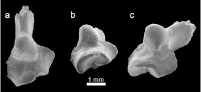
The first specimen described is a fragment of tooth which Gelfo et al. ascribe to an unknown Sparnotheriodont Liptotern. The Liptoterns were a group of herbivorous Placental Mammals indigenous to South America, which appeared in the Palaeocene and persisted until the closure of the Panama Seaway, when they rapidly went extinct, apparently unable to cope with competition from invading Horses, Tapirs and Deer and predation by invading Placental Carnivores. The Sparnotheriodonts were medium to large members of this group, which are known from the Palaeocene and Eocene of Brazil, Mendoza Province in Argentina, Patagonia and Antarctica. Gelfo et al. note that the tooth very closely resembles that of Notiolofos arquinotiensis, although this species is recorded only from a single specimen on James Ross Island, dated to 37.8 million years old, making a 17.5 million year gap between the two specimens; an improbable period of time for a Mammal species to persist. They suggest instead that the tooth may have come from a closely related species, potentially in the same genus.

Notiolofos cf. Notiolofos arquinotiensis. Occlusal view of the talonid fragment of the lower right molar, andreconstruction of the complete tooth based on the Ross Island Specimen adjusted to fit with the fragmentary specimen. Scale bar represents10 mm. Abbreviations: c, crest; co, cristidobliqua; ect, ectoflexid; hyp, hypoconid; hyptid, hypocristid; me, metaconid; tb, talonid basin. Top and left of the figure, mesial and lingual side, respectively. Gelfo et al. (2014).

The second specimen described is a phalanx from an unknown Mammal. This is well preserved and 20.45 mm wide (quite big). While it is not generally thought possible to identify Mammal species from isolated phalanxes, Gelfo et al. suggest that this is most likely from a large Placental Mammal with South American affinities such as a Xenarthran or Liptotern; while Marsupial Mammals are more common than Placentals in Antarctic faunal assemblages, the largest phalanx recorded from an Antarctic Marsupial is only 5.58 mm wide, making it unlikely that this bone had a Marsupial origin.

The Seymour Island Phalanx, in (A) plantar; (B) dorsal; (C) distal; (D) proximal views. Scale bar represents 10 mm. Abbreviations. app, articular surfaces for proximal phalanges; con, condyle; pe, Processusextensorius; pf, proximal foramina; pp, plantal projections. Gelfo et al. (2014).

See also…

South America was effectively an island continent for much of the Cenozoic, connected only to Antarctica in the south. As such it was not colonised by carnivorous Placental Mammals until the closure of the Panama...

http://sciencythoughts.blogspot.co.uk/2013/05/a-south-american-marsupial-from-early.html A 'South American' Marsupial from the early Eocene of Australia.                                Modern Australian Marsupials are generally held to be a distinct evolutionary lineage, distinct from South...

http://sciencythoughts.blogspot.co.uk/2012/07/new-caviomorph-rodents-from-early.html New Caviomorph Rodents from the Early Oligocene Tinguiririca Fauna of the Andean Main Range of central Chile                                      All South American Rodents are classified as members of a single monophyletic group, the Caviomorpha. This appears to be most closely related to Rodents with African and...
Follow Sciency Thoughts on Facebook.

Interpreting the relationship between Ants and Termites from a single piece of Mexican Amber.


Ants and Termites are eusocial Insects that have dominated many tropical ecosystems since at least the Early Cretaceous. Relationships between the two groups (which are not closely related) are complex, with some species able to tolerate one-another and even share nests, while others are deeply hostile, typically with Ants feeding on Termites or Termites fighting to keep all Ants away from their territories. However, while these relationships have been extensively studied in modern representatives of the group, understanding how these Insects may have interacted in the remote past is much more difficult.

In a paper published in the journal PLoS One on 20 August 2014, David Coty of the Muséum National d’Histoire Naturelle in Paris, Cédric Aria of the Department of Natural History-Palaeobiology at the Royal Ontario Museum and the Department of Ecology & Evolutionary Biology at the University of Toronto, and RomainGarrouste, Patricia Wils, Frédéric Legendre and André Nel, also of the of the Muséum National d’Histoire Naturelle in Paris, describe a single piece of amber from the Salt River Amber Mine in Chiapas State, Mexico, which includes several different Ants and Termites, and discuss the implications of this for relationships between the groups.

General configuration of the Mexican amber. (A) Overview of the amber piece, under optical microscope. Scale bar is 3 mm. (B) Three dimensional replica of the same; colours define taxonomic groups, viz. purple for Azteca Ants, blue for Nasutitermes Termites, red for Neivamyrmex Ant, green for small Psocodea. Labels: Az1, Azteca Ant nearest to predation scene; Az2 and Az3, two other Azteca Ants, both trapped in a flow distinct to that of the others inclusions and whose physical density matches that of the Nasutitermes soldier; Na1, Nasutitermes worker trapped between the Neivamyrmex mandibles; Na2, isolated Nasutitermes termite closest to predation scene; Na3, Nasutitermes worker with damaged gaster; Na4, Nasutitermes soldier; Ne, Neivamyrmex Ant; Ps, Psocodea; Scale bar is 3 mm. Aria et al. (2014).

The Amber contains four Termites assigned to a single (unknown) species of the genus Nasutitermes, three workers and a soldier, as well as three Ants of the genus Azteca and one Neivamyrmex Army Ant.

Neivamyrmex, like all Army Ants, are voracious predators, attacking invertebrate and invertebrate prey alike, typically with a sudden attack by a large number of Army Ants overwhelming the prey. Other Ants and Termites are favoured prey for such Insects, and nests will always be attacked when encountered. The Neivamyrmex Ant in the amber has a small worker Termite in its jaws, which suggests that it was engaged in such an attack when it became stuck in, and overwhelmed by, the plant resin which later became preserved as amber. Potentially the Termite could have been seized reflexively by the trapped, dying Ant, or the Ant could have become trapper while trying to scavenge a Termite previously caught in the resin, but one of the other worker Termites shows signs of damage typical of an Army Ant attack suggesting that the two species were fighting before becoming overwhelmed. The presence of a soldier Termite also supports this hypothesis, as in modern members of the genus Nasutitermes, members of this caste remain inside the nest for most of the time, only emerging when the nest is threatened or attacked, and never moving far from the nest.

Details of the Termites. (A) General side view of the NeivamyrmexAnt holding a Nasutitermes Termite (Na1) between its mandibles, under optical microscope, scale bar is 1 mm. (B) Detail of damaged gaster of Nasutitermes worker (Na3) closely contiguous to a Nasutitermes soldier (Na4), scale bar is 1 mm. (C) Side view of closely contiguous Nasutitermes soldier (Na4) and worker (Na3), black arrow: digestive tube of Nasutitermes worker scale bar is 1 mm.Aria et al. (2014).

The situation with the AztecaAnts is more complex. Some modern members of this genus are able to co-occupy the nests of some species of Nasutitermes, on either a seasonal or permanent basis. This is not an entirely benign relationship, as the Ants do consume some of the Termites, but the benefits to the Termites appear to outweigh the costs of removing the Ants; the Termites consume dead Ants, which are a rich source of nitrogen, although they do not actively predate the Ants, and the two species appear to cooperate when defending the nest against mutual threats, such as Army Ants. Aria et al. suggest that the presence of Azteca Ants alongside Nasutitermes Termites that were apparently engaged in defending their nest against an Army Ant attack strongly suggests that such a relationship was occurring in this instance, and that the Ants and Termites were engaged in a defence of a mutual nest against the invading Army Ants.

The precise age of amber from the Salt River Mine has yet to be determined, however it is thought to be at least Middle Miocene in age, suggesting that the mutualistic relationship between Azteca Ants and Nasutitermes Termites is at least that old.

See also…

Leafcutter Ants harvest vegetation from the tropical rainforests of South and Central America, which they then carry back to their nests and use as feed in fungal farms. Each species of Ant has its own unique...


Thief Ants of the genus Solenopsis are one of the most numerous and widespread Ant groups, particularly in the tropics. However they are not greatly studied or understood, despite the fact that some species are...


Ants are among the most widespread and abundant of Insect groups, with over 13 000 described species. They play a major role in the shaping of modern ecosystems and landscapes, and many other species of animals, plants and even fungi have commensal...


Follow Sciency Thoughts on Facebook.